Abstract
Background. Distal femoral cartilage is the most commonly deteriorated articular cartilage during metabolic and inflammatory processes. Musculoskeletal ultrasound (MSUS) is a widely accessible imaging modality to assess distal femoral cartilage thickness (DFCT). This study aims to explore DFCT and associated factors in healthy adolescents.
Methods. Healthy adolescents aged between 12-18 years were eligible for the study. The central points of the medial and lateral femoral condyles and the intercondylar area were measured on bilateral knees by using B-mode MSUS according to a predefined scanning protocol. The average of DFCT measurements was evaluated according to age, sex, anthropometric measurements, exercise habits, and vitamin D levels.
Results. A total of 150 adolescents participated in the study, with a mean age of 15 year; 67% were female. Age and sex were two factors showing significant effects on mean DFCT (p≤0.001). A negative mild correlation was observed between age and mean DFCT (r=-0.252, p=0.002). Weight, height, body mass index, and exercise frequency were not related to DFCT. Participants with severe vitamin D deficiency had similar DFCT when compared with others, and no correlation was observed between their levels (r=0.109, p=0.191).
Conclusion. DFCT varies during adolescence, with age and sex identified as the primary associated factors. Anthropometric measurements, exercise frequency, and vitamin D levels did not show any effect on DFCT in healthy adolescents.
Keywords: knee, cartilage, adolescence, musculoskeletal ultrasound, vitamin
Introduction
Articular cartilage is important for easy and compact movement of diarthrodial joints, ensuring optimal joint function and mobility. Various factors, including chronic diseases, mechanical overload, metabolic syndrome, aging, and genetic factors can significantly impact the structure, quantity, and quality of cartilage.1 Notably, distal femoral cartilage (DFC) is one of the most commonly deteriorated articular cartilages during metabolic and inflammatory processes.2
Musculoskeletal ultrasound (MSUS) is increasingly used in imaging of joint structures in the pediatric population with rheumatic diseases.3 Recent efforts have been made on normative data of the MSUS findings of joints in children and adolescents.4,5 Most of these studies were mainly focused on cartilage thickness.6,7 Yet, B-mode MSUS is an excellent, reliable, and feasible tool with a high level of agreement with magnetic resonance imaging to assess the DFC thickness (DFCT).8,9 The studies in adults without chronic conditions reported that sex, height, vitamin D level, and professional sports are contributors to DFCT.10-12 Despite the increased accessibility in the use of MSUS in the pediatric population, there remains a paucity of studies investigating DFCT and the associated factors to DFCT.
This study aims to explore DFCT according to age and sex and to examine its relationship with anthropometric measurements, exercise habits, and vitamin D levels during adolescence.
Materials and Methods
Study population and design
Adolescents aged between 12-18 years were included in this cross-sectional study between January-April 2023 in Tekirdağ City Hospital in the city of Tekirdağ located on the Northwestern coastline of Turkiye. Demographic and clinical characteristics, exercise routines, and routine laboratory data were noted. The participants were grouped according to their ages as follows: group 1: ≥12 and <14, group 2: ≥14 and <16, and group 3: ≥16 years. Standard deviation scores (SDS) of the anthropometric measurements (weight, height, and body mass index [BMI]) were calculated according to the growth charts of Turkish children.13 Sex- and age-specific BMI ≥95th percentile defined obesity.14 Serum 25-hydroxy-vitamin D (25-OH vitamin D) levels were considered deficient if <20 ng/mL and severely deficient if <10 ng/mL. Health-related quality of life (HRQoL) was evaluated using the Pediatric Quality of Life Inventory Generic Core Scales (PedsQL-GC) with young adult self- and parent proxy-reports15, which have been translated and validated in the Turkish adolescent population.16 The scale consists of 23 items encompassing four domains: physical, emotional, social, and school functioning. Domain scores were calculated as the mean of the transformed item scores within each domain. The Health Summary Score (HSS) in PedsQL is a composite measure that reflects a child’s overall HRQoL by combining key functional domains. The Physical HSS was calculated by the physical functioning domain and the Psychosocial HSS by the emotional, social, and school functioning domains. Higher scores indicate better HRQoL.15 Adolescents were excluded if they had a chronic disease, a history of knee trauma in the preceding six months or knee surgery. Adolescents with abnormal laboratory tests suggesting chronic diseases (e.g. hyperthyroidism) and those involved in professional sports were also excluded. The participants were recruited after their routine well-child visit for an ultrasonographic examination.
Ultrasonographic examination
A pediatric rheumatologist (POAA) and a physiatrist (EGK), blinded to the clinical/laboratory findings, measured DFCT by MSUS within 10 days of the clinical visit of the participants. Both clinicians had at least three years of experience in MSUS. A senior physiatrist (LO) technically controlled MSUS images and measurements.
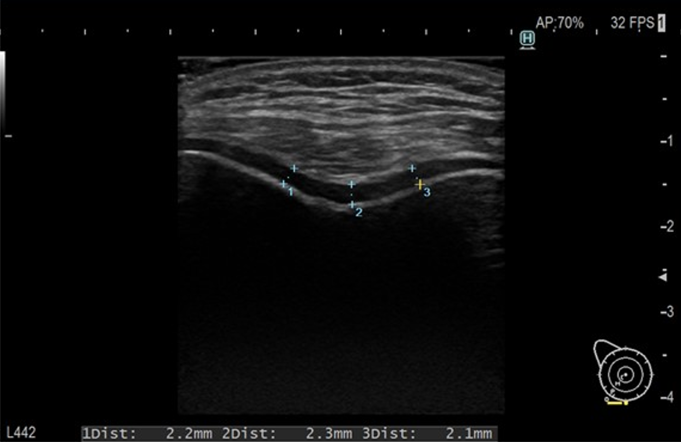
During the measurements, the participants lay supine with their knees maximally flexed and the probe positioned just superior to the upper border of the patella in the transverse plane. The greatest DFCT (hyaline/articular cartilage) perpendicular to the bony surface was visualized.17,18 The central points of the medial and lateral femoral condyles (right lateral condyle: RLC, left lateral condyle: LLC, right medial condyle: RMC, left medial condyle: LMC), and the intercondylar area (RICA and LICA) were measured bilaterally (Fig. 1). At least three consecutive measurements of each were taken and the average was noted. Likewise, the average of the measured RLC, LLC, RMC, LMC, RICA, and LICA was then defined as ‘mean DFCT’. Intra- and inter-reader reliability of both physicians among the measurements taken from the same area on the same day were also assessed. All measurements were performed using either a 5-12 MHz linear transducer (Siemens Acuson S3000) or a 12-2 MHz linear transducer (Hitachi Arietta 65).
The study complied with the Declaration of Helsinki and it was approved by the local Human Research Ethics Committee. Written parental permission and assent were obtained from all participants.
Statistical analysis
Descriptive statistics were defined by the frequency (n) and percentage (%) for categorical and mean ± standard deviation (SD) or median with interquartile range (IQR; 25th-75th percentile) for continuous variables. The Shapiro-Wilk test with distribution graphs was performed to demonstrate the normality of the data distribution. Categorical variables between groups were compared by the chi-square test while the Mann-Whitney U test was used for continuous variables. The effects of age group and sex on femoral cartilage thickness were evaluated using a two-way analysis of variance (two-way ANOVA) for each cartilage measurement (mean DFCT, RLC, RICA, RMC, LLC, LICA, and LMC). Age group (three levels: ≥12-14, ≥14-16, and ≥16-18 years) and sex (male, female) were entered as fixed factors, and each cartilage thickness parameter was analyzed as a separate dependent variable. For each model, main effects of age group and sex, as well as the interaction effect (age*sex), were examined. Assumptions of normality and homogeneity of variances across groups was assessed using the Shapiro-Wilk and Levene’s test to ensure the validity of the model. Correlations between clinical, laboratory, and ultrasonographic measurements were evaluated by Pearson coefficients (r), whereby r ≥0.2 was considered significant, 0.4 - 0.6 as moderate, and r ≥0.6 as strong. Intra- and inter-reader reliability of two physicians was determined by intraclass correlation coefficient (ICC) with the 95% confidence interval (CI) and ICC ≥0.75 was considered an excellent level of reliability. All data were analyzed using SPSS version 21.0.0 and statistical significance was set at p <0.05.
Results
A total of 150 adolescents between the ages of 12-18 years participated in the study. The characteristics of the participants are shown in Table I. DFCT measurements of the right and left knee were strongly correlated (r for RLC and LLC=0.862, p<0.001; r for RMC and LMC=0.861, p<0.001; r for RICA and LICA=0.854, p<0.001). Regarding MSUS measurements of the two physicians, the ICC for intra- and inter-reader reliability were 0.96 (0.95-0.97) and 0.86 (0.81-0.90), respectively.
| BMI: body mass index, HSS: Health Summary Score, IQR: interquartile range, PedsQL-GC: Pediatric Quality of Life Inventory-Generic Core, SD: standard deviation, SDS: standard deviation score. | |
| Table I. Characteristics of the study population (N=150). | |
| Age, years, mean ±SD |
|
| Age groups, n (%) | |
| Group 1: ≥12-14 |
|
| Group 2: ≥14-16 |
|
| Group 3: ≥16-18 |
|
| Female sex, n (%) |
|
| Weight, SDS, mean ±SD |
|
| Height, SDS, mean ±SD |
|
| BMI, SDS, mean ±SD |
|
| Obesity, n (%) |
|
| 25-OH vitamin D level, ng/mL, mean ±SD |
|
| 25-OH vitamin D status, n (%) | |
| Deficiency group: <20 |
|
| Severe deficiency group: <10 |
|
| Regular exercise, n (%) |
|
| PedsQL-GC, median (IQR) | |
| Physical HSS, self-report |
|
| Physical HSS, parent proxy-report |
|
| Psychosocial HSS, self-report |
|
| Psychosocial HSS, parent proxy-report |
|
Comparison of the DFCT according to the age groups and sex is given in Table II. Two-way ANOVA revealed significant main effects of age and sex on mean DFCT (p≤0.001 for age and sex). Overall, DFCT decreased with increasing age and was consistently thicker in males compared with females. The age*sex interaction was significant for mean DFCT (p=0.040), indicating that the age-related decrease in cartilage thickness differed between males and females. There was a negative mild correlation between age and mean DFCT (Table III). Weight, height, and BMI were not significantly correlated with DFCT measurements. Serum 25-OH vitamin D levels, weight, height, BMI, exercise habits, and HRQoL did not differ between the age groups (all p>0.05).
|
*Two-way ANOVA; values are presented as mean ± SD (mm). DFCT: distal femoral cartilage thickness, LICA: left intercondylar area, LLC: left lateral condyle, LMC: left medial condyle, RICA: right intercondylar area, RLC: right lateral condyle, RMC: right medial condyle, SD: standard deviation. |
|||||||
| Table II. Comparison of distal femoral cartilage thickness measurements according to age groups and sex.* | |||||||
|
|
|
|
|
|
|
|
|
| Mean DFCT |
|
|
|
|
|
|
|
| RLC |
|
|
|
|
|
|
|
| RICA |
|
|
|
|
|
|
|
| RMC |
|
|
|
|
|
|
|
| LLC |
|
|
|
|
|
|
|
| LICA |
|
|
|
|
|
|
|
| LMC |
|
|
|
|
|
|
|
|
*Pearson correlation coefficients [r (p-value)]; mild: 0.2–0.39, moderate: 0.4–0.59, strong: ≥ 0.6; p-value <0.05 BMI: body mass index, DFCT: distal femoral cartilage thickness, SDS: standard deviation score. |
|||
| Table III. Correlation of distal femoral cartilage thickness with age and anthropometric measurements according to sex.* | |||
|
|
|||
|
|
|
|
|
| Age, years |
|
|
|
| Weight, SDS |
|
|
|
| Height, SDS |
|
|
|
| BMI, SDS |
|
|
|
| 25-OH vitamin D, ng/mL |
|
|
|
The participants with obesity had similar mean DFCT as those with normal weight (2.16 mm, IQR 1.97-2.43 vs. 2.04 mm, IQR 1.80-2.32, p=0.074). The mean DFCT of the participants doing exercise regularly did not differ from the others (2.10 mm, IQR 2.03-2.28 vs. 2.07 mm, IQR 1.87-2.37, p=0.673). The participants with severe vitamin D deficiency demonstrated similar mean DFCT when compared with others (1.96 mm, IQR 1.77-2.37 vs. 2.10 mm, IQR 1.88-2.34, p=0.111) and there were no correlations between vitamin D levels and mean DFCT (r=0.109, p=0.191).
Discussion
This cross-sectional study demonstrated that age and sex were the major associated factors of DFCT in healthy adolescents. On the other hand, anthropometric measurements, exercise, and vitamin D levels did not show any relation with DFCT during adolescence.
Cartilage thickness has been reported to be thicker during early childhood, decreasing till the ages of 13-15 years, and almost stabilizing thereafter.4,19 In line with this finding reported by a few studies available in the literature, we found that DFC was thicker in participants below 14 years of age. Furthermore, we showed that an inverse correlation was present between age and DFCT during adolescence. The second significant factor affecting DFCT was found to be sex in our study, i.e., males had significantly thicker DFC than females. The observed interaction patterns suggest that the decrease in DFCT with age during adolescence does not progress uniformly between sexes. This result is not only consistent with postpubertal and adult reports but also with those of prepubertal children, which suggests factors other than sex hormones affect cartilage thickness.5,7,11 Of note, the reason for different cartilage volumes between males and females even in the prepubertal period remains unexplained in the current literature.
A recent study reported that height, weight, and BMI were not effectors of cartilage thickness in school-aged children.7 Another study found that thinner cartilage was observed with increasing height and weight in healthy children. Notably, height was suggested to be the best predictor of cartilage thickness with age.5 Moreover, a longitudinal study demonstrated a significant positive correlation between the cartilage volume accrual with changes in height but not with weight. Indeed, children who were overweight had similar cartilage volume as children with normal weight.20 We found no relationship between DFCT and height, weight, and BMI. Also, obesity appeared not to affect DFCT. Although obesity is a well-known risk factor for osteoarthritis, it seems that its effect on cartilage becomes apparent in older ages.1 Lastly, we did not observe any impact of regular exercise on DFCT. However, the number of children doing regular exercise was limited in our study and we excluded those performing professional sports to homogenize our sample. A study in healthy young university students showed that cartilage thickness was higher in sportsmen and there was a direct relationship between the muscle percentage and cartilage thickness.21 Moreover, children undertaking more vigorous sports showed higher amounts of cartilage accrual.20
Vitamin D is an essential mediator in skeletal health and there is a very well-known association between vitamin D deficiency and rickets, osteomalacia, and osteoporosis, particularly in newborns, the elderly, and high-risk patient populations. Besides, diverse studies suggest that vitamin D has many extraskeletal functions and that its deficiency is related to several other diseases.22 Despite its prevalent deficiency worldwide, there is no consensus on the routine measurement of its level and vitamin D supplementation in healthy children.23 Strikingly, vitamin D deficiency was present in almost 90% of our study population and one-third had severe vitamin D deficiency despite living in a coastline city of Turkiye with high numbers of sunny days. We showed that the DFCT was not affected by severe vitamin D deficiency. Fortunately, its potential detrimental effects on cartilage are not clearly evident during adolescence; however, opposite reports in adults and the elderly delineate further attention to implementing guidelines on screening and supplementation of vitamin D deficiency in this critical period of life.10,24,25
This study has several limitations, including its cross-sectional design and single-center setting. It investigated only DFC, although it is one of the most commonly deteriorated articular cartilages that is easily measured. Other cartilages can also be evaluated in the future. Unfortunately, the majority of the study population had low vitamin D levels and they mostly demonstrated a sedentary lifestyle. Other environmental and hormonal factors that might affect cartilage thickness might have been included. Further, it would be interesting to have follow-up visits with longitudinal ultrasonographic evaluations of cartilage thickness. Also, the structural features that can be observed during MSUS, such as surface irregularity or echogenicity, might be taken into consideration in future studies. Advanced methods for functional and biomechanical assessment can be used to support our findings. On the other hand, the use of a standardized approach with excellent reliability in MSUS examination represents an important strength of our study.
In conclusion, DFCT shows variations during adolescence, whereby age and sex are the main associated factors, while weight, height, BMI, and exercise seem to have no effects. Vitamin D deficiency does not seem to affect DFCT in adolescents; however, given its negative effect on cartilage health in adults, longitudinal data are needed.
Ethical approval
The study was approved by the Clinical Research Ethics Committee of Tekirdag City Hospital (date: 25.11.2022, number: TSH-2023-017).
Source of funding
The authors declare the study received no funding.
Conflict of interest
The authors declare that there is no conflict of interest.
References
- Rose BJ, Kooyman DL. A tale of two joints: the role of matrix metalloproteases in cartilage biology. Dis Markers 2016; 2016: 4895050. https://doi.org/10.1155/2016/4895050
- Martel-Pelletier J, Boileau C, Pelletier JP, Roughley PJ. Cartilage in normal and osteoarthritis conditions. Best Pract Res Clin Rheumatol 2008; 22: 351-384. https://doi.org/10.1016/j.berh.2008.02.001
- Vega-Fernandez P, Ting TV, Pratt L, Bacha CM, Oberle EJ. Ultrasonography in pediatric rheumatology. Rheum Dis Clin North Am 2022; 48: 217-231. https://doi.org/10.1016/j.rdc.2021.09.009
- Windschall D, Trauzeddel R, Haller M, et al. Pediatric musculoskeletal ultrasound: age- and sex-related normal B-mode findings of the knee. Rheumatol Int 2016; 36: 1569-1577. https://doi.org/10.1055/s-0036-1587723
- Wittoek R, Decock C, Dewaele N, et al. Structural ultrasound of joints and tendons in healthy children: development of normative data. Pediatr Rheumatol Online J 2023; 21: 105. https://doi.org/10.1186/s12969-023-00895-8
- Samanta M, Mitra S, Samui PP, Mondal RK, Hazra A, Sabui TK. Evaluation of joint cartilage thickness in healthy children by ultrasound: an experience from a developing nation. Int J Rheum Dis 2018; 21: 2089-2094. https://doi.org/10.1111/1756-185X.13374
- Gau CC, Yao TC, Gan ST, et al. Age, gender, height and weight in relation to joint cartilage thickness among school-aged children from ultrasonographic measurement. Pediatr Rheumatol Online J 2021; 19: 71. https://doi.org/10.1186/s12969-021-00554-w
- Spannow AH, Stenboeg E, Pfeiffer-Jensen M, et al. Ultrasound and MRI measurements of joint cartilage in healthy children: a validation study. Ultraschall Med 2011; 32(Suppl 1): 110-116. https://doi.org/10.1055/s-0029-1245374
- Pradsgaard DØ, Fiirgaard B, Spannow AH, Heuck C, Herlin T. Cartilage thickness of the knee joint in juvenile idiopathic arthritis: comparative assessment by ultrasonography and magnetic resonance imaging. J Rheumatol 2015; 42: 534-540. https://doi.org/10.3899/jrheum.140162
- Malas FU, Kara M, Aktekin L, Ersöz M, Ozçakar L. Does vitamin D affect femoral cartilage thickness? An ultrasonographic study. Clin Rheumatol 2014; 33: 1331-1334. https://doi.org/10.1007/s10067-013-2432-y
- Bedewi MA, Elsifey AA, Naguib MF, et al. Sonographic assessment of femoral cartilage thickness in healthy adults. J Int Med Res 2020; 48: 300060520948754. https://doi.org/10.1177/0300060520948754
- Azami P, Ashraf A, Yousefi O, Hosseinpour A, Nasiri A. Impact of treadmill running on distal femoral cartilage thickness: a cross-sectional study of professional athletes and healthy controls. BMC Sports Sci Med Rehabil 2024; 16: 104. https://doi.org/10.1186/s13102-024-00896-4
- Neyzi O, Günöz H, Furman A, et al. Weight, height, head circumference and body mass index references for Turkish children. Çocuk Sağlığı ve Hastalıkları Dergisi 2008; 51: 1-14.
- Centers for Disease Control and Prevention. Defining childhood obesity. Available at: https://web.archive.org/web/20200714053456/https:// http://www.cdc.gov/obesity/childhood/defining.html
- Varni JW, Burwinkle TM, Seid M. The PedsQL 4.0 as a school population health measure: feasibility, reliability, and validity. Qual Life Res 2006; 15: 203-215. https://doi.org/10.1007/s11136-005-1388-z
- Cakin Memik N, Ağaoğlu B, Coşkun A, Uneri OS, Karakaya I. The validity and reliability of the Turkish Pediatric Quality of Life Inventory for children 13-18 years old. Turk Psikiyatri Derg 2007; 18: 353-363.
- Özçakar L, Kara M, Chang KV., et al. EURO-MUSCULUS/USPRM basic scanning protocols for knee. Eur J Phys Rehabil Med 2015; 51: 641-646.
- Pirri C, Stecco C, Güvener O, et al. EURO-MUSCULUS/USPRM dynamic ultrasound protocols for knee. Am J Phys Med Rehabil 2023; 102: e67-e72. https://doi.org/10.1097/PHM.0000000000002173
- Sidharthan S, Yau A, Almeida BA, et al. Patterns of articular cartilage thickness in pediatric and adolescent knees: a magnetic resonance imaging-based study. Arthrosc Sport Med Rehabil 2021; 3: 381-390. https://doi.org/10.1016/j.asmr.2020.09.029
- Jones G, Ding C, Glisson M, Hynes K, Ma D, Cicuttini F. Knee articular cartilage development in children: a longitudinal study of the effect of sex, growth, body composition, and physical activity. Pediatr Res 2003; 54: 230-236. https://doi.org/10.1203/01.PDR.0000072781.93856.E6
- Herrera H GA, Llinás PJ, Flórez L, et al. Ultrasound measurement of femoral cartilage thickness in the knee of healthy young university students. Rev Esp Cir Ortop Traumatol 2020; 64: 244-250. https://doi.org/10.1016/j.recot.2020.04.001
- Bouillon R, Marcocci C, Carmeliet G, et al. Skeletal and extraskeletal actions of vitamin d: current evidence and outstanding questions. Endocr Rev 2019; 40: 1109-1151. https://doi.org/10.1210/er.2018-00126
- Santos HO, Martins CEC, Forbes SC, Delpino FM. A scoping review of vitamin D for nonskeletal health: a framework for evidence-based clinical practice. Clin Ther 2023; 45: 127-150. https://doi.org/10.1016/j.clinthera.2023.03.016
- Joseph GB, McCulloch CE, Nevitt MC, et al. Associations between vitamins c and d intake and cartilage composition and knee joint morphology over 4 years: data from the osteoarthritis initiative. Arthritis Care Res (Hoboken) 2020; 72: 1239-1247. https://doi.org/10.1002/acr.24021
- Wang R, Wang ZM, Xiang SC, et al. Relationship between 25-hydroxy vitamin D and knee osteoarthritis: a systematic review and meta-analysis of randomized controlled trials. Front Med (Lausanne) 2023; 10: 1200592. https://doi.org/10.3389/fmed.2023.1200592
Copyright and license
Copyright © 2026 The Author(s). This is an open access article distributed under the Creative Commons Attribution License (CC BY), which permits unrestricted use, distribution, and reproduction in any medium or format, provided the original work is properly cited.