Abstract
Background. Brain abscesses (BA) are a medical emergency in all age groups and require early diagnosis and treatment. Childhood BA constitute 25% of all abscesses and may have a different clinical course than adults. This article aims to investigate patients followed up for adult and child BA over a 10-year period.
Materials and Methods. A retrospective analysis was performed on adult and pediatric patients diagnosed with BA. Demographic, clinical, predisposing factors, imaging, treatment modalities, and prognosis of the patients, and clinical differences were examined.
Results. Of a total of 46 patients; 54.3% of the patients were children and the mean age was 10.9±6.3 years for the pediatric group and 46.2±16.7 years for the adult group. Fever, vomiting, and seizures were significantly higher in children (p<0.05). When the differences between the groups were examined, headache was the most common finding in the adult group, while the most common finding in children was fever and vomiting. The classic triad of fever, headache, and focal neurological deficits was present in 12 (48%) of the children, compared to only 4 (19.1%) of the adults. Trauma history (21.7%), immunosuppression (19.5%), and local spread (mastoiditis, sinusitis, and otitis) (32.6%) were among the most common predisposing factors. Previous otorhinolaryngogenic infections were the most common cause in children (36%), while immunosuppression was the most common cause in adults (28.5%). The frontal lobe was most frequently involved among the patients (47.8%). BA was more commonly localized in the left hemisphere in both groups. Culture growth was detected in 23.9% of all operated patients, with a positivity rate of 20% in children and 28% in adults. The most common microorganism was Staphylococcus aureus (8.6%). The average hospitalization time was significantly longer in children than in adults (46 ± 18 days vs 34 ± 13 days; p=0.024). 26.1% of the patients recovered with sequelae, and the mortality rate was 8.6%.
Conclusion. Our findings suggest that a high index of suspicion should be maintained for BA in children, especially in the presence of fever, vomiting, and seizures. This study highlights significant clinical and etiological differences between pediatric and adult BA, underscoring the importance of age-specific diagnostic and management strategies. The differences observed in clinical presentation and underlying causes between the two groups warrant further investigation to improve outcomes.
Keywords: adult, brain abscess, child
Introduction
Although the prognosis of central nervous system infections and their complications has improved significantly with advanced imaging techniques and antibiotics, they continue to be a significant cause of mortality and morbidity nowadays.1,2 Brain abscesses (BA) are life-threatening intracranial infections characterized by localized suppuration in the intracranial parenchyma. They occur when pustular material is surrounded by a capsule and organized following a focus of cerebri.3,4 Its incidence is 8% of intracranial masses in developing countries and 1-2% in Western societies and can be seen in almost all ages.4 The mortality rate of BA is higher in the fourth decade of life.
Although BA predominantly affects adults, children comprise approximately one quarter of cases.5 Within the pediatric population, the peak incidence occurs between 4 and 7 years of age. It is particularly important because of the neurological sequelae it may cause in children.6,7 The most common causes of BA are; spread of infections from anatomically adjacent areas (otitis media, dental infections, mastoiditis, sinusitis), hematogenous spread (secondary to cyanotic congenital heart disease, lung abscess or empyema), direct inoculation after trauma or surgery. Between 4.6% and 43.4% of cases, no etiology can be found, these called cryptogenic.8,9
BA may not present clinical findings in the early stages, but may also present with nonspecific findings or a variable clinical course ranging from mild to severe. The most important factors determining the patient’s clinical picture are the patient’s age, stage, localization of the abscess, immune status and presence of meningitis.5,7,10 In the early stages of the abscess, the patient may be asymptomatic. The classical triad of BA; fever, headache, and focal neurological deficits, is seen in only 9-28% of children.5,10
The microorganisms responsible for BA are aerobic and anaerobic streptococci and staphylococci. Other agents are Bacteroides species, Proteus species, Haemophilus influenzae, Escherichia coli, Citrobacter, Nocardia, Aspergillus, Corynebacterium and Mycobacterium tuberculosis.11-14 The literature has shown that cultures are negative in 10-56% of cases.14
Treatment typically involves a combination of antibiotics for 6-8 weeks and, in most cases, surgical drainage. Antimicrobial therapy alone is recommended for small lesions (<2.5 cm), multiple abscesses, and deep-seated lesions.8,10,12,15,16 While several studies in the literature evaluate either pediatric or adult BA, there is a scarcity of comparative studies that investigate the distinct features of these two age groups. This retrospective study aims to address this gap by comparing pediatric and adult BA over a 10-year period, discussing risk factors, clinical courses, and outcomes in light of the available literature. This work seeks to provide valuable insights into the age-specific differences in BA, contributing to improved diagnostic and management strategies.
Materials and Methods
In this retrospective study, a total of 46 patients followed at the Pediatric Infection and Neurosurgery Clinics of Adana City Training and Research Hospital between 2012 and 2022 were investigated. All patients under the age of 18 were defined as the pediatric group (n=25), while patients aged 18 and older were included in the adult group (n=21).
BA was defined as a localized parenchymal lesion with perilesional brain edema and contrast enhancement on cranial imaging or the presence of purulent material in the cranial cavity on biopsy/surgery. Patients with subdural empyema, epidural abscesses, or secondary abscesses due to shunt infection were excluded from the study. Ten patients were excluded from the study: six with subdural empyema, three with epidural abscess, and one with a secondary abscess related to shunt infection.
Differences between pediatric and adult patients were evaluated with respect to demographic characteristics, presenting complaints, underlying risk factors, and outcomes. The indications for treatment modality were defined as follows: Patients with abscesses smaller than 2.5 cm, multiple lesions, or lesions in deep/critical locations received exclusive medical treatment (long-term intravenous antibiotics). All other patients (lesions >2.5 cm, accessible lesions) were considered for surgical drainage (stereotactic aspiration or excision) in addition to medical treatment. The hospital records were reviewed to obtain information on patient demographics, clinical presentation, laboratory findings, imaging results, treatment methods, and clinical outcomes. Surgical samples were immediately sent for aerobic and anaerobic culture and Gram staining. The hospital laboratory’s capacity for anaerobic culture was noted to be limited, which may have contributed to the overall low culture yield observed in the study.
Ethics approval
The study’s ethics committee approval was obtained by the Adana City Training and Research Hospital Clinical Research Ethics Committee (January 12, 2023, meeting number 120, decision number 2371).
Statistical analysis
Statistical analysis was performed using the Statistical Package for Social Sciences version 20 (IBM Corp., Armonk, NY, USA) software package. Descriptive statistics of numerical parametric variables were calculated as mean±standard deviation (SD); categorical variables were expressed as percentage (%). Chi-square test was used to compare categorical variables between groups, independent samples T-test was used to compare numerical variables between groups if assumptions were met, otherwise Mann-Whitney U test and analysis of variance (ANOVA) were used to compare more than one group. P value <0.05 was considered statistically significant.
Results
Of the 46 patients followed, 25 (54.3%) were children and 21 (45.7%) were adults. The mean age of the patients was 27± 20.6 years (median 18 years, min-max: 3 months - 74 years) (Table I). There was a male predominance in the study group, with 71.8% of the patients being male and a male-to-female ratio of 2.6:1.
| *Triad: Association of fever, headache and neurological dysfunction. CRP: C-reactive protein, SD: standard deviation, WBC: white blood cell |
|||
| Table I. Comparison of clinical, demographic, radiological, and prognostic characteristics of patients. | |||
| Characteristic |
|
|
|
| Demographic characteristics | |||
| Age, yrs, mean±SD (min-max) |
(3 months-18 years) |
(18-74 years) |
|
| Male gender, n (%) |
|
|
|
| Clinical symptoms and signs, n (%) | |||
| Fever |
|
|
|
| Vomiting |
|
|
|
| Feeding intolerance |
|
|
|
| Headache |
|
|
|
| Classic triad* |
|
|
|
| Seizure |
|
|
|
| Status epilepticus |
|
|
|
| Hydrocephalus |
|
|
|
| Shift |
|
|
|
| Hemiplegia |
|
|
|
| Altered state of consciousness |
|
|
|
| Radiological and laboratory findings | |||
| Frontal localization of abscess, n (%) |
|
|
|
| Located in left cerebral hemisphere, n (%) |
|
|
|
| Abscess size, cm, |
|
|
|
| WBC, /mm3, mean±SD |
|
|
|
| CRP, mg/dL, mean±SD |
|
|
|
| Outcomes | |||
| Length of hospital stay, days, mean±SD |
|
|
|
| Sequelae development, n (%) |
|
|
|
| Mortality, n (%) |
|
|
|
The most common complaints of the patients were headache (73.9%), vomiting (73.9%) and fever (54.3%). The triad of fever, headache and neurological dysfunction was present in 34.7% of the patients. Other reported conditions included feeding intolerance (54.3%), seizure (26.1%), confusion (26.1%), hydrocephalus (15.2%) and hemiplegia (15.2%). Increased intracranial pressure (ICP) findings were present in 7 patients (15.2%). When the two groups were compared, headache was the most common finding in the adult group, while vomiting was the most common finding in children. Vomiting history was present in 22 (88%) of the pediatric patients, while it was present in 12 (57.1%) of the adults (p=0.038). Fever history was present in 20 (80%) of the pediatric patients, while it was present in 5 (23.8%) of the adults, and the difference was statistically significant (p=0.001). The classic triad of fever, headache and neurological dysfunction was present in 12 (48%) of the children, compared to 4 (19.1%) of the adults. Although the triad appeared more frequent in children than adults, this difference was not statistically significant (p=0.065). Hydrocephalus was documented more often in children (24%) than adults (4.7%), and this difference was not statistically significant (p=0.119). Shift appeared more frequent in adults (38%) than in children (16%), and this difference was not statistically significant (p=0.059) (Table I). At the time of diagnosis, seizures were present in 10 (40%) of the children, while a history of seizures was present in 2 (9.5%) of the adults, and the difference was significant (p=0.029). Loss of consciousness was present in 36% of the children and 14.2% of the adults (Table I).
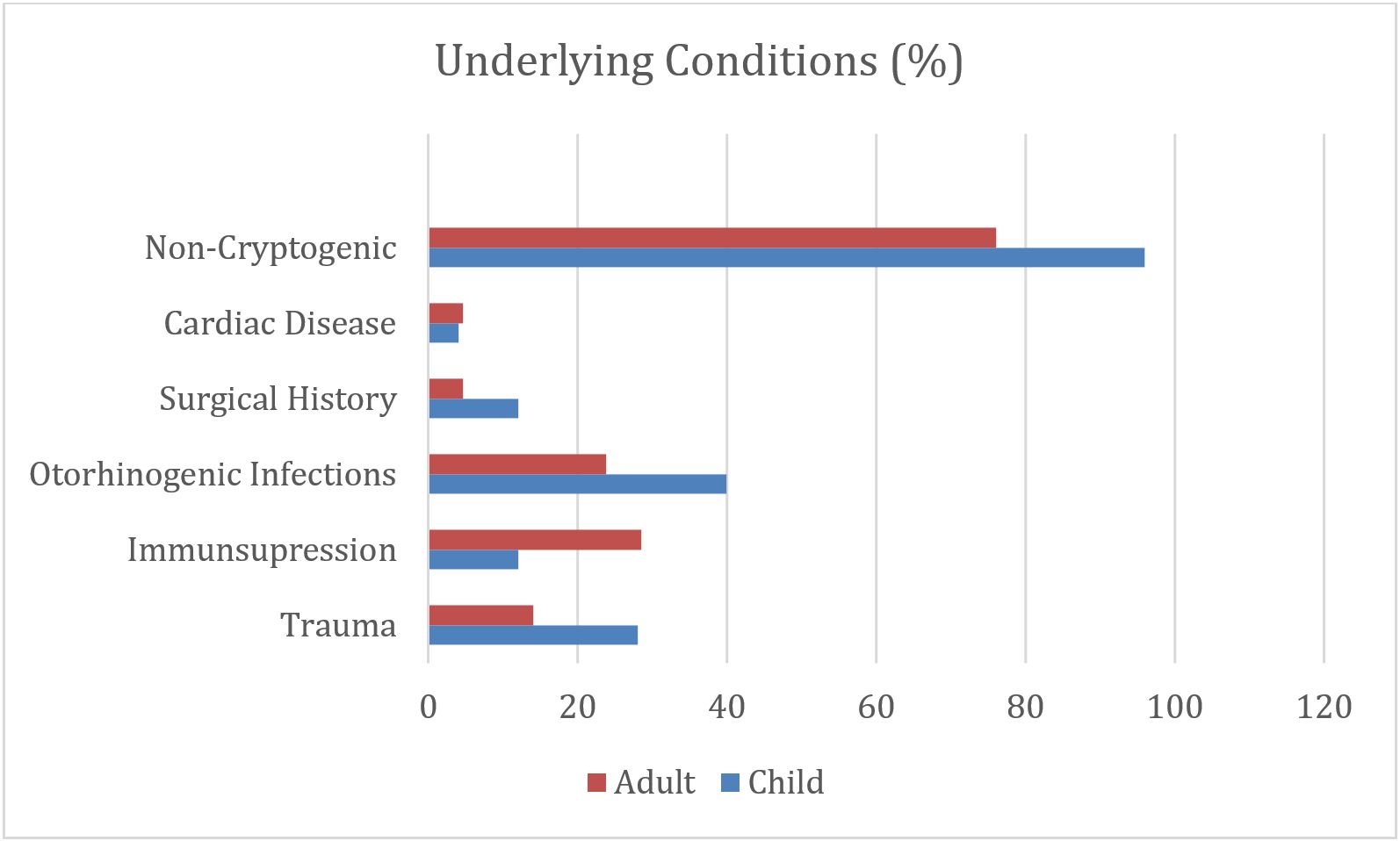
Regarding the underlying causes, trauma history was present in 10 (21.7%), immunosuppression (acute lymphoblastic leukemia [ALL], chronic granulomatous disease (CGD) in 9 (19.5%), local spread (mastoiditis, sinusitis and otitis history) in 15 (32.6%), previous surgery in 4 (8.6%) and congenital heart disease in 2 (4.3%) patients. Previous otorhinolaryngogenic infections were the most common cause in pediatric cases (36%), while immunosuppression was the most common cause in adult cases (28.5%). In total, no underlying cause could be detected in 6 (13%) patients. The underlying cause was detected in 24 (96%) of pediatric patients and 16 (76.1%) of adults (p=0.040) (Fig. 1).
Cranial computed tomography (CT) and magnetic resonance imaging (MRI) were the most commonly used imaging methods for diagnosis, with MRI being preferred more frequently in pediatric cases. The mean abscess size was similar in both groups (3.6 cm in children vs. 3.8 cm in adults), with no statistically significant difference (Table I). The frontal lobe was most frequently involved (n=22, 47.8%). BA was more commonly localized in the left hemisphere in both groups (47.8%), while abscesses were observed in both hemispheres in 23.9% of patients. There was no significant difference between the groups in terms of localization (Table I). Initial white blood cell (WBC) count and C-reactive protein (CRP) level were elevated in both children and adults, but no significant difference was observed between the groups (p=0.118, p=0.682 respectively).
Regarding surgical intervention, the timing of aspiration/drainage was generally performed within 24-48 hours of diagnosis for large or symptomatic lesions. No statistically significant difference in surgical timing was observed between the pediatric and adult groups (p > 0.05). Three of the patients (6.5%) received only medical treatment, while the others underwent surgical drainage in addition to medical treatment. The duration of medical treatment was 6-8 weeks. The most frequently initiated empirical treatment was ceftriaxone, vancomycin, and metronidazole (95%). Antibiotic therapy was adjusted according to the results of patients with positive cultures. Corticosteroid (dexamethasone) treatment was given to patients with shifts or significant cerebral edema (26.1%). Electrolyte imbalance developed in 10 of the patients (21.7%), 6 of whom were children, and one of these children also developed renal failure (2.1%). Culture growth was detected in 23.9% of all patients who underwent surgery, with a positivity rate of 20% in children and 28% in adults. The most common microorganism was Staphylococcus aureus (8.6%).
12 of the patients (26.1%) recovered with sequelae, 7 of whom were children. The most common sequelae were hemiparesis, epilepsy and hearing loss. The mean hospitalization day of the patients was 40.6 days. The hospitalization duration was longer in children than in adults (p=0.024) (Table I). Four of the patients (8.6%) died, the underlying diseases were immunosuppression (ALL), meningitis and intracranial hemorrhage in children and trauma in adults.
Discussion
BA continues to be an important cause of mortality and morbidity worldwide, especially in developing countries, despite advanced diagnostic methods and modern treatments. In this study, we found a male/female ratio of 2.6/1, which aligns with the general male dominance reported in the literature. While female dominance was noted in a pediatric BA series from Türkiye, another study reported male dominance, highlighting regional variations.5,17 In our study, 25 cases were younger than 18 years. We observed that 69.4% of all cases were under 40 years of age. According to a meta analysis report, the average age was reported as 34 years.18
In children, especially at young ages, the presenting complaints are usually nonspecific. In studies, headache and fever were found to be the most common presenting complaints.14 Although seizure is a less common finding, Lee et al. observed seizure in 48% of their patients in their study.19 Again, Atiq et al. observed seizures in 45% of patients.20 In our study, headache, neurologic dysfunction and fever were present in 34.7% of all cases and 48% of children. Fever and vomiting were the most common symptoms in children. Mental status changes and seizures were present in 26.1% of all cases, while seizures were more common in pediatric cases compared to adults and were statistically significant (p=0.029).
The underlying etiology is known in many cases. Cryptogenic abscesses have been reported in 4.6-43.4% of cases.5 Identifiable causes were present in 86.9% of our cases. While previous otorhinogenic infections were the most common cause in pediatric cases, immunosuppression was found to be the most common cause in adult cases. Several predisposing factors, including immunosuppression, underlying disease, and contiguous spread from adjacent structures, have been implicated in the development of BA. In developing countries, BA most commonly develops secondary to otitis media. Sharma et al. found neighboring infections such as sinusitis, otitis or meningitis in 42.5% of patients followed up for BA.21 Nathoo et al., on the other hand, demonstrated abscess development after otorhinogenic infection in 38.5% and after trauma in 32.5%.15 Otorhinogenic infections, which are the most important predisposing factor in the development of BA, are observed especially in the first decade of life.21,22 In our study, the rate of otorhinogenic infection was 32.6%. This finding supports the literature on the role of adjacent infections, particularly in the pediatric age group. Immunodeficiency was present in 19.5% of our patients and these were mostly adult cases. This highlights a crucial etiological difference, with systemic factors being more dominant in adults. Cryptogenic cases were present in only 4.1% of pediatric cases and 23.8% of adult cases. The significantly lower rate of cryptogenic BA in children suggests a more readily identifiable underlying cause in the pediatric population, which can aid in timely diagnosis.
Of the patients’ abscesses, 84.8% were single localized and 15.2% were multiple localized. Multiple localization was approximately similar in pediatric and adult cases (p=0.476). BA was mostly located in the left hemisphere in both pediatric and adult cases. There was no significant difference between the mean abscess sizes. A study showed that adult BA were mostly single localized in the temporal and temporoparietal region and only 2.6% cases were multiple localized.16 In a study including 93 adult and pediatric patients, Dhar et al. reported that 77.4% of abscesses were 2.5 cm or more in size and mostly supratentorial.23 In our patients, frontal lobe involvement was predominant in both groups. Evaluation of blood CRP level and WBC count can indicate whether the pathologic mass in the brain has an infectious character. Since, inflammatory parameters may be within normal ranges in 30-40% of patients; normal levels may not rule out BA. They do not contribute to the diagnosis of BA, however, they should be routinely included in the initial diagnostic process.24,25 In our patients of all ages, baseline CRP and WBC values were elevated similar to the literature, but no significant difference was observed between age groups.
In the literature, it has been suggested that CT and MRI are the most preferred radiologic diagnostic modalities. MRI is significantly more specific (91%) and sensitive (92%) than CT, and should therefore be the first choice, but in cases where MRI is not available, cranial CT should be performed in the foreground.26 In our study, cranial CT and MRI were the most commonly used imaging modalities, although MRI was preferred more frequently in pediatric cases. In the same study, it was stated that the empirical antibiotic treatment used may vary according to the patient’s underlying cause of BA or antimicrobial resistance pattern. In community-acquired abscesses, the combination of 3rd generation cephalosporin and metronidazole, in immunosuppressed hosts, the addition of trimethoprim - sulfamethoxazole and voriconazole in addition to 3rd generation cephalosporin and metronidazole, and in abscesses developing after surgery, the combination of meropenem and vancomycin/linezolid were recommended.26 They argued that the duration of treatment should be considered according to the balance between the absence of drug side effects and the risk of relapse. Although it has been suggested that the duration of antibiotic treatment should be at least 6-8 weeks, another study showed that patients who were given antibiotic treatment for 3-4 weeks and who underwent surgical abscess drainage and antimicrobial installation both had a good response to treatment and did not develop abscess again in a 26-month follow-up.27 In our study, medical treatment was given for at least 6-8 weeks and surgery was performed in 93.4% of the patients.
The rate of culture positivity obtained from drained abscess material has been reported as 33-80% in the literature.5,22,26,28 Culture was positive in 23.9% of our cases, which is substantially lower than most published series. This low culture yield is a key limitation of our study. Our findings are in line with other reports showing a low yield in centers with limited resources or high pre-admission antibiotic use. We believe it may be attributed to several factors, including the high likelihood of antibiotic use prior to diagnosis, which can inhibit bacterial growth. Additionally, some microorganisms can be difficult to isolate, and potential issues with sample handling or the hospital’s limited anaerobic culture capacity may have contributed to this low rate.14
The hospitalization day was longer in children than in adults (p=0.024). This finding is clinically important and may be due to the more non-specific initial presentation in children, which can lead to delayed diagnosis. The need for prolonged inpatient care for comprehensive follow-up and management of potential complications could also contribute to longer hospital stays. The observed difference warrants further investigation into age-specific care pathways.
Of the patients, 26.1% recovered with sequelae and the mortality rate was 8.6%. Our findings are consistent with the literature. In a cohort study examining a total of 6027 cases, the hospital mortality rate was 4.22% in the 0-14 years of age group, while it was 17.34% in individiuals over 60 years of age.29 The presence of sequelae in over a quarter of our patients emphasizes the long-term impact of BA, particularly on the pediatric population who comprised the majority of cases with sequelae.25
Conclusion
This single-center retrospective study reveals significant differences between pediatric and adult BA cases. It should not be overlooked that the presenting complaints and underlying etiological causes differ between age groups, and this directly affects diagnostic and treatment approaches. Notably, symptoms such as fever, vomiting, and seizures are significantly more frequent in children (p<0.05), while the most common underlying cause is otorhinolaryngological infections in children and immunosuppression in adults. These findings underscore the importance of early diagnosis and age-specific treatment strategies.
Our study has certain limitations, including its retrospective design and single-center setting. However, our findings demonstrate the existence of clinical differences that lead to a significantly longer hospital stay in children (p<0.05). This carries important clinical implications for treatment management and the planning of hospital resources. BA remains a serious clinical condition with high rates of morbidity and mortality.
In conclusion, the findings of this study support the necessity of age-specific approaches. Future multicenter and prospective studies would be beneficial to broadly validate our findings.
Acknowledgements
We would like to thank Dr. İlker Ünal for his help in the statistical part of the article.
Ethical approval
The study was approved by Adana City and Education Research Hospital Clinical Research Ethics Committee (date: January 12, 2023, number: 2371).
Source of funding
The authors declare the study received no funding.
Conflict of interest
The authors declare that there is no conflict of interest.
References
- Muzumdar D. Central nervous system infections and the neurosurgeon: a perspective. Int J Surg 2011; 9: 113-116. https://doi.org/10.1016/j.ijsu.2010.11.001
- Alvis Miranda H, Castellar-Leones SM, Elzain MA, Moscote-Salazar LR. Brain abscess: current management. J Neurosci Rural Pract 2013; 4: S67-S81. https://doi.org/10.4103/0976-3147.116472
- Wiwanitkit S, Wiwanitkit V. Pyogenic brain abscess in Thailand. N Am J Med Sci 2012; 4: 245-248. https://doi.org/10.4103/1947-2714.97200
- Muzumdar D, Jhawar S, Goel A. Brain abscess: an overview. Int J Surg 2011; 9: 136-144. https://doi.org/10.1016/j.ijsu.2010.11.005
- Ozsürekci Y, Kara A, Cengiz AB, et al. Brain abscess in childhood: a 28-year experience. Turk J Pediatr 2012; 54: 144-149.
- Shachor-Meyouhas Y, Bar-Joseph G, Guilburd JN, Lorber A, Hadash A, Kassis I. Brain abscess in children - epidemiology, predisposing factors and management in the modern medicine era. Acta Paediatr 2010; 99: 1163-1167. https://doi.org/10.1111/j.1651-2227.2010.01780.x
- Sáez-Llorens X. Brain abscess in children. Semin Pediatr Infect Dis 2003; 14: 108-114. https://doi.org/10.1053/spid.2003.127227
- Patel K, Clifford DB. Bacterial brain abscess. Neurohospitalist 2014; 4: 196-204. https://doi.org/10.1177/1941874414540684
- Kao KL, Wu KG, Chen CJ, et al. Brain abscesses in children: analysis of 20 cases presenting at a medical center. J Microbiol Immunol Infect 2008; 41: 403-407.
- Yogev R, Bar-Meir M. Management of brain abscesses in children. Pediatr Infect Dis J 2004; 23: 157-159. https://doi.org/10.1097/01.inf.0000110272.67271.a2
- Frazier JL, Ahn ES, Jallo GI. Management of brain abscesses in children. Neurosurg Focus 2008; 24: E8. https://doi.org/10.3171/FOC/2008/24/6/E8
- Hakan T, Ceran N, Erdem I, Berkman MZ, Göktaş P. Bacterial brain abscesses: an evaluation of 96 cases. J Infect 2006; 52: 359-366. https://doi.org/10.1016/j.jinf.2005.07.019
- Tsou TP, Lee PI, Lu CY, et al. Microbiology and epidemiology of brain abscess and subdural empyema in a medical center: a 10-year experience. J Microbiol Immunol Infect 2009; 42: 405-412.
- Kanu OO, Ojo O, Esezobor C, et al. Pediatric brain abscess - etiology, management challenges and outcome in Lagos Nigeria. Surg Neurol Int 2021; 12: 592. https://doi.org/10.25259/SNI_605_2021
- Nathoo N, Nadvi SS, Narotam PK, van Dellen JR. Brain abscess: management and outcome analysis of a computed tomography era experience with 973 patients. World Neurosurg 2011; 75: 716-26; discussion 612-7. https://doi.org/10.1016/j.wneu.2010.11.043
- Sarmast AH, Showkat HI, Bhat AR, et al. Analysis and management of brain abscess; a ten year hospital based study. Turk Neurosurg 2012; 22: 682-689. https://doi.org/10.5137/1019-5149.JTN.5458-11.3
- Sahbudak Bal Z, Eraslan C, Bolat E, et al. Brain abscess in children: a rare but serious infection. Clin Pediatr (Phila) 2018; 57: 574-579. https://doi.org/10.1177/0009922817733301
- Brouwer MC, Coutinho JM, van de Beek D. Clinical characteristics and outcome of brain abscess: systematic review and meta-analysis. Neurology 2014; 82: 806-813. https://doi.org/10.1212/WNL.0000000000000172
- Lee CG, Kang SH, Kim YJ, et al. Brain abscess in Korean children: a 15-year single center study. Korean J Pediatr 2010; 53: 648-652. https://doi.org/10.3345/kjp.2010.53.5.648
- Atiq M, Ahmed US, Allana SS, Chishti KN. Brain abscess in children. Indian J Pediatr 2006; 73: 401-404. https://doi.org/10.1007/BF02758560
- Sharma R, Mohandas K, Cooke RPD. Intracranial abscesses: changes in epidemiology and management over five decades in Merseyside. Infection 2009; 37: 39-43. https://doi.org/10.1007/s15010-008-7359-x
- Helweg-Larsen J, Astradsson A, Richhall H, Erdal J, Laursen A, Brennum J. Pyogenic brain abscess, a 15 year survey. BMC Infect Dis 2012; 12: 332. https://doi.org/10.1186/1471-2334-12-332
- Dhar S, Pal B. Analysis of 93 brain abscess cases to review the effect of intervention to determine the feasibility of the management protocol: a tertiary care perspective. Asian J Neurosurg 2021; 16: 483-487. https://doi.org/10.4103/ajns.AJNS_467_20
- Antkowiak Ł, Putz M, Mandera M. Clinical features, microbiology, and management of pediatric brainstem abscess. Childs Nerv Syst 2020; 36: 2919-2926. https://doi.org/10.1007/s00381-020-04835-9
- Brouwer MC, van de Beek D. Epidemiology, diagnosis, and treatment of brain abscesses. Curr Opin Infect Dis 2017; 30: 129-134. https://doi.org/10.1097/QCO.0000000000000334
- Bodilsen J, D’Alessandris QG, Humphreys H, et al; ESCMID Study Group for Infections of the Brain (ESGIB). European society of Clinical Microbiology and Infectious Diseases guidelines on diagnosis and treatment of brain abscess in children and adults. Clin Microbiol Infect 2024; 30: 66-89. https://doi.org/10.1016/j.cmi.2023.08.016
- Yu X, Liu R, Wang Y, et al. CONSORT: May stereotactic intracavity administration of antibiotics shorten the course of systemic antibiotic therapy for brain abscesses? Medicine (Baltimore) 2017; 96: e6359. https://doi.org/10.1097/MD.0000000000006359
- Udayakumaran S, Joseph T. A proposal for a tailored protocol for focal suppurative infection of the central nervous system: analysis of an institutional experience in pediatric patients. Neurosurg Focus 2019; 47: E11. https://doi.org/10.3171/2019.5.FOCUS19277
- Ong CT, Tsai CF, Wong YS, Chen SCC. Epidemiology of brain abscess in Taiwan: a 14-year population-based cohort study. PLoS One 2017; 12: e0176705. https://doi.org/10.1371/journal.pone.0176705
Copyright and license
Copyright © 2026 The Author(s). This is an open access article distributed under the Creative Commons Attribution License (CC BY), which permits unrestricted use, distribution, and reproduction in any medium or format, provided the original work is properly cited.