Abstract
Background. Oculodentodigital dysplasia (ODDD) is a rare autosomal dominant disorder caused by pathogenic variants in the GJA1 gene and characterized by variable craniofacial, dental, digital, and ocular abnormalities. Reported ocular manifestations include microcornea, microphthalmia, glaucoma, cataract, and retinal abnormalities. We report a genetically confirmed case of ODDD with bilateral megalopapilla, a finding that has not previously been described in association with this disorder.
Case Presentation. An 11-year-old boy presented with decreased vision and was found to have characteristic craniofacial, dental, and digital features suggestive of ODDD, including a narrow pinched nose, enamel hypoplasia, dental caries, and fifth-finger clinodactyly. Ophthalmologic examination revealed bilateral microcornea, myopia, persistent pupillary membrane, and markedly enlarged optic discs. Fundus photography demonstrated optic disc areas of 6.95 mm2 and 6.07 mm2 in the right and left eyes, respectively, consistent with megalopapilla. Optical coherence tomography showed choroidal thinning, while retinal nerve fiber layer thickness and visual field testing were normal. Genetic analysis identified a heterozygous NM_000165.5(GJA1): c.119C>T; (p.Ala40Val) variant in the GJA1 gene, confirming the diagnosis.
Conclusion. This report describes megalopapilla as a previously unrecognized ocular finding in genetically confirmed ODDD, which may expand the phenotypic spectrum of the disease. Comprehensive ophthalmologic evaluation, including optic nerve head assessment, is essential in patients with ODDD to better characterize its full phenotypic spectrum.
Keywords: oculodentodigital dysplasia, GJA1, connexin 43, megalopapilla, optic disc anomaly, microcornea
Introduction
Oculodentodigital dysplasia (ODDD) is a highly penetrant autosomal dominant congenital disorder characterized by developmental anomalies of the craniofacial structures, eyes, limbs, and dentition. The condition exhibits significant phenotypic variability both within and between affected families.1 Mutations in the GJA1 gene, which encodes the transmembrane protein connexin 43 (Cx43), are responsible for the disease. Under normal conditions, six Cx43 subunits assemble to form a hemichannel known as a connexon. This connexon aligns with another connexon on a neighboring cell membrane, creating a gap junction that enables direct communication between adjacent cells.2
Clinically, ODDD presents with a distinct combination of features. Craniofacial abnormalities may include a thin nose with hypoplastic alae nasi, a prominent columella, small nares, and microcephaly. Dental findings typically consist of enamel hypoplasia, microdontia, and premature tooth loss. Digital anomalies frequently involve syndactyly, particularly between the fourth and fifth fingers, as well as clinodactyly of the fifth finger.2
Ocular abnormalities are also well recognized and may include microphthalmia, microcornea, iris atrophy, glaucoma, and cataracts. Glaucoma, in particular, can have a significant impact on visual prognosis if not identified and managed early.3 Additionally, neurological involvement has been increasingly reported, with manifestations such as spasticity, ataxia, and white matter changes on neuroimaging found in approximately 30% of patients.4
Herein, we present a genetically confirmed case of ODDD with megalopapilla, a previously unreported ocular finding, occurring in the absence of neurocognitive impairment. By documenting this case, we aim to contribute to the expanding pleiotropic phenotypic spectrum of ODDD and emphasize the relevance of comprehensive ophthalmologic evaluation in affected individuals.
Case Presentation
An 11-year-old male patient with a known diagnosis of congenital hypothyroidism presented to our clinic with complaints of decreased vision in both eyes. He was born at 41 weeks of gestation via spontaneous vaginal delivery after an uneventful pregnancy. At the time of birth, both the mother and father were 31 years old. He is the third child in the family; his two older siblings are healthy, phenotypically and developmentally normal. Congenital hypothyroidism was identified at 18 days of age during routine newborn screening. The patient was started on thyroid hormone replacement therapy, which was continued until the previous year, when treatment was discontinued due to normalization of thyroid function. He remains under regular follow-up by the endocrinology department.
At three months of age, he was diagnosed with benign extracranial hydrocephalus, based on cranial ultrasonography findings of non-ventricular dilation without compression of the cerebral gyri. The condition was managed conservatively, with serial transcranial ultrasonography confirming complete resolution by the age of one year, without the need for surgical intervention. Neurological development has since proceeded within normal age-appropriate milestones, and the patient currently demonstrates normal cognitive function and satisfactory academic performance.
The patient exhibited a normocephalic head shape with sparse, wispy scalp hair. At the time of presentation, his anthropometric measurements were as follows: weight 38 kg (50th – 75th percentile), height 152 cm (50th – 75th percentile), and head circumference 55 cm (50- 75 thth percentile), based on the reference values reported by Neyzi et al.5 for Turkish children.
Craniofacial examination revealed a narrow, pinched nose with hypoplastic alae nasi and a prominent columella. The ears were low-set and anteverted. The patient had no history of hearing loss, and routine otolaryngologic evaluations have been normal. His orthopedic examination was normal except for clinodactyly of the fifth fingers of hands. The patient was noted to be under regular dental follow-up for ongoing management of his dental anomalies. Dental evaluation revealed multiple dental caries, yellow discoloration of the teeth, and enamel hypoplasia (Fig. 1).
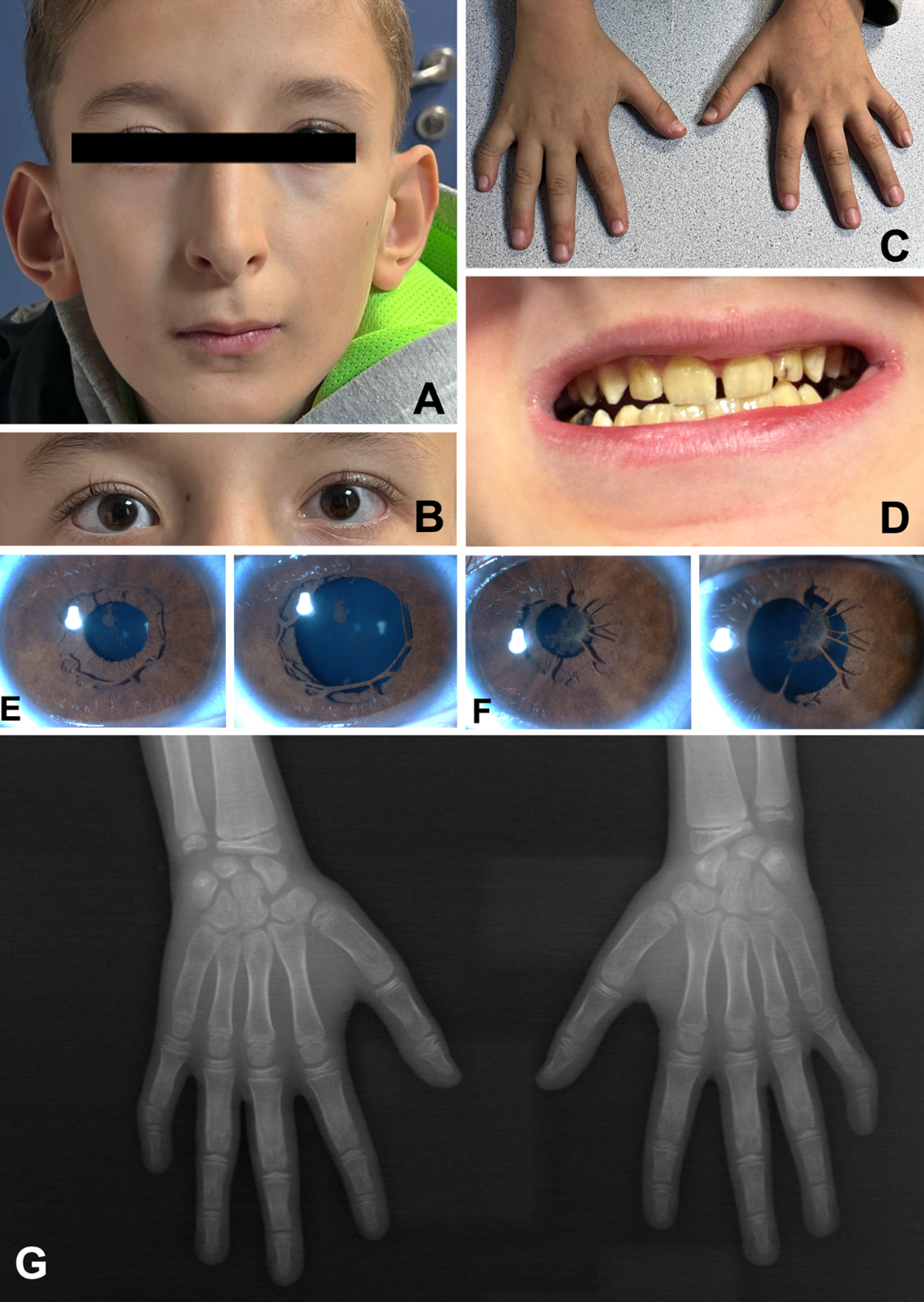
The best-corrected visual acuity at presentation was 0.1 logMAR in the right eye (OD) (spherical equivalent: -3.75) and 0.7 logMAR in the left eye (OS) (spherical equivalent: -3.25). The eyes were orthophoric in primary position, with no signs of strabismus.
Anterior segment examination revealed irregular pupil margins, iris strands adherent to the anterior lens capsule compatible with mild persistent pupillary membrane, and posterior embryotoxon in the cornea; the lenses appeared clear. Horizontal corneal diameters of 10.8 mm (OD) and 10.6 mm (OS), met the diagnostic criteria for mild microcornea. Intraocular pressure measurements were within normal limits, recorded as 19 mmHg (OD) and 21 mmHg (OS). Pachymetric analysis revealed central corneal thicknesses of 572 µm OD and 590 µm OS, and anterior chamber angles (ACA) measured 34° in both eyes. Additionally, axial lengths were 25.0 mm OD and 25.4 mm OS. Fundus examination revealed bilaterally enlarged optic discs, a tigroid retinal background, and prominent choroidal vessels. Fundus photography confirmed marked optic disc enlargement. Device-integrated software calculated the optic disc areas of 6.95 mm2 in the right eye and 6.07 mm2 in the left eye. Optical coherence tomography (OCT) showed choroidal thinning and retinal nerve fiber layer (RNFL) thickness was within normal limits in all four quadrants in both eyes, and standard automated perimetry demonstrated no glaucomatous visual field defects (Fig. 2).
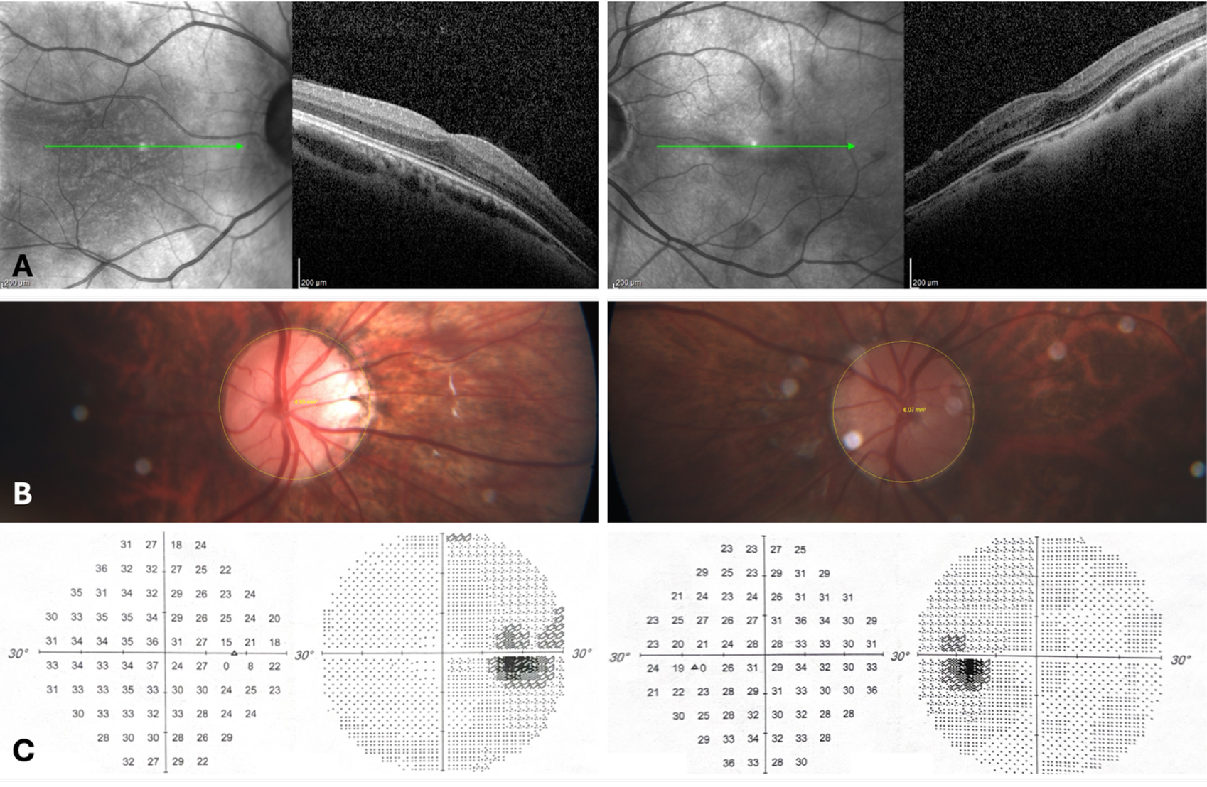
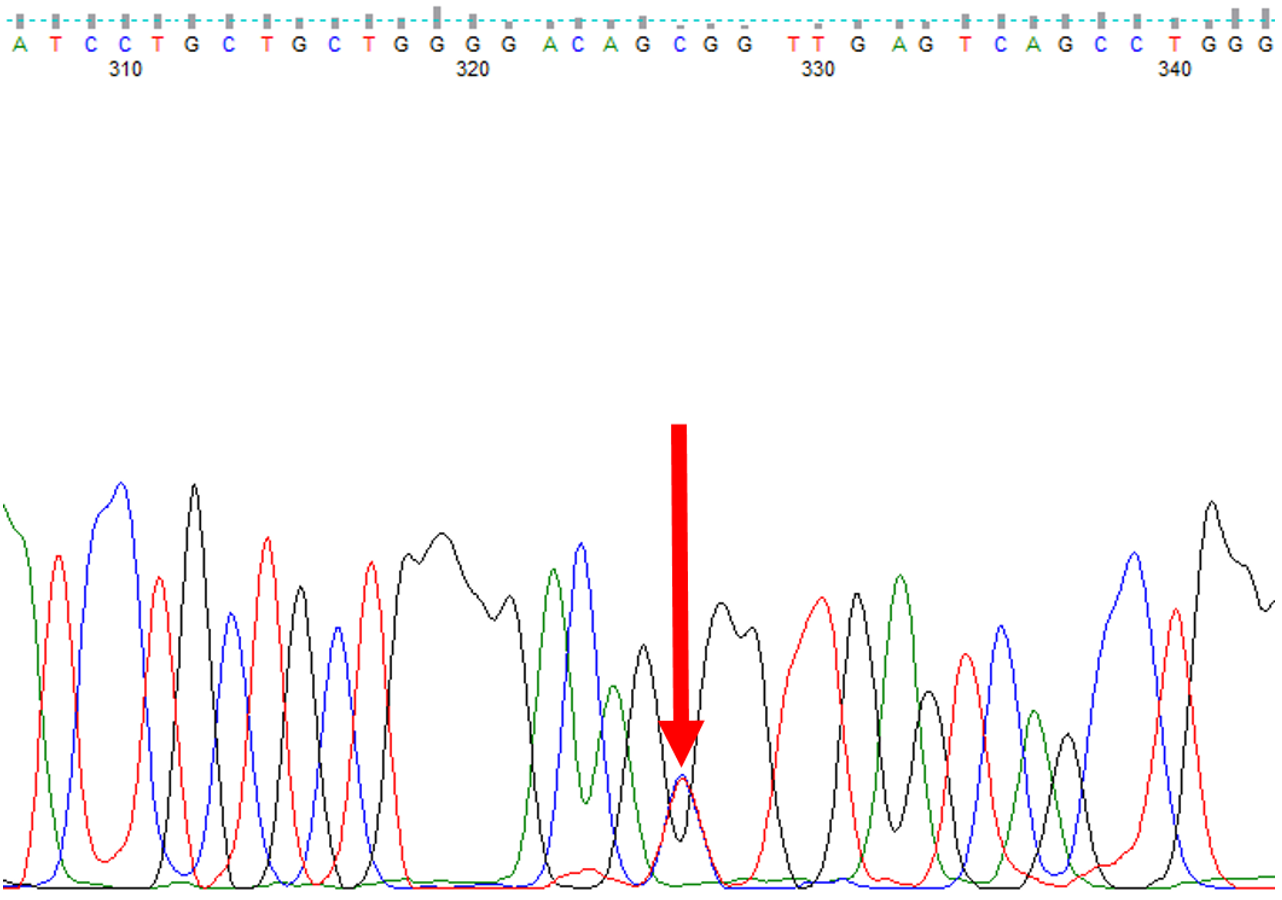
Based on the constellation of craniofacial anomalies (narrow pinched nose with hypoplastic alae nasi and prominent columella), dental abnormalities (enamel hypoplasia, tooth discoloration, and multiple dental caries), and digital findings (clinodactyly of the fifth fingers), a syndromic condition was suspected. Given the characteristic combination of these findings, ODDD was considered the most likely clinical diagnosis, and the patient was referred to the Division of Pediatric Genetics for further evaluation. Genetic analysis was conducted using Sanger sequencing, covering all coding exons and exon–intron boundaries of the GJA1 gene. A heterozygous NM_000165.5(GJA1): c.119C>T; (p.Ala40Val) variant (rs1554200992) was identified. The Sanger sequencing electropherogram demonstrating the heterozygous variant in the index patient is provided in Fig. 3.
According to the American College of Medical Genetics and Genomics and Association for Molecular Pathology (ACMG/AMP) guidelines 6, the detected GJA1 variant was evaluated for pathogenicity. The variant fulfills the criteria PM2 (absence from large population databases), PP3 (multiple in silico prediction tools suggesting a deleterious effect), and PP4 (patient’s phenotype highly specific for a disease with a single known genetic etiology) and published functional data (PP5), the variant may be classified as likely pathogenic according to ACMG/AMP 2015 criteria. Parental segregation analysis was recommended; however, genetic testing of the parents could not be performed due to the unavailability of parental DNA samples. Neither parent exhibited craniofacial, dental, digital, or ocular features suggestive of OCDD, and the patient’s siblings were phenotypically and developmentally normal. There was no known consanguinity between the parents.
Based on these findings, the variant provides supportive evidence of pathogenicity according to ACMG/AMP 2015 criteria. Written informed consent was obtained from the patient’s parents for genetic testing and publication.
Discussion
Oculodentodigital dysplasia is a rare autosomal dominant disorder caused by variants in the GJA1 gene, which encodes the gap junction protein connexin 43. Mutations in the GJA1 gene, which encodes connexin 43 and is located on chromosome 6q22–q24, impair Cx43-mediated intercellular communication. This disruption interferes with normal morphogenetic patterning during development and alters cellular function in differentiated tissues.7 The syndrome presents with a spectrum of craniofacial, dental, digital, and ocular anomalies, with considerable phenotypic variability.1 With an estimated prevalence of 1 in 10 million, ODDD is exceptionally rare, and around 300 cases have been reported worldwide.7
In a comprehensive review by Kumar et al.3, 295 ODDD cases were compiled, including 165 genetically confirmed cases encompassing 73 distinct GJA1 variants. Microcornea and microphthalmia were the most frequently observed ocular findings, present in 111 and 110 patients, respectively, followed by short palpebral fissures and glaucoma.3
Recent analyses have suggested that the location of the GJA1 variant within the Cx43 protein may influence the pattern of ocular involvement. In particular, variants located within the first intracellular domain of Cx43, as in our case, have been associated with posterior segment abnormalities, including dysplastic optic discs, macular hypoplasia, and dysplastic fundus.3 Gabriel et al.8 reported a patient with a variant in this domain who exhibited a dysplastic optic disc, poorly developed retinal pigment epithelium (RPE), and prominent choroidal vessels, further supporting this structure -function relationship.
Our patient harbored the NM_000165.5(GJA1): c.119C>T; (p.Ala40Val) variant in GJA1, one of the three most frequently reported mutations in the literature, along with c.605G>A (p.Arg202His) and c.389T>C (p.Ile130Thr). While ocular findings such as microcornea (n=9) and myopia (n=4) have been reported in this group, both were also present in our patient, posterior segment anomalies, including optic disc abnormalities, have not been documented in association with this specific variant.3 To date, 19 patients with the c.119C>T variant have been reported, systemic and ocular findings, when available, are summarized in Table I. The pathogenicity of the detected GJA1 variant was evaluated in accordance with the ACMG guidelines. The c.119C>T; (p.Ala40Val) variant affects a highly conserved amino acid residue located within the N-terminal intracellular domain of connexin 43, a region known to be critical for gap junction assembly and function. The variant has been reported in multiple individuals with clinically confirmed ODDD and shows strong phenotype–genotype concordance. In addition, the variant is absent or extremely rare in large population databases, and multiple in silico prediction tools support a deleterious effect on protein function. Functional studies further support the biological relevance of this variant. Specifically, mutations affecting the N-terminal and transmembrane domains of connexin 43, including A40V, have been shown to result in preserved gap junction plaque formation but complete loss of intercellular electrical coupling, indicating significant functional impairment.9 Based on these findings, the variant fulfills ACMG criteria including PM2, PP3, PP4 and PP5 providing supportive evidence of likely pathogenicity.6 Additionally, because parental molecular testing was not available, the variant could not be confirmed as de novo, and parental mosaicism or very mild parental expression cannot be excluded.
| Table I. Previously reported ocular and systemic findings in oculodentodigital dysplasia patients with the GJA1: c.119C>T (p.Ala40Val) variant. | ||||
| First author, year | Country | Age (yr) and sex | Ocular findings | Extraocular findings |
| Park et al.13 2019 (3 patients of 1 family) |
Korea | 32, F | Bilateral microcornea, glaucoma, shallow anterior chamber | Narrow, pinched nose; hypoplastic alae nasi; thin anteverted nares; narrow nasal bridge; prominent epicanthic; folds; and hypertelorism |
| Korea | 37, F | Bilateral microcornea, glaucoma, shallow anterior chamber | Narrow, pinched nose; hypoplastic alae nasi; thin anteverted nares; narrow nasal bridge; prominent epicanthic folds; and hypertelorism together with small and carious teeth and yellow palms | |
| Korea | 10, M | Tigroid fundus, tilted disc, esotropia, shallow anterior chamber | Narrow, pinched nose; hypoplastic alae nasi; thin anteverted nares; narrow nasal bridge; prominent epicanthic folds; hypertelorism; and small and carious teeth | |
| Paznekas et al.2, 2009 (11 patients of 2 family) |
USA | 8 patients | Not reported | Not reported |
| USA | 3 patients | Glaucoma | Not reported | |
| Hayashi et al.14, 2014 | Japan | 4, M | Strabismus, glaucoma, macular hypoplasia of retina | Sparse scalp hair, low-set ear, narrow nose, brittle nails of fingers and toes, brachydactyly, enamel hypoplasia, hypodontia |
| Debeer et al.15, 2005 (2 patients of 1 family) |
Belgium | Not reported | Not reported | Brachydactyly of the fifth fingers, camptodactyly of fingers 4 and 5 |
| Belgium | 9, M | Hypotelorism | Poor dentition (microdontia, caries, enamel hypoplasia), bilateral 4–5 syndactyly, thin sparse hair, small mandibula, thin nose | |
| Richardson et al.16, 2004 | UK | Not reported | Not reported | Not reported |
| Paznekas et al.17, 2003 | USA | Not reported | Not reported | Not reported |
| Current study, 2025 | Türkiye | 11, M | Bilateral microcornea, myopia, persistent pupillary membrane, megalopapilla, choroidal thinning | Sparse scalp hair, narrow, pinched nose with hypoplastic alae nasi, prominent columella, low-set, anteverted ears, clinodactyly, dental caries, yellow discoloration and enamel hypoplasia |
Megalopapilla is characterized by an enlarged optic disc with a surface area greater than 2.5–3.0 mm2 in the absence of glaucomatous damage10,11, and is often considered a benign anatomical variant. In our case, optic disc areas were measured as 6.95 mm2 and 6.07 mm2 in the right and left eyes, respectively, significantly larger than the defined threshold. While megalopapilla is a nonspecific finding, its presence in this genetically confirmed ODDD case broadens the posterior segment spectrum associated with the disorder. However, a direct genotype–phenotype association between megalopapilla and the c.119C>T variant cannot be established based on a single observation, and this finding should be interpreted with caution. Although posterior segment abnormalities have been described in ODDD, including optic disc and retinal findings, megalopapilla has not, to our knowledge, been specifically reported in association with ODDD or with the recurrent GJA1 p.Ala40Val variant. In addition, persistent pupillary membrane, a relatively uncommon anterior segment finding noted in only 13 previously reported cases, was also observed.3
The differential diagnosis encompassed glaucoma, morning glory disc anomaly, optic nerve hypoplasia, and optic nerve coloboma.12 In this case, although the optic disc was enlarged, it retained normal color, prominent margins, and typical anatomical vascular configuration.
The patient had a medical history of congenital hypothyroidism and transient benign extracranial hydrocephalus, raising the question of whether these comorbidities might have influenced optic nerve morphology. However, the hypothyroidism was diagnosed through neonatal screening and treated promptly until age 10, with normal neurodevelopmental milestones and cognitive function throughout childhood. The hydrocephalus, diagnosed at three months of age, was extracranial in nature (non-ventricular and non-compressive) and completely resolved without intervention by age one, as confirmed by serial transcranial ultrasounds. There was no history of increased intracranial pressure or visual developmental delay. Given this clinical context and in the absence of any optic atrophy or other signs of optic nerve compromise, the observed megalopapilla was unlikely to be secondary to these early-life conditions.
Although intraocular pressures were in the upper-normal range (19 mmHg OD, 21 mmHg OS), corrected values based on central corneal thickness (572 µm OD, 590 µm OS) remained below the threshold for ocular hypertension. Anterior chamber angles were wide open in both eyes (34°), effectively ruling out angle-closure mechanisms. Axial lengths measured 25.0 mm in the right eye and 25.4 mm in the left eye, which are consistent with moderate myopia. While increased axial length can be associated with certain anatomical variations in the posterior segment, it does not necessarily explain the marked optic disc enlargement observed in this patient. Furthermore, structural evaluation with OCT showed normal RNFL thickness in all quadrants, and standard automated perimetry revealed no glaucomatous visual field defects, supporting the diagnosis of megalopapilla without evidence of glaucoma as a novel optic disc phenotype for the NM_000165.5(GJA1):c.119C>T; (p.Ala40Val) variant.
This case highlights the importance of comprehensive ophthalmologic evaluation, including posterior segment imaging and optic nerve head assessment, in patients with ODDD. Given the emerging evidence linking variant localization within Cx43 to phenotypic expression, further genotype–phenotype studies are warranted to better define the full spectrum of ocular involvement in ODDD.
Acknowledgement
The authors would like to thank Pelin Özlem Şimşek Kiper and Gizem Ürel Demir from the Division of Pediatric Genetics, Department of Pediatrics, Hacettepe University Medical Faculty, Ankara, Türkiye for their valuable contribution to the genetic evaluation and interpretation in this case.
Ethical approval
Written informed consent was obtained from the patient’s parents for genetic testing and publication.
Source of funding
The authors declare the study received no funding.
Conflict of interest
The authors declare that there is no conflict of interest.
References
- Musa FU, Ratajczak P, Sahu J, et al. Ocular manifestations in oculodentodigital dysplasia resulting from a heterozygous missense mutation (L113P) in GJA1 (connexin 43). Eye (Lond) 2009; 23: 549-555. https://doi.org/10.1038/eye.2008.77
- Paznekas WA, Karczeski B, Vermeer S, et al. GJA1 mutations, variants, and connexin 43 dysfunction as it relates to the oculodentodigital dysplasia phenotype. Hum Mutat 2009; 30: 724-733. https://doi.org/10.1002/humu.20958
- Kumar V, Couser NL, Pandya A. Oculodentodigital dysplasia: a case report and major review of the eye and ocular adnexa features of 295 reported cases. Case Rep Ophthalmol Med 2020; 2020: 6535974. https://doi.org/10.1155/2020/6535974
- Loddenkemper T, Grote K, Evers S, Oelerich M, Stögbauer F. Neurological manifestations of the oculodentodigital dysplasia syndrome. J Neurol 2002; 249: 584-595. https://doi.org/10.1007/s004150200068
- Neyzi O, Bundak R, Gökçay G, et al. Reference values for weight, height, head circumference, and body mass index in Turkish children. J Clin Res Pediatr Endocrinol 2015; 7: 280-293. https://doi.org/10.4274/jcrpe.2183
- Richards S, Aziz N, Bale S, et al. Standards and guidelines for the interpretation of sequence variants: a joint consensus recommendation of the American College of Medical Genetics and Genomics and the Association for Molecular Pathology. Genet Med 2015; 17: 405-424. https://doi.org/10.1038/gim.2015.30
- Parashari UC, Khanduri S, Bhadury S, Qayyum FA. Radiographic diagnosis of a rare case of oculodentodigital dysplasia. S Afr J Radiol 2011; 15(4): 44-46. https://doi.org/10.4102/sajr.v15i4.359
- Gabriel LAR, Sachdeva R, Marcotty A, Rockwood EJ, Traboulsi EI. Oculodentodigital dysplasia: new ocular findings and a novel connexin 43 mutation. Arch Ophthalmol 2011; 129: 781-784. https://doi.org/10.1001/archophthalmol.2011.113
- Shibayama J, Paznekas W, Seki A, et al. Functional characterization of connexin43 mutations found in patients with oculodentodigital dysplasia. Circ Res 2005; 96: e83-e91. https://doi.org/10.1161/01.RES.0000168369.79972.d2
- Raffa LH, Basalem EA. Optic disc characteristics on digital fundus photographs in Saudi children. Neurosciences (Riyadh) 2024; 29: 161-167. https://doi.org/10.17712/nsj.2024.3.20230124
- Hellström A, Svensson E. Optic disc size and retinal vessel characteristics in healthy children. Acta Ophthalmol Scand 1998; 76: 260-267. https://doi.org/10.1034/j.1600-0420.1998.760302.x
- Lee HS, Park SW, Heo H. Megalopapilla in children: a spectral domain optical coherence tomography analysis. Acta Ophthalmol 2015; 93(4): e301-305. https://doi.org/10.1111/aos.12545
- Park KW, Ryu HS, Kim J, Chung SJ. Oculodentodigital dysplasia presenting as spastic paraparesis: the first genetically confirmed Korean case and a literature review. J Mov Disord 2017; 10: 149-153. https://doi.org/10.14802/jmd.17050
- Hayashi R, Bito T, Taniguchi-Ikeda M, Farooq M, Ito M, Shimomura Y. Japanese case of oculodentodigital dysplasia caused by a mutation in the GJA1 gene. J Dermatol 2014; 41: 1109-1110. https://doi.org/10.1111/1346-8138.12656
- Debeer P, Van Esch H, Huysmans C, et al. Novel GJA1 mutations in patients with oculo-dento-digital dysplasia (ODDD). Eur J Med Genet 2005; 48: 377-387. https://doi.org/10.1016/j.ejmg.2005.05.003
- Richardson R, Donnai D, Meire F, Dixon MJ. Expression of GJA1 correlates with the phenotype observed in oculodentodigital syndrome/type III syndactyly. J Med Genet 2004; 41: 60-67. https://doi.org/10.1136/jmg.2003.012005
- Paznekas WA, Boyadjiev SA, Shapiro RE, et al. Connexin 43 (GJA1) mutations cause the pleiotropic phenotype of oculodentodigital dysplasia. Am J Hum Genet 2003; 72: 408-418. https://doi.org/10.1086/346090
Copyright and license
Copyright © 2026 The Author(s). This is an open access article distributed under the Creative Commons Attribution License (CC BY), which permits unrestricted use, distribution, and reproduction in any medium or format, provided the original work is properly cited.