Abstract
Background. Arteriovenous malformations (AVMs) are rare causes of lower gastrointestinal bleeding in the pediatric population.
Case Presentation. We report two female children aged 3 and 7 years old with multiple episodes of hematochezia and severe anemia who underwent computed tomography angiography. The first case demonstrated diffuse concentric wall thickening with small calcifications (phleboliths) involving the sigmoid colon and rectum. The affected segments demonstrated moderate progressive enhancement, along with an arcade of prominent vascular channels in the pericolic fat. The second case demonstrated hyperdense wall thickening involving the descending colon and proximal sigmoid colon with intense homogeneous contrast enhancement and an arcade of prominent vessels along the left colon. In both cases, the feeding arteries arose from the inferior mesenteric artery and the venous drainage was into the inferior mesenteric vein. Our first case was subjected to ligation of inferior mesenteric vascular pedicle feeding and draining the AVM, which succeeded in controlling bleeding episodes, while the second patient, who presented with relatively milder symptoms, underwent embolization therapy.
Conclusion. Vascular malformations of the bowel should be considered in pediatric age group in cases of lower gastrointestinal bleed.
Keywords: radiology, pediatric surgery, gastrointestinal bleeding, gastroenterology, gastrointestinal surgery
Introduction
Pediatric lower gastrointestinal (GI) bleed is most commonly caused by intussusception, Meckel’s diverticulum, infectious enterocolitis, juvenile polyposis and anal fissures.1
Angiodysplasias are vascular malformations more commonly seen in elderly patients, frequently involving the right colon, and are well recognized causes of lower intestinal bleeding in the elderly.2 Their development in the elderly population is attributed most commonly to degenerative changes. Conversely, vascular malformations of the GI tract in the pediatric population are rarely documented, and many of them may be asymptomatic, or present with non-specific abdominal pain. Rarely, they may clinically present as obstruction or intestinal perforation.3 However, they may present as massive lower gastrointestinal bleed, associated with severe anemia requiring repeated blood transfusions. Such cases may produce catastrophic hemorrhage and prove to be life-threatening. The severity of this clinical scenario warrants a definitive management in the form of embolization of the feeding artery, ligation of the vascular pedicle or surgical resection of the involved bowel segments, as decided by the treating physician. The physician takes into consideration the age and clinical condition of the patient, as well as the available facilities and infrastructure.
In this report, we present two pediatric cases of arteriovenous malformation (AVM) of the distal large bowel, predominantly involving the sigmoid colon and rectum, who underwent computed tomography (CT) angiography to localize the cause of recurrent fresh bleeding per rectum.
Case Presentations
Case 1
A 3-year-old girl was admitted to our hospital with recurrent episodes of massive fresh lower GI bleed and intermittent abdominal pain. She was severely anemic and had previously undergone repeated blood transfusions in a local primary care facility. Diagnostic colonoscopy and biopsy had been performed at another health center, which reportedly revealed bluish venous congestion in the walls of the rectosigmoid colon and anal canal. The colonoscopic biopsy showed evidence of colonic wall edema, non-specific lympho-plasmocytic infiltrate and focal crypt formation. A differential diagnosis of hemangioma or venous malformation was considered based on the colonoscopic findings. Radionucleotide scan for Meckel’s diverticulum was normal. The child was being managed conservatively with iron, calcium and multivitamin supplements, and propranolol. However, she suffered from intermittent rectal bleeding and resultant chronic anemia.
There was no history otherwise suggestive of any bleeding dyscrasias. All the developmental milestones were appropriately attained. The child was born at term, assisted by Cesarean delivery, with good APGAR scores at birth. Past history was also significant for not having received any vaccinations.
At presentation to our center, physical examination was remarkable for evident pallor, a heart rate of 90 per minute and a respiratory rate of 18 per minute. She had a body weight of 13.5 kg (10th to 25th percentiles) and a height of 100 cm (50th percentile for age). Her abdomen was soft, non-tender and non-distended. Rectal examination revealed no growths or polyps, however, blood staining was observed. The child had a hemoglobin level of 3.9 g/dL at the time of presentation, which, after blood transfusions, increased to 6 g/dL and subsequently to 10.6 g/dL. Other blood investigations including liver function, renal function, electrolytes, platelet count and coagulation profile were within normal limits.
Transabdominal sonography revealed mild concentric rectal wall thickening (maximum thickness of 7mm). Other abdominal organs appeared normal on sonographic evaluation. As magnetic resonance imaging (MRI) was not available at the health facility, CT angiography was performed for further and more definitive evaluation. The study was done on a 16-slice helical CT scanner (Philips®). Multiphase scanning was done for proper characterization of the lesion and accurate determination of arterial supply and venous drainage. Arterial, venous and delayed phase images were acquired at 15, 60 and 180 seconds respectively. Subsequently, intravenous non-ionic contrast was administered, at the rate of 3 mL/min, with a pressure pump injector. Non-contrast images revealed diffuse concentric hyperdense thickening of the walls of anal canal, rectum, and sigmoid colon. Few phleboliths, appearing as small nodular calcific foci, were also noted in the walls of these segments of the large bowel (Fig. 1a and Fig. 1b). Following administration of the contrast material, the thickened walls of the affected segments of the large bowel demonstrated moderate enhancement in the arterial phase, which also increased in the venous phase and persisted into the delayed phase. Prominent vascular channels in the pericolic / perirectal fat were also noted (Fig. 1c and Fig. 1d). Early opacification of mesenteric veins was noted on the arterial phase images (obtained at 15 seconds), which indicated arteriovenous shunting. The lesion received its arterial supply from branches of the inferior mesenteric artery. The venous drainage of the lesion was predominantly into the inferior mesenteric vein (Fig. 1e and Fig. 1f). The portal vein, superior and inferior mesenteric veins were dilated. The imaging-based diagnosis was a high flow vascular malformation. Adjuncts to the diagnosis of AVM were the moderate mural enhancement of the involved bowel segments, early opacification of draining veins on the arterial phase, prominent pericolic and perirectal vessels, and dilated mesenteric and portal veins.
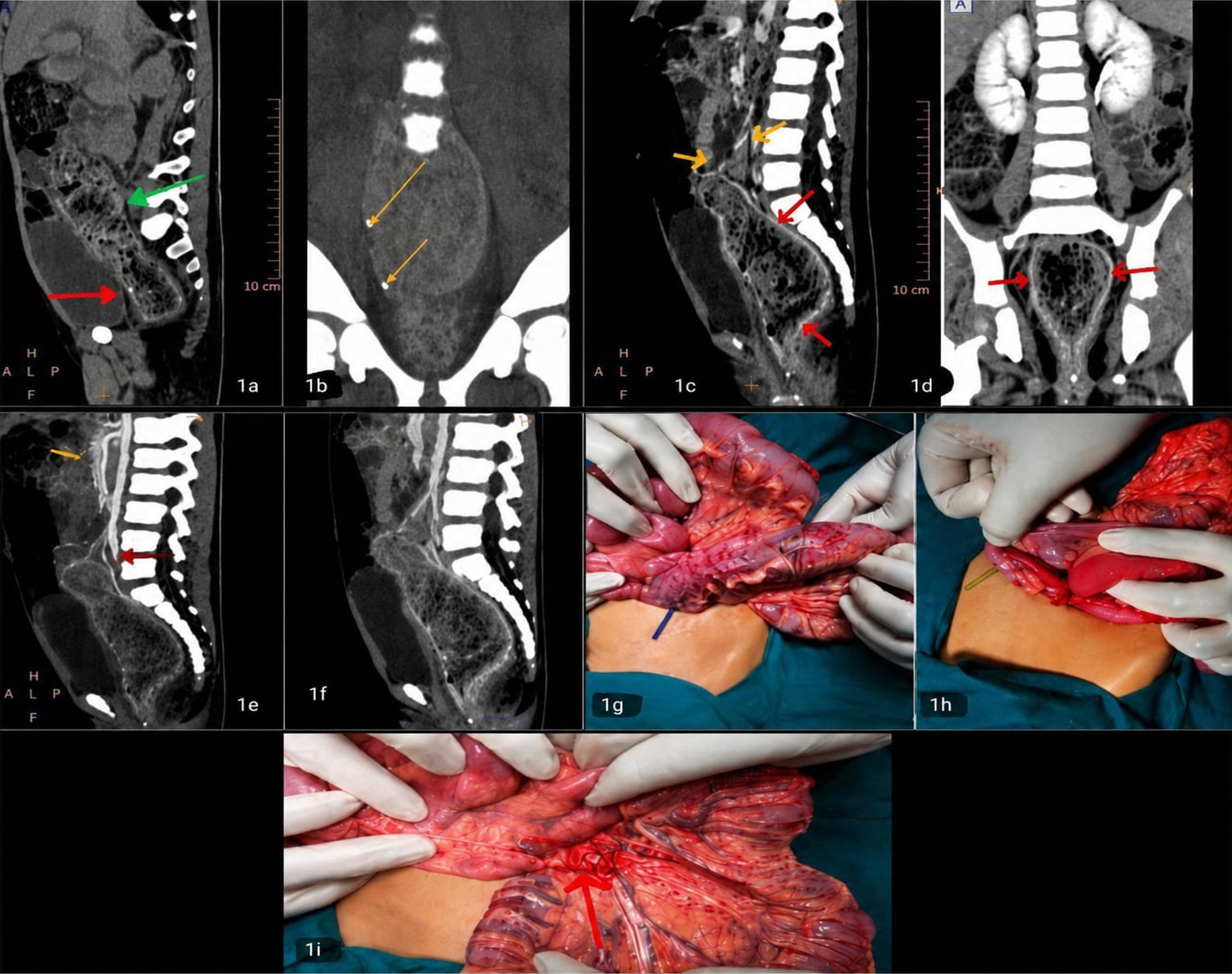
Thereafter, an exploratory laparotomy was performed, which revealed a vascular lesion, associated with prominent bright red vessels, involving the anal canal, rectum & sigmoid colon, extending up to the descending-sigmoid colonic junction (Fig. 1g). Increased pulsatility of the feeding arteries to the affected segment was noted. The draining veins were also found to be dilated and surgical diagnosis of AVM of the large bowel was considered. Another similar vascular lesion was noted involving the cecum and appendix (Fig. 1h) so an appendectomy was performed. However, as it was deemed difficult to excise the diseased segment without affecting anal continence in the young child, the inferior mesenteric vascular pedicle (including both the inferior mesenteric artery and vein) was ligated (Fig. 1i). Histopathological evaluation of the appendectomy specimen confirmed the diagnosis of an AVM, with evidence of congestion and hemorrhage on the appendiceal surface, and numerous dilated and congested vessels in the submucosa, serosa and adherent mesentery of the appendix. The child is doing well with an uneventful follow-up period of the last 30 months.
Case 2
A 7-year-old girl presented to the pediatric outpatient clinic with the chief complaint of a few episodes of recurrent rectal bleeding over the past four weeks. The patient had no relevant history suggestive of any infectious cause for the bleeding. At presentation, the child was hemodynamically stable, with no clinically evident pallor, heart rate of 89 beats per minute, and a blood pressure of 106/76 mm Hg. Physical examination was normal, except for mild tenderness in the left flank on deep palpation. Digital rectal examination was within normal limits with no evidence of any polyps, fissures, or hemorrhoids. Blood count revealed hemoglobin of 11.2 g/dL, with normal total leucocyte and platelet counts. The peripheral smear and prothrombin time were also within normal limits.
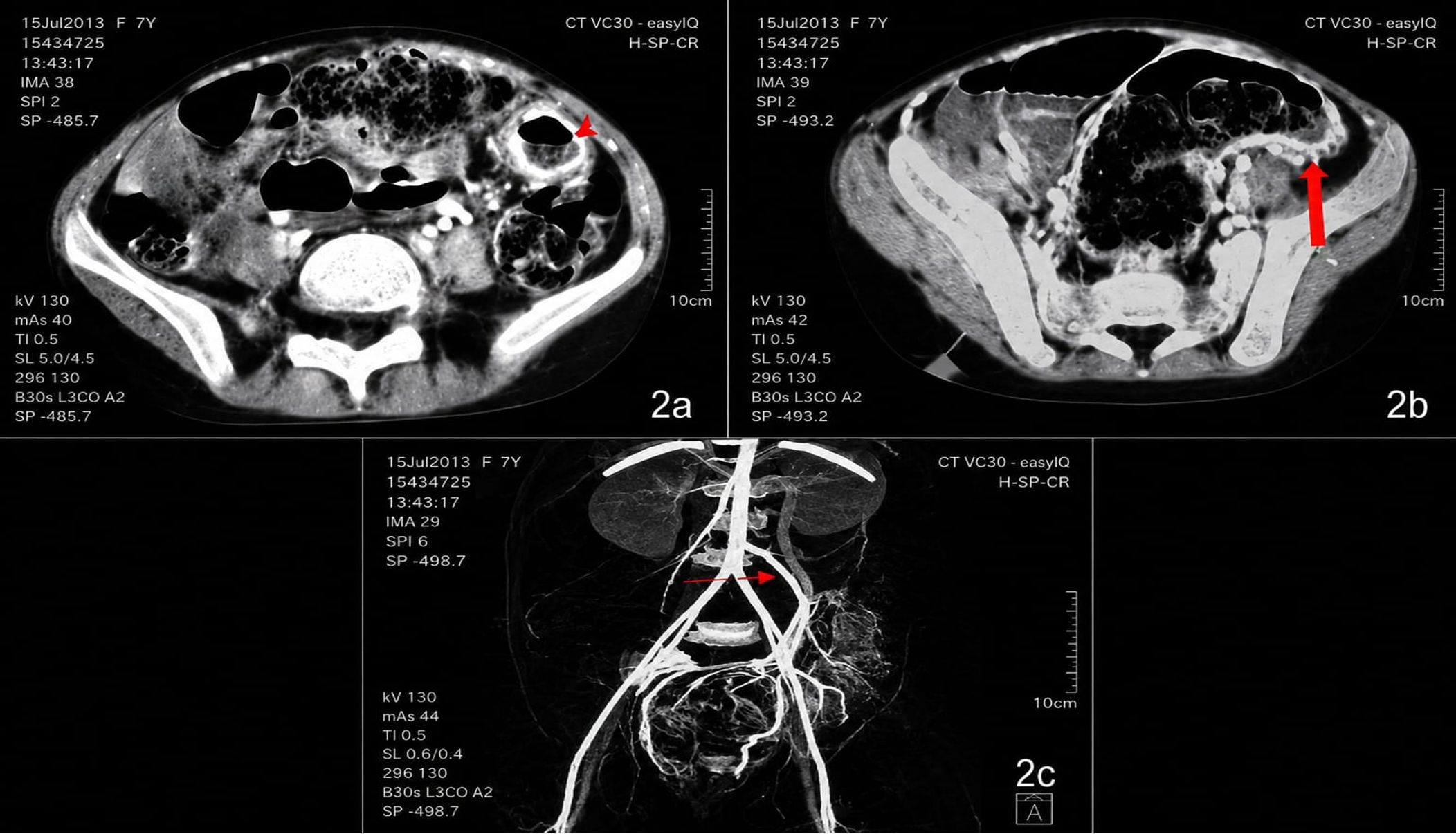
Colonoscopic evaluation revealed congested hyperemic mucosa of the descending colon, with a few bleeding spots. The child underwent CT angiography on a 64-slice CT scanner (Siemens®) with contrast enhancement, including arterial and venous phase imaging for definitive characterization and determination of the extent of the lesion. The study demonstrated diffuse concentric wall thickening of the distal descending colon and proximal sigmoid colon, with intense homogeneous mural enhancement of the affected bowel on post contrast scans (Fig. 2a and Fig. 2b). An arcade of vessels surrounding the involved segment, with arterial supply from a hypertrophied inferior mesenteric artery, and early opacifying veins draining into the inferior mesenteric vein was noted. The inferior mesenteric vein was dilated and coursed superiorly, parallel to the inferior mesenteric artery, and drained at the junction of the splenic and superior mesenteric veins (Fig. 2c). The child was initially treated conservatively. Subsequently, she underwent embolization of the AVM at a referral center, after which the symptoms resolved.
Informed consent was obtained from the parents of both patients for this publication and use of intra-operative and radiological images.
Discussion
The ISSVA (International Society for the Study of Vascular Anomalies) classification divides vascular anomalies into 2 broad groups- vascular tumors and vascular malformations.4 Hemangiomas (infantile and congenital), tufted angiomas and angiosarcomas are the vaso-proliferative tumors. Vascular malformations include slow flow i.e., venous (previously erroneously called cavernous hemangiomas), lymphatic and veno-lymphatic (or lymphangioma) and high flow (arteriovenous malformations or arteriovenous fistulae).5 The vascular anomalies may be a rare cause of bleeding per rectum in children and need proper work up and management.
Venous malformations are one of the most common low- flow vascular malformations. They are most often located superficially within the head and neck or limbs and can rarely involve the viscera. The most common venous lesions found on CT evaluation for obscure GI bleed are venous angiomas and small bowel varices. Albeit rare, venous angiomas of the small bowel may be sporadic or associated with Klippel-Trenaunay or blue rubber nevus syndrome.6 Calcified phleboliths are characteristic of venous stasis along with typical monophasic flow on Doppler sonography.7 In the setting of mesenteric venous hypertension, varices form within the adhesions between adjacent bowel loops or between the bowel and peritoneum.8 Such adhesions provide a pathway for the varices to form. On multiphase CT enterography, venous lesions show progressive enhancement; i.e., they are non-enhancing on the arterial phase and intensely enhancing on enteric and delayed phases.9 Treatment of venous malformations is determined by their extent and location. Although many are managed expectantly, involvement of vital organs or extensive symptoms may necessitate treatment in the form of sclerotherapy and surgical excision.10,11
Arterio- venous malformations (AVMs) are the fast-flow vascular malformations. They usually involve the brain and peripheral extremities and comprise a feeding artery, central nidus, and an early draining vein, and exhibit an extensive vascular network.12 The feeding artery and the vascular lake may vary from being extremely obvious to very subtle. The early draining vein is usually very prominent, and tends to be parallel to the feeding artery. It is almost invariably visualized in the late arterial phase, when the peripheral branches of the mesenteric arteries and the vasa recta still exhibit the contrast material-induced opacification.13 Extensive vascularity with arterial flow waveforms is seen on Doppler sonography and MRI exhibits prominent flow voids.
AVM of the bowel in the pediatric population is considered to be a developmental anomaly - mostly sporadic, or may be associated with syndromes such as hereditary haemorrhagic telangiectasia (HHT), also known as Osler-Weber-Rendu syndrome. Therapy for high-flow malformations is preceded by angiography for complete mapping of vessels. The first-line therapy for AVMs and arteriovenous fistulas (AVF) is embolization. Surgery, or a combination of therapies, may be needed in some children.14 Angiodysplasia may be a differential diagnosis, which is characterized by dilation and proliferation of blood vessels in the submucosal and mucosal layers. However, angiodysplasia is a disease of the elderly, that predominates in the right colon. It is postulated to be a degenerative change in the basement membrane of the colon under high tension. Angiographically, it presents as focal nodular areas of enhancement in anintramural location, which appears brightest on venous phase and gradually fades away in delayed phase.9 They generally do not show an enlarged feeding artery or prominent draining vein. These lesions are thus different from arterial lesions or AVMs which enhance most in the arterial phase.
CT angiography is a very useful tool in suspected cases and can accurately depict lesions in the bowel wall, along with their vascular communications, with high spatial resolution and fast scanning time. It is thus better than the conventional, more invasive selective mesenteric angiographic studies. The use of 3D maximum intensity projection (MIP) reconstructions can provide better mapping of abnormal mural vessels, and their arterial supply and venous drainage, thus providing helpful guidance for the surgeon.15,16 Although low-dose pediatric CT techniques and protocols are available in all modern CT scanners, magnetic resonance angiography (MRA) should be used where available, in view its non-ionizing nature for pediatric patients. However, the need for prolonged sedation is an important limitation with MRI studies. Intensely enhancing bowel wall thickening, early opacification of draining veins, and dilated arterial and venous channels, are all classical diagnostic features of an AVM of the bowel.
Both our described cases presented with bleeding per rectum (more severe in the first case), displayed early moderate to markedly enhancing large bowel wall thickening, early opacification of draining veins in the arterial phase signifying arteriovenous shunting, as well as significantly dilated draining veins. All these findings were pointers to the radiological diagnosis of an AVM in both cases.
However, a few points in the CT study of the first case including persistent enhancement of the lesion in the delayed phase and presence of phleboliths, may need discussion, but they may be explained by the possibility of more numerous venous channels as part of the AVM, which was ultimately confirmed as the final diagnosis on surgical and histopathological findings. Also, the presence of phleboliths in AVM is a known entity, though less common, and suggests a chronic venous component. The enhancement pattern in venous malformation is more of gradual as compared to AVM, where it persists throughout the phases. The early opacification of mesenteric veins in the arterial phase is the most definitive sign of arteriovenous shunting, which rules in AVM. The presence of phleboliths and prominent venous structures may suggest a mixed lesion or chronic venous changes secondary to AVM, but not a pure venous malformation. Differential diagnosis of an enhancing lesion involving the bowel wall with increased mesenteric vasculature includes congenital / infantile hemangioma, AVM (high flow), venovenous malformation (slow flow), and angiodysplasia (largely a degenerative disease of the elderly).
Conclusion
Vascular malformations of the bowel should be considered in the pediatric age group in cases of lower GI bleed where the usual suspects such as Meckel’s diverticulum and enterocolitis have been ruled out. Although most patients present with mild or insignificant symptoms, an accurate diagnosis and definitive management are imperative for symptomatic patients, as it may present as a life-threatening condition with massive lower GI bleed and severe anemia.
Acknowledgements
Acknowledgments should be extended to those individuals or institutions whose contributions to the study were limited or minimal.
Ethical approval
The parent’s consent taken for publication and using radiological and intra-operative pictures.
As per our hospital policy, Ethics approval not applicable as two cases presented with discussion focused on rare anomaly and were retrospective in nature.
Source of funding
The authors declare the study received no funding.
Conflict of interest
The authors declare that there is no conflict of interest.
References
- Fox VL. Gastrointestinal bleeding in infancy and childhood. Gastroenterol Clin North Am 2000; 29: 37-66. https://doi.org/10.1016/s0889-8553(05)70107-2
- Foutch PG. Angiodysplasia of the gastrointestinal tract. Am J Gastroenterol 1993; 88: 807-818.
- Fishman SJ, Burrows PE, Leichtner AM, Mulliken JB. Gastrointestinal manifestations of vascular anomalies in childhood: varied etiologies require multiple therapeutic modalities. J Pediatr Surg 1998; 33: 1163-1167. https://doi.org/10.1016/s0022-3468(98)90552-8
- Steiner JE, Drolet BA. Classification of Vascular Anomalies: An Update. Semin Intervent Radiol 2017; 34: 225-232. https://doi.org/10.1055/s-0037-1604295
- Lowe LH, Marchant TC, Rivard DC, Scherbel AJ. Vascular malformations: classification and terminology the radiologist needs to know. Semin Roentgenol 2012; 47: 106-117. https://doi.org/10.1053/j.ro.2011.11.002
- Jin XL, Wang ZH, Xiao XB, Huang LS, Zhao XY. Blue rubber bleb nevus syndrome: a case report and literature review. World J Gastroenterol 2014; 20: 17254-17259. https://doi.org/10.3748/wjg.v20.i45.17254
- Trop I, Dubois J, Guibaud L, et al. Soft-tissue venous malformations in pediatric and young adult patients: diagnosis with Doppler US. Radiology 1999; 212: 841-845. https://doi.org/10.1148/radiology.212.3.r99au11841
- Tang SJ, Jutabha R, Jensen DM. Push enteroscopy for recurrent gastrointestinal hemorrhage due to jejunal anastomotic varices: a case report and review of the literature. Endoscopy 2002; 34: 735-737. https://doi.org/10.1055/s-2002-33448
- Huprich JE, Barlow JM, Hansel SL, Alexander JA, Fidler JL. Multiphase CT enterography evaluation of small-bowel vascular lesions. AJR Am J Roentgenol 2013; 201: 65-72. https://doi.org/10.2214/AJR.12.10414
- Pappas DC Jr, Persky MS, Berenstein A. Evaluation and treatment of head and neck venous vascular malformations. Ear Nose Throat J 1998; 77: 914-922. https://doi.org/10.1177/014556139807701110
- Pascarella L, Bergan JJ, Yamada C, Mekenas L. Venous angiomata: treatment with sclerosant foam. Ann Vasc Surg 2005; 19: 457-464. https://doi.org/10.1007/s10016-005-4656-z
- Hyodoh H, Hori M, Akiba H, Tamakawa M, Hyodoh K, Hareyama M. Peripheral vascular malformations: imaging, treatment approaches, and therapeutic issues. Radiographics 2005; 25(Suppl 1): S159-S171. https://doi.org/10.1148/rg.25si055509
- Lee EJ, Park YC, Lee YH, Jung KJ, Kim HK. Intestinal arteriovenous malformation involving the descending colon: a case report. Journal of the Korean Radiological Society. 2007; 57: 151-154. https://doi.org/10.3348/jkrs.2007.57.2.151
- Legiehn GM, Heran MKS. Classification, diagnosis, and interventional radiologic management of vascular malformations. Orthop Clin North Am 2006; 37: 435-74, vii-viii. https://doi.org/10.1016/j.ocl.2006.04.005
- Okamoto S, Matsui Y, Sakae H, Oshima K, Hiraki T. A Case of Small Bowel Arteriovenous Malformation Diagnosed Using Multiphase CT Angiography and Digital Subtraction Angiography. Cureus 2023; 15: e42644. https://doi.org/10.7759/cureus.42644
- Kanna ST, Rajavel K, Nayyer S, Gnanasekaran P, Ramesh P, Panda AK. Advanced CT angiography in detecting rare arterio-venous malformations: Addressing diagnostic challenges in obscure gastrointestinal bleeding. Bioinformation 2024; 20: 1917-1921. https://doi.org/10.6026/9732063002001917
Copyright and license
Copyright © 2026 The Author(s). This is an open access article distributed under the Creative Commons Attribution License (CC BY), which permits unrestricted use, distribution, and reproduction in any medium or format, provided the original work is properly cited.