Abstract
Background. Noonan syndrome (NS) is a multisystemic genetic disorder primarily affecting the cardiovascular system. While pulmonary valve stenosis and hypertrophic cardiomyopathy are common cardiac manifestations, coronary artery anomalies (CAAs) are rare but potentially life-threatening complications.
Case Presentation. A 12-year-old boy with genetically confirmed Noonan syndrome due to a PTPN11 mutation (p.Asn308Asp, c.922A>G) had been followed since infancy. He initially had mild pulmonary valve stenosis that regressed spontaneously. He received intermittent growth hormone therapy, which was discontinued for two years and restarted 1.5 years prior to presentation. Routine echocardiography revealed newly detected coronary artery ectasia involving the left main coronary artery (LMCA) and the left anterior descending (LAD) artery. Coronary computed tomography angiography confirmed ectatic dilation of the LMCA and LAD, with an intramyocardial course of the mid-LAD. Further evaluation with cardiopulmonary exercise testing, myocardial perfusion scintigraphy, and selective coronary angiography demonstrated normal myocardial perfusion, an absence of ischemia, and diffuse coronary ectasia without discrete aneurysm formation. The patient remained asymptomatic, and no flow-limiting lesions were identified.
Conclusion. This case highlights a rare vascular manifestation of NS and underscores the importance of vigilant cardiovascular surveillance. Due to the rarity of such cases and the lack of standardized monitoring or treatment protocols, individualized risk stratification remains essential. Reporting such cases is critical for raising awareness and guiding future management strategies for coronary anomalies in Noonan syndrome.
Keywords: Noonan syndrome, coronary ectasia, heart diseases, protein-tyrosine phosphatase non-receptor type 11
Introduction
Noonan syndrome is an autosomal dominant genetic disorder that affects multiple organ systems, particularly the cardiovascular system. It is characterized by distinctive facial features, short stature, developmental delay, and multisystem involvement. The syndrome is caused by pathogenic variants in genes involved in the RAS/MAPK signaling pathway, most commonly PTPN11.1-3
Cardiovascular abnormalities are among the most important clinical manifestations of Noonan syndrome and occur in approximately 50–80% of affected individuals. Pulmonary valve stenosis and hypertrophic cardiomyopathy are the most frequently observed cardiac findings.1-3 Although advances in genetic testing and cardiac imaging have improved the diagnosis of cardiovascular involvement in Noonan syndrome, uncommon cardiovascular manifestations remain insufficiently characterized in the literature. Recognition of these rare findings is important because they may influence clinical surveillance, therapeutic decision-making, and long-term patient outcomes.
Among these uncommon manifestations, coronary artery anomalies (CAAs) have been reported only rarely. Nevertheless, their presence may be associated with serious complications, including thrombotic or ischemic events and even cardiac arrest.1-3 Owing to the limited number of reported cases, the clinical characteristics, management strategies, and prognosis of coronary artery involvement in Noonan syndrome remain incompletely understood. This report discusses a patient with Noonan syndrome and coronary ectasia, highlighting its clinical implications.1,2
Case presentation
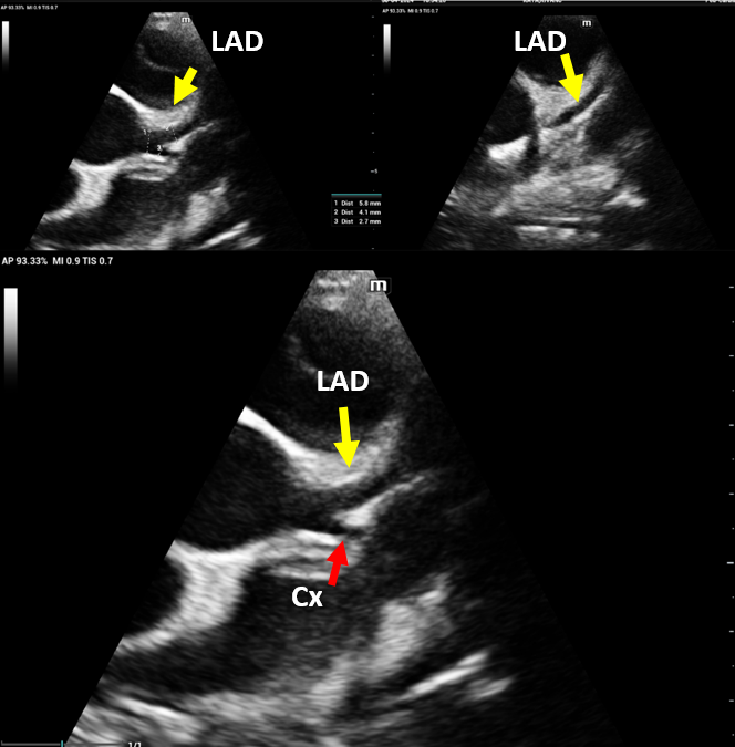
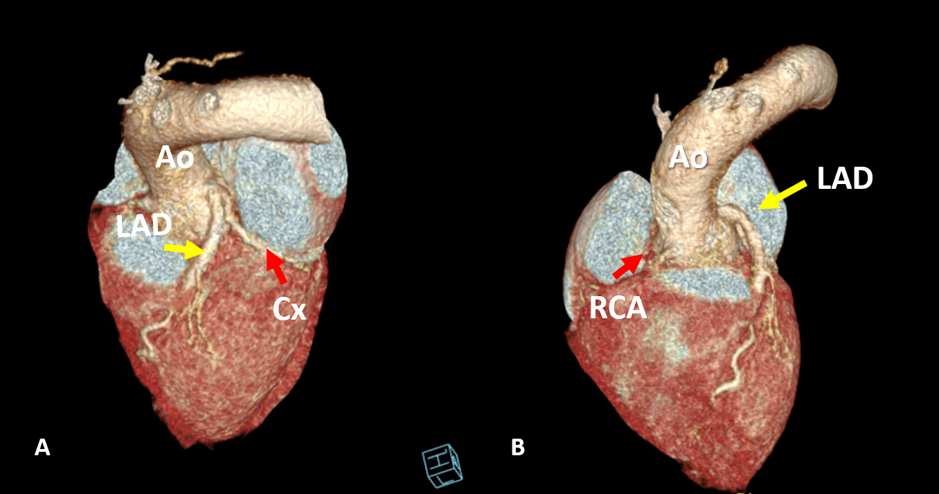
A 12-year-old boy with Noonan syndrome, carrying the PTPN11 mutation p.Asn308Asp (c.922A>G), had been under medical surveillance since infancy. Initially, he presented with mild pulmonary valve stenosis, which regressed spontaneously. He underwent intermittent growth hormone (GH) therapy, which was paused for two years before being resumed 1.5 years before presentation. On examination, he displayed characteristic stigmata of Noonan syndrome. The patient had been followed annually from 6 months of age to 8 years of age with a diagnosis of mild pulmonary stenosis without right or left ventricular hypertrophy. At the time of the diagnosis of coronary ectasia, the patient had not attended follow-up visits for four years. Previous echocardiographic examinations showed no evidence of coronary artery ectasia. Routine echocardiography revealed newly detected coronary ectasia in the left main coronary artery (LMCA; 5.7 mm, z-score +5.4) and the left anterior descending (LAD; 4.1 mm, z-score +4.8) (Fig. 1). Coronary computerized tomography angiography (CTA) demonstrated ectatic dilation of the LMCA and LAD, with the midportion of the LAD exhibiting an intramyocardial course, narrowing during systole (Fig. 2).
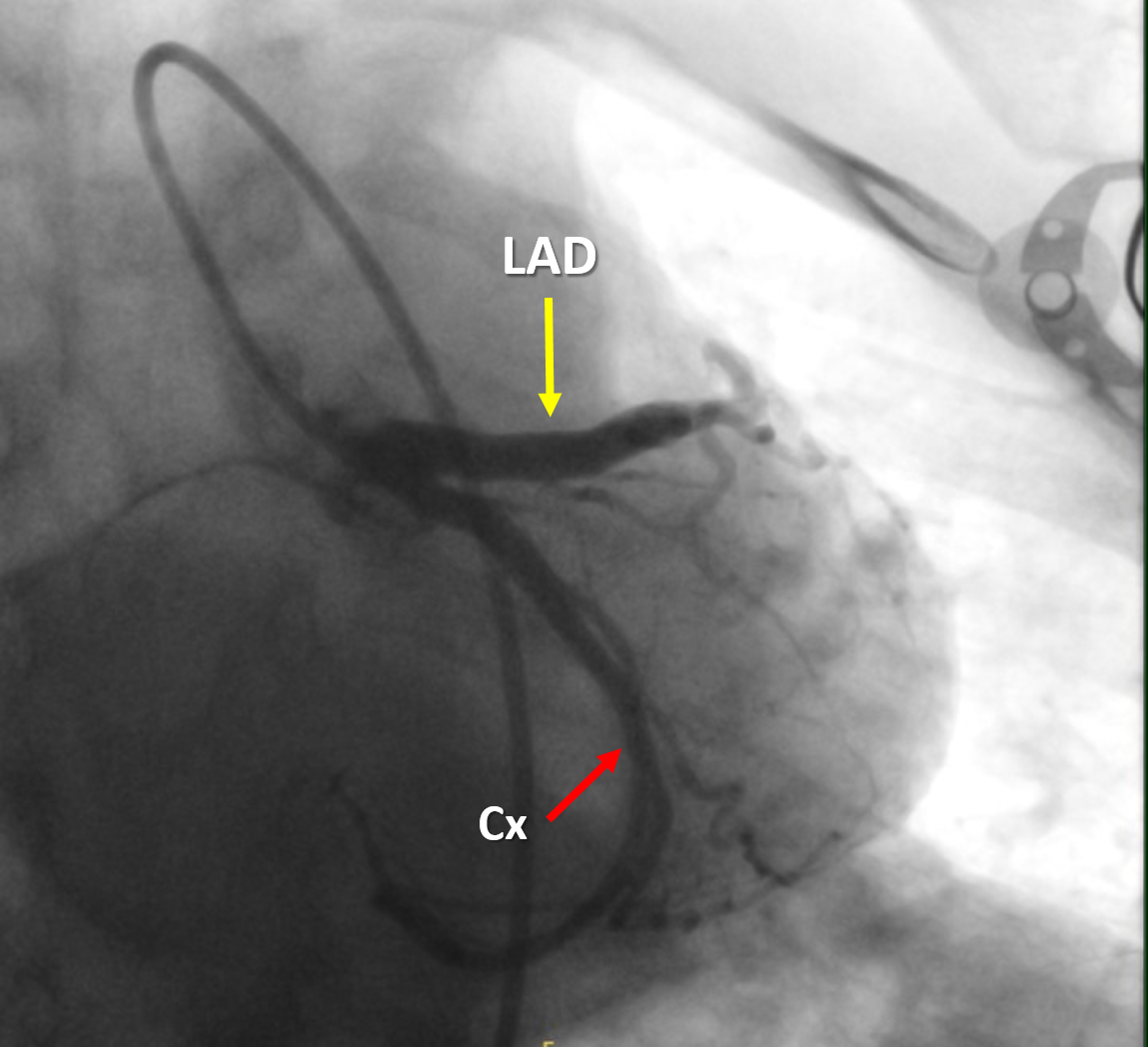
Therefore, a cardiopulmonary exercise stress test, myocardial perfusion scintigraphy and selective coronary angiography were planned for further evaluation. Myocardial perfusion scintigraphy with 99mTc-MIBI showed no anginal symptoms or ST-T changes on ECG during maximal exertion. Myocardial perfusion was normal in all segments of the left ventricle, with no evidence of ischemia or scarring. Cardiopulmonary exercise stress testing was also normal. Selective coronary angiography revealed moderate dilation of the LMCA, along with ectasia of the LAD and left circumflex (Cx) artery (Fig. 3, and Video 1, which is available online in the Supplementary Materials). No discrete aneurysms were detected. The LMCA diameter measured 5.7 mm (z-score: +6.16), while the LAD diameter was 4.5 mm (z-score: +5.39). Additionally, echocardiography indicated mild dilation of the Cx artery, measuring 3.5 mm (z-score: +2.66). Hemodynamic assessment during catheterization revealed the following findings: Right ventricular systolic pressure: 34 mmHg, Main pulmonary artery pressure: 26/11 mmHg (mean 17 mmHg) indicating mild pulmonary valvar stenosis; and aortic pressure: 112/72 mmHg (mean 93 mmHg). The patient remained asymptomatic, and no flow-limiting lesions were identified. At the one-year follow-up after the diagnosis of coronary ectasia, no evidence of progression was detected. Informed consent was obtained from the patient’s parents for the publication of this case report.
Discussion
Coronary artery anomalies in Noonan syndrome are rare but clinically significant.1,2 Fewer than 20 cases of coronary aneurysms or ectasia have been reported in the literature, mostly in adults.1 Most cases were detected incidentally during routine imaging or after patients presented with nonspecific symptoms such as chest pain or exertional fatigue. There are no clear reports of myocardial infarction, sudden cardiac death, or thrombotic occlusion directly attributed to coronary artery ectasia in Noonan syndrome, although these remain potential risks especially in adult patients.2,4
The precise etiology of CAAs in Noonan syndrome remains unclear. Mutations in the PTPN11 gene, which encodes the SHP2 phosphatase, are thought to increase SHP2 activity, thereby weakening arterial wall structure through dysregulation of the RAS-MAPK signaling pathway.2,5,6 Moreover, histopathological studies have identified significant inflammatory changes within the walls of coronary aneurysms, suggesting the involvement of unknown inflammatory triggers in disease progression.2,6-9 Cases associated with SOS1 mutations have also been reported.10
Detecting CAAs in Noonan syndrome is challenging, as many patients remain asymptomatic. When present, symptoms are typically vague and may include fatigue or mild chest discomfort.2 In our case, the patient was asymptomatic, and the coronary ectasia was discovered incidentally during routine echocardiographic screening. This underscores the importance of regular cardiovascular evaluations in Noonan syndrome. Nevertheless, transthoracic echocardiography (TTE) may not always be adequate for detecting subtle coronary anomalies. Contrast-enhanced CTA is recommended for early detection and longitudinal monitoring. However, there remains uncertainty regarding the optimal timing and frequency of CTA or selective coronary angiography in the diagnostic algorithm. Additionally, the fact that most cases in the literature were incidentally identified further complicates surveillance strategies. Consequently, a consensus on the appropriate monitoring approach is lacking, and further studies are needed to define this clinical spectrum and inform standardized guidelines.
The presence of coronary ectasia or aneurysms raises concerns regarding thrombotic and ischemic risks, especially since these abnormalities can remain subclinical for extended periods.2,5 In asymptomatic patients with smooth, non-tortuous ectatic vessels, conservative management with routine follow-up is generally favored.2,5 In our patient, antiplatelet or anticoagulant therapy was not indicated due to the absence of irregular or tortuous coronary anatomy, as confirmed by both CTA and coronary angiography. Coronary angiography revealed no stenosis in the myocardial bridge segment. However, cases in the literature have documented the use of antiplatelet therapy in asymptomatic individuals with more complex coronary anatomy.1 In cases in which thrombotic events occurred, warfarin therapy was instituted.2
The rarity of these cases contributes to uncertainty in diagnosis and management, emphasizing the need for individualized, case-based decisions. Current evidence does not support a clear algorithm regarding the timing of CTA or coronary angiography, or the initiation or antiplatelet/anticoagulant therapy. Thus, reporting such cases is essential for expanding the knowledge base and guiding clinical practice.
Moreover, the incidental nature of most reported cases calls for heightened awareness and comprehensive cardiovascular evaluation in patients with Noonan syndrome. Further research is required to better characterize the clinical course and establish evidence-based monitoring and treatment strategies.
Supplementary materials
Supplementary materials for this article are available online at https://cdn.turkjpediatr.org/html-files/TJP-6871-Suppl-Video-1.mp4
Acknowledgements
The authors would like to express their sincere gratitude to the patient and his family for their cooperation and consent to the publication of this case.
Ethical approval
Ethics committee approval was not required for this case report. Informed consent was obtained from the patient’s legal guardian for publication of this case report.
Source of funding
The authors declare the study received no funding.
Conflict of interest
The authors declare that there is no conflict of interest.
References
- Ly R, Soulat G, Iserin L, Ladouceur M. Coronary artery disease in adults with Noonan syndrome: case series and literature review. Arch Cardiovasc Dis 2021; 114: 598-605. https://doi.org/10.1016/j.acvd.2021.06.006
- Ogihara Y, Fujimoto N, Ohashi H, et al. Case of Noonan syndrome with an expanding coronary arterial aneurysm. Circ Cardiovasc Imaging 2019; 12: e009429. https://doi.org/10.1161/CIRCIMAGING.119.009429
- Sun L, Xie YM, Wang SS, Zhang ZW. Cardiovascular abnormalities and gene mutations in children With Noonan syndrome. Front Genet 2022; 13: 915129. https://doi.org/10.3389/fgene.2022.915129
- Grotti S, Dall’Ara G, Tarantino F, Bachetti C, Ottani F, Galvani M. Cardiac arrest in a 31-year-old man with Noonan syndrome. J Invasive Cardiol 2019; 31: E40. https://doi.org/10.25270/jic/19.3102.E40
- Jain S, Ravindra MS, Sathe YC, Kulkarni SM, Banpurkar A. Coronary arteriopathy in a patient with Noonan phenotype: case report. Ann Pediatr Cardiol 2024; 17: 70-73. https://doi.org/10.4103/apc.apc_145_23
- Gulati GS, Gupta A, Juneja R, Saxena A. Ectatic coronary arteries in Noonan syndrome. Tex Heart Inst J 2011; 38: 318-319.
- Tartaglia M, Mehler EL, Goldberg R, et al. Mutations in PTPN11, encoding the protein tyrosine phosphatase SHP-2, cause Noonan syndrome. Nat Genet 2001; 29: 465-468. https://doi.org/10.1038/ng772
- Tartaglia M, Gelb BD, Zenker M. Noonan syndrome and clinically related disorders. Best Pract Res Clin Endocrinol Metab 2011; 25: 161-179. https://doi.org/10.1016/j.beem.2010.09.002
- Uçar T, Atalay S, Tekin M, Tutar E. Bilateral coronary artery dilatation and supravalvular pulmonary stenosis in a child with Noonan syndrome. Pediatr Cardiol 2005; 26: 848-850. https://doi.org/10.1007/s00246-005-0935-2
- Mauro DM, Flors L, Hoyer AW, Norton PT, Hagspiel KD. Development of bilateral coronary artery aneurysms in a child with Noonan syndrome. Pediatr Radiol 2016; 46: 422-425. https://doi.org/10.1007/s00247-015-3472-z
Copyright and license
Copyright © 2026 The Author(s). This is an open access article distributed under the Creative Commons Attribution License (CC BY), which permits unrestricted use, distribution, and reproduction in any medium or format, provided the original work is properly cited.