


Abstract
Background. Myocarditis is frequently suspected in pediatric patients presenting with fever, elevated cardiac enzymes, and sudden cardiovascular collapse. However, inherited arrhythmia syndromes such as Brugada syndrome may mimic myocarditis and lead to misdiagnosis. Prompt differentiation is critical, as therapeutic strategies and outcomes differ significantly.
Case Presentation. We report a previously healthy 5-year-old girl who suffered sudden cardiac arrest during a febrile illness and had markedly elevated troponin levels. Initial evaluation suggested myocarditis, but cardiac MRI revealed no abnormalities. Retrospective analysis of the event electrocardiogram (ECG) recorded during resuscitation demonstrated a diagnostic type 1 Brugada pattern that was absent on baseline ECGs. Ajmaline provocation testing confirmed Brugada syndrome, and an implantable cardioverter-defibrillator (ICD) was recommended for secondary prevention.
Conclusion. This case underscores the diagnostic challenge of distinguishing Brugada syndrome from myocarditis in pediatric patients presenting with cardiac arrest. Event ECG recordings obtained during or immediately after cardiac arrest are crucial for accurate diagnosis. Clinicians should maintain a high index of suspicion for Brugada syndrome in pediatric patients experiencing cardiac arrest during a febrile illness to guide timely management. Reporting such cases contributes to clinical awareness and may prevent delays in life-saving interventions.
Keywords: myocarditis, Brugada syndrome, pediatric cardiac arrest, electrocardiography, ajmaline test, implantable cardioverter-defibrillator
Introduction
Fever accompanied by cardiovascular collapse or arrhythmia in pediatric patients often lead clinicians to suspect myocarditis as the primary diagnosis. The presence of mild cardiac enzyme elevation further reinforces this assumption, steering many pediatric specialists toward this seemingly plausible diagnosis. However, these findings may also mask rarer conditions that mimic similar presentations, leading to potential misdiagnosis. For instance, genetic cardiac disorders such as Brugada syndrome can present with arrhythmias triggered by fever and may easily lead to cardiovascular decompensation or ventricular arrhythmias that can be mistaken for myocarditis.1,2 This report describes the diagnostic journey in a pediatric patient presenting with fever and cardiovascular collapse, initially suspected as myocarditis but subsequently revised to Brugada syndrome based on event electrocardiogram (ECG) findings.
This report emphasizes the critical importance of preserving and accessing event ECG recordings obtained during acute episodes.
Case presentation
A previously healthy 5-year-old girl presented to an external medical facility with fever and sudden loss of consciousness. Cardiac arrest was promptly recognized, and cardiopulmonary resuscitation (CPR) was initiated, resulting in the return of spontaneous circulation. According to the records from the referring center, the patient developed wide-QRS tachycardia during the initial resuscitation. This rhythm was initially interpreted as supraventricular tachycardia and treated with adenosine; however, it did not terminate. The duration of the first cardiopulmonary resuscitation, which occurred prior to arrival at the emergency department, was unknown. Initial echocardiographic evaluation revealed a left ventricular ejection fraction (LVEF) of 50% with a shortening fraction indicative of borderline myocardial function and minimal mitral regurgitation. Because wide-QRS tachycardia had preceded the cardiovascular collapse, an amiodarone infusion was recommended. However, the patient subsequently experienced a second episode of cardiac arrest. CPR lasted approximately 3 minutes.
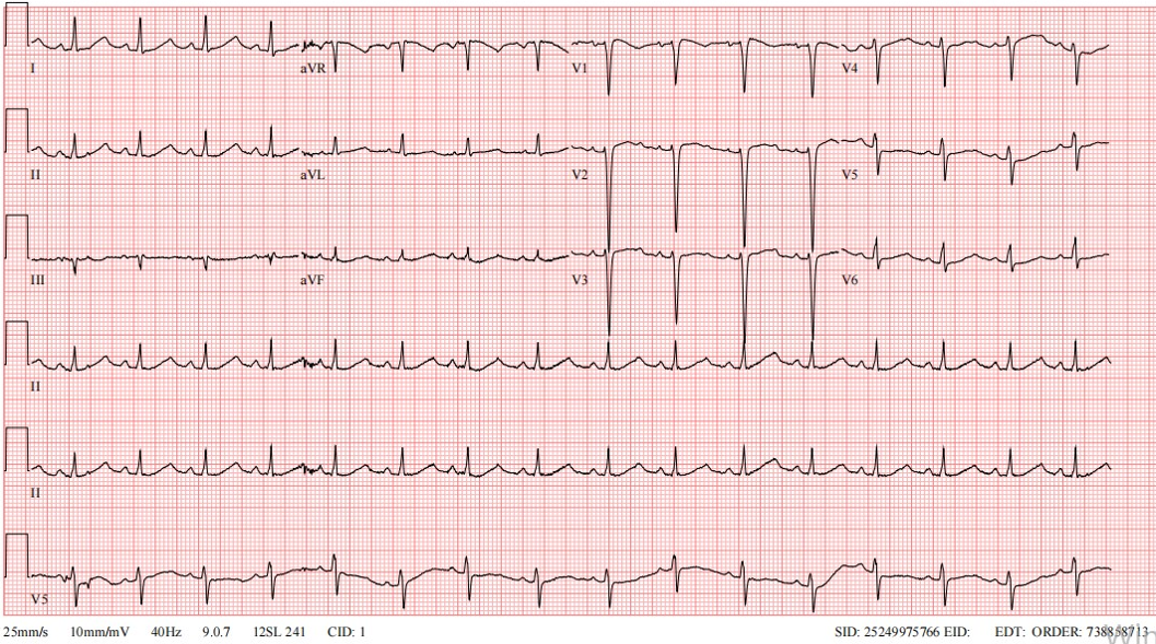
Post-arrest laboratory investigations revealed significantly elevated troponin levels, raising suspicion of myocarditis (Troponin I: 2269 ng/L reference range: 8.4–18.3 ng/L). Consequently, intravenous immunoglobulin (IVIG) therapy was initiated. The patient was transferred to the intensive care unit for management of persistent clinical instability. Follow-up echocardiographic assessment revealed borderline normal left ventricular function. However, cardiac magnetic resonance imaging (MRI) did not reveal any findings consistent with myocarditis including involvement of the right ventricular outflow tract (RVOT). Troponin I levels returned to the normal range within a week. The QTc prolongation observed on admission was transient and likely related to hypoxia during cardiac arrest, as it is normalized on subsequent ECG recordings. Mild ST-segment elevation in leads V1–V2 was noted; however, the changes were subtle and not clearly diagnostic, although they were potentially suggestive of a possible type 3 Brugada pattern. (Fig. 1). No clear correlation was observed between troponin levels and ECG findings. Although the ECG abnormalities resolved rapidly following clinical stabilization, troponin I levels gradually normalized over approximately one week. The elevated cardiac biomarkers were considered secondary to the preceding cardiac arrest and resuscitation rather than indicative of myocarditis. The patient was tested for COVID-19 and the result was negative.
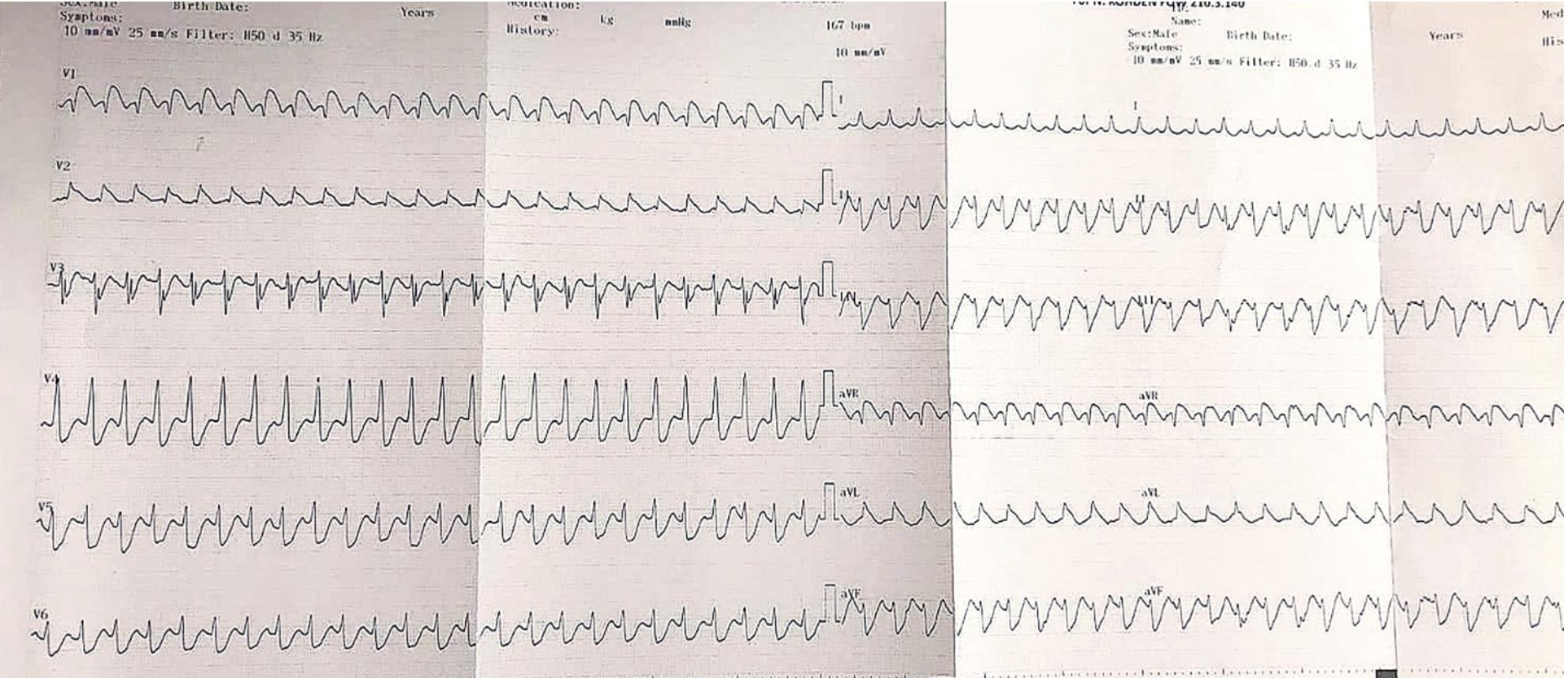
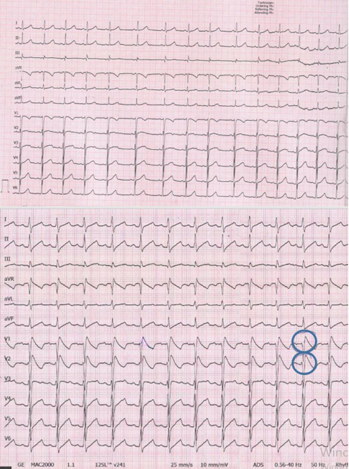
Retrospective analysis of the ECG recordings obtained during the cardiac arrest episodes revealed a diagnostic Brugada pattern type 1 (Fig. 2). Based on the available ECG findings and clinical course, the cardiac arrest was attributed to ventricular tachycardia. Based on this finding, amiodarone therapy was discontinued. An ajmaline provocation test was subsequently performed four months later and conclusively confirmed the diagnosis of Brugada syndrome (Fig. 3). Given the high risk of recurrent life-threatening arrhythmias, an implantable cardioverter-defibrillator (ICD) was recommended. Genetic testing did not reveal a pathogenic mutation. The patient had a prior episode of presyncope. Family history was negative for sudden cardiac death or known arrhythmias. ECG screening including high precordial lead recordings was performed in both parents and the results were normal. The patient achieved full neurological recovery and remains clinically stable under regular outpatient follow-up. Informed consent was obtained from the patient’s parents for this publication.
Discussion
This case highlights the paramount importance of ECG interpretation during and after cardiac arrest in pediatric patients. The identification of a Brugada pattern on the event ECG significantly altered the diagnostic trajectory and guided appropriate management strategies (Fig. 2). Fever has been well-documented as a potent arrhythmogenic trigger in Brugada syndrome, emphasizing the need for aggressive temperature control in affected patients.1 In patients with Brugada syndrome, routine ECGs may not always reveal diagnostic findings, as the characteristic Brugada pattern can be transient or concealed under baseline conditions.2
Although the ECG obtained during the cardiac event revealed Brugada syndrome, ajmaline provocation testing played a crucial role in confirming the diagnosis in this patient (Fig. 3). While Brugada syndrome remains rare in pediatric populations, its presentation with cardiac arrest necessitates a high index of clinical suspicion.3 Early diagnosis, appropriate therapeutic strategies, and ICD implantation are essential for preventing recurrent episodes of sudden cardiac arrest and optimizing clinical outcomes.4 In patients with Brugada syndrome presenting with refractory ventricular arrhythmias, isoproterenol infusion may be considered as a therapeutic option.5
Distinguishing Brugada phenocopy from true Brugada syndrome is essential, particularly in patients presenting with conditions such as myocarditis. Brugada phenocopy is typically associated with reversible clinical conditions and resolves with treatment of the underlying cause, whereas true Brugada syndrome reflects an underlying channelopathy and persists independently of transient clinical factors. In our case, the absence of structural abnormalities on cardiac MRI and the confirmation of a type 1 Brugada pattern through ajmaline testing supported the diagnosis of true Brugada syndrome.6,7
Brugada-like electrocardiographic patterns may be observed across three distinct but sometimes overlapping clinical contexts. First, true Brugada syndrome represents an inherited cardiac channelopathy, typically characterized by persistent or inducible type 1 ECG patterns. Second, similar ECG findings may arise in the setting of reversible conditions such as myocarditis, electrolyte disturbances, or metabolic abnormalities, a phenomenon described as Brugada phenocopy, which resolves with treatment of the underlying cause. Third, emerging evidence suggests the existence of a more complex spectrum in which channelopathies and myocardial inflammation may coexist or interact, creating a diagnostic “gray zone.” In such cases, distinguishing among these entities can be challenging and requires careful integration of the clinical presentation, imaging findings, temporal evolution of ECG changes, and, when appropriate, pharmacological provocation testing.8,9
Ajmaline provocation testing can unmask a diagnostic type 1 Brugada ECG pattern when the pattern is not consistently present. Because sodium channel blockers may induce malignant ventricular arrhythmias, the test should be performed under close monitoring with resuscitation equipment readily available. Establishing the diagnosis is particularly important in diagnostically challenging pediatric cases with suspected Brugada syndrome, as it may influence further management and the consideration of preventive strategies such as ICD. However, it should also be noted that positive responses to ajmaline may occasionally occur in otherwise healthy individuals; therefore, test results should always be interpreted in conjunction with the clinical presentation and ECG findings.4
ECG abnormalities, including Brugada-specific patterns, are frequently overlooked during acute resuscitation efforts, underscoring the need for meticulous ECG assessment in all cardiac arrest scenarios. Febrile illnesses are well-recognized as potent triggers of arrhythmogenic events in Brugada syndrome, further highlighting the importance of temperature regulation and arrhythmia prevention in such clinical contexts.2
In addition, emerging evidence suggests that Brugada syndrome may not be purely an electrical disorder but may also involve subtle structural myocardial abnormalities, particularly in the RVOT region. This overlap further complicates the distinction among true Brugada syndrome, Brugada phenocopy, and myocarditis-related presentations. Therefore, careful clinical evaluation, imaging, and provocation testing are essential for establishing an accurate diagnosis.10
Conclusion
This case underscores the importance of recognizing Brugada syndrome as a potential cause of sudden cardiac arrest in pediatric patients, particularly in the context of febrile illnesses. ECG recordings obtained during cardiac events remain indispensable for accurate diagnosis and management, highlighting the need for careful evaluation of similar clinical presentations.
Ethical approval
Ethics committee approval was not required for this case report. Informed consent was obtained from the patient’s parents.
Source of funding
The authors declare the study received no funding.
Conflict of interest
The authors declare that there is no conflict of interest.
References
- Antzelevitch C, Patocskai B. Brugada syndrome: clinical, genetic, molecular, cellular, and ionic aspects. Curr Probl Cardiol 2016; 41: 7-57. https://doi.org/10.1016/j.cpcardiol.2015.06.002
- Brugada P, Brugada J. Right bundle branch block, persistent ST segment elevation and sudden cardiac death: a distinct clinical and electrocardiographic syndrome. A multicenter report. J Am Coll Cardiol 1992; 20: 1391-1396. https://doi.org/10.1016/0735-1097(92)90253-j
- Probst V, Denjoy I, Meregalli PG, et al. Clinical aspects and prognosis of Brugada syndrome in children. Circulation 2007; 115: 2042-2048. https://doi.org/10.1161/CIRCULATIONAHA.106.664219
- Priori SG, Blomström-Lundqvist C, Mazzanti A, et al. 2015 ESC guidelines for the management of patients with ventricular arrhythmias and the prevention of sudden cardiac death: the Task Force for the Management of Patients with Ventricular Arrhythmias and the Prevention of Sudden Cardiac Death of the European Society of Cardiology (ESC). Endorsed by: Association for European Paediatric and Congenital Cardiology (AEPC). Eur Heart J 2015; 36: 2793-2867. https://doi.org/10.1093/eurheartj/ehv316
- AlKendi F, Jamil M, Qureshi A, Jamil G. Brugada syndrome with elevated cardiac biomarkers. BMJ Case Rep 2013; 2013: bcr2013009918. https://doi.org/10.1136/bcr-2013-009918
- Saplaouras A, Kariki O, Mililis P, et al. Diagnostic and therapeutic dilemmas in a patient with myocarditis, Brugada syndrome and arrhythmic syncope. J Electrocardiol 2023; 80: 45-50. https://doi.org/10.1016/j.jelectrocard.2023.04.006
- Talishinsky A, Bachour K, Lin C, et al. Immune checkpoint inhibitor myocarditis masquerading as Brugada syndrome. JACC Case Rep 2025; 30: 105434. https://doi.org/10.1016/j.jaccas.2025.105434
- Kawauchi D, Yunoki K, Yoshino T, Oka T. Brugada phenocopy in fulminant eosinophilic myocarditis: a case series. Eur Heart J Case Rep 2024; 8: ytae646. https://doi.org/10.1093/ehjcr/ytae646
- Bueno-Gomez A, Castro FJ, Gran F, Garrido-Pontnou M, Dolader P, Rosés-Noguer F. Brugada phenocopy in a child with parvovirus B19 myocarditis. JACC Case Rep 2025; 30: 105283. https://doi.org/10.1016/j.jaccas.2025.105283
- De Raffele M, Di Domenico A, Balla C, et al. Structural abnormalities in Brugada syndrome and non-invasive cardiac imaging: a systematic review. Biology (Basel) 2023; 12: 606. https://doi.org/10.3390/biology12040606
Copyright and license
Copyright © 2026 The Author(s). This is an open access article distributed under the Creative Commons Attribution License (CC BY), which permits unrestricted use, distribution, and reproduction in any medium or format, provided the original work is properly cited.


