Abstract
Introduction. Wilms tumor is the most common renal malignancy in children. Survival rates now exceed 90% with multimodal therapy. This study aims to retrospectively analyze 104 pediatric Wilms tumor patients diagnosed over a 47-year period at our institution, examining demographic characteristics, tumor staging, treatment modalities, complications, recurrence rates, and survival outcomes.
Materials and Methods. This retrospective single-center study included 104 pediatric patients diagnosed with Wilms tumor between January 1, 1978, and January 31, 2025. Patients were stratified into early (1978–2005) and late (2006–2025) periods. Survival analysis was performed using the Kaplan–Meier method and multivariable Cox proportional hazards regression.
Results. The 5-year overall survival (OS) and event-free survival rates were 61.1% and 57.3% in the early era, 86.8% and 81.5% in the late era, and 73.6% and 65.8% in the overall cohort. OS was significantly lower in patients with anaplasia (p=0.035), advanced-stage disease (p=0.004), and in those treated in the early era (p=0.009). In multivariable analysis, treatment era and metastatic disease at diagnosis were independently associated with poorer survival, whereas anaplasia did not retain statistical significance after adjustment.
Conclusion. Survival outcomes improved substantially over time. Metastatic disease at diagnosis and treatment era were the strongest independent predictors of survival, while advanced stage and anaplasia were associated with outcome in univariable analyses.
Keywords: Wilms tumor, pediatric oncology, long-term outcomes, prognostic factors
Introduction
Wilms tumor is the most common renal malignancy in children, representing approximately 7.1% of all pediatric cancers in Türkiye.1,2 Advances in multimodal therapy, incorporating surgery, chemotherapy, and radiotherapy, have significantly improved prognosis, with overall survival rates now often exceeding 90%.3 However, managing Wilms tumor remains a challenge that requires a precise balance between achieving high cure rates and minimizing long-term treatment-related morbidity.
Clinically, Wilms tumor typically presents as an asymptomatic abdominal mass, often discovered incidentally by parents or during routine physical examinations. Although many cases remain silent at presentation, some patients may exhibit symptoms such as abdominal pain, hematuria, or hypertension.4 The stage at presentation and histologic subtype remain the most significant predictors of survival.
Current clinical practice is primarily guided by standardized protocols from the Children’s Oncology Group (COG) and the International Society of Pediatric Oncology (SIOP).1,5 Despite high survival rates, recurrence still occurs in approximately 15% of cases, and survivors remain at risk for late complications such as nephrotoxicity, cardiotoxicity, and secondary malignancies.6,7 Analyzing long-term institutional data is essential to understanding how evolving treatment strategies influence patient outcomes over several decades.
This study presents a retrospective analysis of 104 pediatric Wilms tumor patients treated at our institution over a 47-year period. We evaluate demographic characteristics, clinical presentation, treatment modalities, and survival outcomes. By documenting nearly five decades of clinical experience, we aim to provide a comprehensive overview of the management and long-term results of pediatric Wilms tumor in a single-center setting.
Materials and Methods
This retrospective single-center study was conducted at the Pediatric Oncology Department of Ondokuz Mayıs University and included 104 pediatric patients diagnosed with Wilms tumor between January 1, 1978, and January 31, 2025. Patients younger than 18 years at the time of diagnosis, with histopathologically confirmed primary renal Wilms tumor and complete diagnostic and treatment records, were included in the study. Patients diagnosed with other tumors involving the renal area, initially considered in the differential diagnosis of Wilms tumor at presentation (7 adrenal neuroblastomas, 3 nephromas, 2 renal cell carcinomas, 1 ganglioneuroma, and 1 malignant mesenchymal tumor), as well as those with insufficient medical records (n=8), were excluded from the analysis.
Demographic characteristics, clinical presentation, physical examination findings, laboratory results, imaging studies, histopathological findings, chemotherapy protocols, treatment-related complications, follow-up duration, long-term sequelae, recurrence, and survival status were retrospectively reviewed. For patients diagnosed prior to the implementation of the hospital’s electronic medical record (EMR) system, data were obtained from archived patient files; for more recent cases, data were extracted from the EMR database.
Histopathological evaluations were performed by institutional pathologists based on the original diagnostic reports. No central pathology review or retrospective re-examination of archival slides was conducted for this study. In accordance with established classification, unfavorable histology was defined by the presence of either focal or diffuse anaplasia, collectively categorized as anaplastic Wilms tumor.8
Initial diagnostic procedures included ultrasonography (USG), computed tomography (CT), and magnetic resonance imaging (MRI). For patients diagnosed before 1985, due to the unavailability of CT, lung metastasis was assessed using chest radiography. Routine laboratory investigations were performed in all patients prior to chemotherapy initiation. Echocardiographic assessment was performed when available; because echocardiography was not routinely accessible before 1990, systematic baseline evaluation could not be obtained for all early-era patients.
Throughout the study period, patients were managed according to evolving treatment protocols influenced by both the National Wilms Tumor Study (NWTS)/COG and SIOP treatment approaches. Because the study spanned several decades, tumor stage was determined according to the protocol in use at the time of diagnosis. Earlier cases were classified using a SIOP-based stage I–V framework, whereas patients diagnosed from 2006 onward were staged according to NWTS/COG criteria.1,7,8 Radiotherapy indications and dose planning were also based on these risk-adapted protocols. Surgical treatment primarily consisted of radical nephroureterectomy (RNU), except in selected cases, and all treatment plans were discussed within multidisciplinary tumor board meetings. In most cases, due to limited availability of advanced pathology infrastructure in earlier years, assessments were based on morphological and immunohistochemical analysis.
The current study integrates two consecutive institutional cohorts of pediatric Wilms tumor: an unpublished series of 56 patients diagnosed between 1978 and 2005 and a more recent cohort of 48 patients diagnosed between 2006 and 2025. For comparative analyses, patients were grouped into an early era (1978–2005) and a late era (2006–2025). Because treatment strategies evolved over time, therapeutic approaches were additionally described according to protocol-defined periods.
Treatment strategies evolved across three sequential periods at our institution. Prior to 1999, patients were managed through an institutional surgery-first approach. Following upfront nephrectomy, children with low-stage, favorable-histology tumors received chemotherapy regimens comprising vincristine and actinomycin D, whereas those with advanced-stage or unfavorable-histology disease were treated with intensified multiagent therapy incorporating doxorubicin and radiotherapy, tailored to the risk-stratification standards of the era. Between 1999 and 2005, management adhered to the Turkish Pediatric Oncology Group (TPOG) Wilms Tumor protocol, which was largely derived from the SIOP treatment approach.8 For patients undergoing primary nephrectomy, postoperative therapy was determined by tumor stage and histological risk. Stage I tumors (regardless of histology) and Stage IIA favorable-histology tumors received vincristine and actinomycin D. In Stage IIB favorable-histology tumors, radiotherapy was added to this two-drug regimen. For Stage III–IV favorable-histology tumors, the regimen expanded to include vincristine, actinomycin D, and doxorubicin plus radiotherapy. Patients with Stage II–IV unfavorable-histology tumors received a more intensive four-drug combination of vincristine, actinomycin D, doxorubicin, and etoposide in addition to radiotherapy. From 2006 onward, the institutional protocol was harmonized with the risk-stratification and treatment strategies established by NWTS and COG. Survival analyses were conducted according to these protocol-defined cohorts, with further stratification by early versus late era for descriptive comparisons.
Statistical analyses were performed using IBM SPSS version 22. The Mann–Whitney U test was applied for comparisons of non-parametric variables, and the chi-square test was used for categorical data. Survival analysis was performed using the Kaplan–Meier method, and intergroup differences were evaluated using the log-rank test. Univariable and multivariable Cox proportional hazards regression analyses were performed to identify prognostic factors associated with overall survival. Results were expressed as hazard ratios (HRs) with 95% confidence intervals (CIs). The Kolmogorov–Smirnov and Shapiro–Wilk tests were used to assess the normality of data distribution. A p-value of <0.05 was considered statistically significant.
The study was approved by the Ondokuz Mayıs University Clinical Research Ethics Committee (Approval No: 2023/115; Date: April 27, 2023).
Results
A total of 104 pediatric patients diagnosed with Wilms tumor were included in the study, consisting of 45 males (43.3%) and 59 females (56.7%). Of these, 56 patients were treated in the early period (1978–2005) and 48 patients in the late period (2006–2025). The detailed demographic and laboratory characteristics of the patients are presented in Table I.
| Table I. Demographic characteristics of the study population. | ||||
| Parameters |
(n=56) |
(n=48) |
(n=104) |
|
| Age (months), median [Q1-Q3] (min-max) |
(5 - 156) |
(4 - 132) |
(4 - 156) |
|
| Sex, n (%) |
|
|||
| Male |
|
|
|
|
| Female |
|
|
|
|
Sixty-five patients (62.5%) were diagnosed before the age of four years. At diagnosis, liver and renal functions were generally within normal limits. Isolated serum creatinine elevation was noted in 8 patients (median 1.35 mg/dL; interquartile range, 1.19–1.58; range, 1.10–2.00), primarily in the early era, although these cases were not associated with elevated blood urea nitrogen (BUN) levels. The most significant laboratory finding was anemia, with hemoglobin levels below 12 g/dL observed in 90 patients (86.5%). Other hematological parameters, including white blood cell and platelet counts, did not show consistent or clinically significant deviations across the cohort.
The initial clinical presentations of the patients are summarized in Table II. When comparing the two study periods, a significant increase in incidentally detected asymptomatic masses was observed in the late era compared to the early era (8.3% vs 0%, p=0.042). While abdominal mass remained the most common finding in both periods, other symptoms such as abdominal pain and hematuria showed no significant difference between eras (p > 0.05).
| Data presented as number (percentage). | |||
| Table II. Presenting complaints of the patients. | |||
| Symptom |
|
|
|
| Abdominal mass |
|
|
|
| Abdominal pain |
|
|
|
| Anorexia & weight loss |
|
|
|
| Fever |
|
|
|
| Macroscopic hematuria |
|
|
|
| Nausea & vomiting |
|
|
|
| Sweating |
|
|
|
| Diarrhea |
|
|
|
| Constipation |
|
|
|
| Seizure |
|
|
|
| Irritability |
|
|
|
| Incidentally detected |
|
|
|
Associated congenital anomalies were detected in 11 patients (10.6%). Among these patients, hemihypertrophy and WAGR syndrome were each observed in 3 patients (27.3%), while 9q deletion, phenylketonuria with panhypopituitarism, hypospadias, horseshoe kidney, and Beckwith-Wiedemann syndrome were each observed in one patient (9.1%). Regarding the stage distribution in this subgroup, eight patients (72.7%) were diagnosed with early-stage disease (Stages I–II), while three patients (27.3%) presented with advanced-stage disease (Stages III–IV).
Regarding hematuria status at diagnosis, macroscopic hematuria was present in 14 patients (13.5%), microscopic hematuria in 36 patients (34.6%), and no hematuria was detected in 54 patients (51.9%). Anemia was observed in all patients with macroscopic hematuria (14/14, 100%). Overall, anemia was present in 45 of 50 patients with hematuria and in 45 of 54 patients without hematuria (90.0% vs. 83.3%, p=0.320).
Regarding metastatic status at diagnosis, CT imaging was not available before 1985, and therefore, CT data were lacking for 16 patients. In the early period, pulmonary metastases were detected in 3 patients by chest radiography, and in an additional 5 patients based on subsequent CT imaging. In the late period, 7 patients presented with pulmonary metastases. No extrapulmonary metastases were identified at diagnosis. Overall, pulmonary metastases were present in 15 patients (14.4%) at initial presentation.
Tumor thrombus was identified in 10 patients (9.6%) at diagnosis, including 5 patients from the early period (8.9%) and 5 from the late period (10.4%). Among patients with tumor thrombus, thrombus extended into the renal vein in 3 patients (30.0%), into the inferior vena cava in 5 patients (50.0%), and reached the right atrium in 2 patients (20.0%).
The distribution of tumor stages and histopathological findings according to the study periods is presented in Table III. For the comparative analysis of stage distribution between the two eras, Stage V cases were excluded. Patients were grouped into early-stage (Stages I-II) and advanced-stage (Stages III-IV) disease. While a numerical shift toward earlier-stage tumors was observed in the later era, this trend did not reach statistical significance (p=0.483).
| Data presented as number (percentage). | |||||
| Table III. Comparison of tumor stage and anaplasia between groups diagnosed in 1978-2005 vs. 2006-2025. | |||||
|
|
|
|
|
|
|
| Stage | I |
|
|
|
|
| II |
|
|
|
||
| III |
|
|
|
||
| IV |
|
|
|
||
| V |
|
|
|
||
| Anaplasia | Present |
|
|
|
|
| Absent |
|
|
|
||
Anaplasia was identified in 8 patients (14.3%) in the early period (1978–2005) and 7 patients (14.6%) in the late period (2006–2025) (p=0.591), yielding an overall anaplasia rate of 14.4% (n=15). Review of pathology reports from the early period showed that diffuse anaplasia was specified in one case and focal anaplasia in one case, whereas six cases were reported as anaplastic without subclassification; in the late period, focal anaplasia was present in three patients and diffuse anaplasia in four patients.
Surgical evaluation revealed that, in the early period, preoperative chemotherapy was administered to 5 patients (8.9%) in accordance with NWTS/COG guidelines for specific indications (one due to tumor thrombus and four due to bilateral tumors), while the remaining 51 patients (91.1%) underwent upfront surgery. Among early-era patients, RNU was performed in 52 cases (92.9%) and partial nephrectomy in 4 cases (7.1%).
In the late period, 7 patients (14.6%) received preoperative chemotherapy due to tumor thrombus or a large inoperable mass. RNU was performed in 45 patients (93.8%), while partial nephrectomy was performed in 2 patients (4.2%) with unilateral Wilms tumor associated with a genetic syndrome. One patient (2.1%) in the late period refused surgical treatment and subsequently died. Additionally, 10 patients (9.6%) underwent two or more surgical procedures due to tumor relapse.
Radiotherapy was administered according to institutional protocols, with dose adjustments based on disease stage and histology. For flank or whole-abdomen irradiation, cumulative doses typically ranged from 9 Gy to 21.6 Gy, delivered in standard fractions. In cases of pulmonary metastases, whole-lung irradiation was performed at doses between 10.5 and 12 Gy. During the early study period, 19 patients (33.9%) underwent radiotherapy at our institution; of these, 13 received abdominal irradiation (68.4%) and three received combined abdominal and lung irradiation (15.8%). Radiotherapy data were unavailable for three patients (15.8%) referred to external centers prior to 1987, when our institutional radiotherapy unit became operational. The stage distribution for this radiotherapy cohort included 5 patients with Stage II disease (26.3%), 10 with Stage III disease (52.6%), and 4 with Stage IV disease (21.1%).
In the late period, 22 patients (45.8%) received radiotherapy, while 26 patients (54.2%) did not. Radiotherapy indications included 3 patients with Stage II disease (13.6%), 13 with Stage III disease (59.1%), and 6 with Stage IV disease (27.3%). Notably, one patient with Stage IV disease in the late period refused both surgical and radiotherapy treatment.
The median follow-up time for the entire cohort was 53 months (interquartile range, 15.0–148.5; range, 1–314). At the time of analysis, 38 of 56 patients (67.9%) in the early period and 42 of 48 patients (87.5%) in the late period were alive, resulting in a crude survival rate of 76.9% (80 of 104 patients) for the entire cohort. A total of 24 patients (23.1%) died during the follow-up period. Of the 24 deaths, 21 (87.5%) were directly attributable to progressive disease and systemic metastases. Treatment-related mortality occurred in 3 cases (12.5%), including two deaths due to neutropenic sepsis and one due to hepatic veno-occlusive disease (VOD). No deaths related to late toxicities were observed during follow-up.
Relapse was observed in 18 patients (17.3%), including 8 patients from the early period (14.3%) and 10 patients from the late period (20.8%). The most common sites of relapse were the liver or diffuse abdominal area in 12 patients (66.7%), followed by the contralateral kidney region in 3 patients (16.7%) and the ipsilateral kidney region in 3 patients (16.7%). All relapsed patients received the ICE (ifosfamide, carboplatin, and etoposide) regimen as salvage therapy. In three patients with suboptimal response (16.7%), treatment was switched to a cyclophosphamide–topotecan protocol. Surgical resection of localized recurrent lesions was performed in 10 patients (55.6%). At the time of final analysis, 8 patients (44.4%) were alive, whereas 10 patients (55.6%) died due to progressive disease. Autologous stem cell transplantation was not utilized in our cohort.
According to the Kaplan-Meier analysis, the estimated 5-year overall survival (OS) and event-free survival (EFS) rates for the entire cohort were 73.6% and 65.8%, respectively. Survival outcomes were significantly associated with the stage at diagnosis (p=0.004); specifically, the 5-year OS rates for Stage I, II, III, and IV were 92.3%, 82.6%, 65.2%, and 41.7%, respectively. When stratified by treatment era, the 5-year OS was 61.1% in the early era and 86.8% in the late era (p=0.009), while the 5-year EFS was 57.3% and 81.5%, respectively (p=0.126). In a further granular analysis, the estimated 5-year OS rates were 70.5% for patients diagnosed during the 1999–2005 period and 86.8% for those diagnosed during the 2006–2025 period; however, the difference in OS between these two periods was not statistically significant (log-rank p=0.333).
Kaplan–Meier analysis showed significantly lower OS in patients with anaplasia (p=0.035), in those with advanced-stage disease compared with early-stage disease (p=0.004), and in patients treated in the early treatment era compared with the late treatment era (p=0.009). No significant difference in OS was observed according to age at diagnosis (<2 years vs ≥2 years) (p=0.840). Kaplan–Meier survival curves illustrating these findings are presented in Fig. 1.
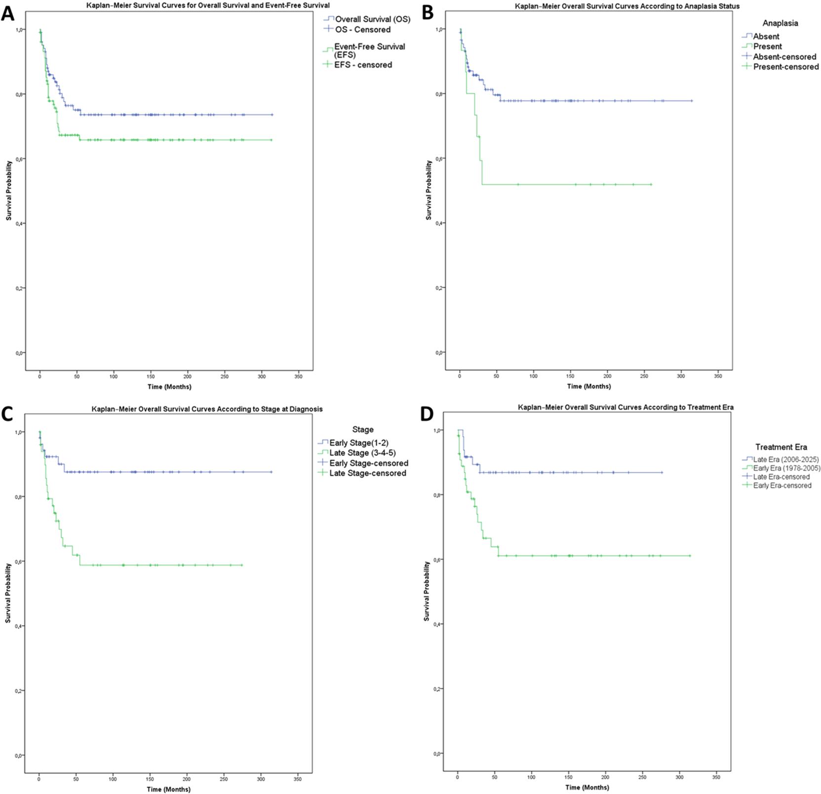
Cox proportional hazards regression analysis was performed to identify prognostic factors associated with overall survival (Table IV). In univariable Cox analysis, early treatment era (HR=3.18; 95% CI: 1.26–8.06; p=0.014), advanced-stage disease (HR=3.60; 95% CI: 1.43–9.09; p=0.007), anaplasia (HR=2.49; 95% CI: 1.03–6.02; p=0.042), and metastatic disease at diagnosis (HR=5.18; 95% CI: 2.29–11.63; p<0.001) were significantly associated with poorer overall survival. In the multivariable Cox regression model including metastatic status, early treatment era (HR=3.12; 95% CI: 1.23–7.94; p=0.017) and metastatic disease at diagnosis (HR=4.17; 95% CI: 1.56–11.11; p=0.004) remained independently associated with poorer overall survival. Advanced stage, anaplasia, sex, and age at diagnosis were not independently associated with overall survival in this model.
| CI: confidence interval, HR: hazard ratio. | |||||||
| Table IV. Univariable and multivariable Cox proportional hazards regression analysis of prognostic factors for overall survival. | |||||||
| Variable | Reference category |
|
|
||||
|
|
|
|
|
|
|
||
| Sex, male | Female |
|
|
|
|||
| Age at diagnosis | Per month increase |
|
|
|
|||
| Early treatment era | Late treatment era |
|
|
|
|
|
|
| Advanced stage | Stage I–II |
|
|
|
|
|
|
| Tumor thrombus | Absent |
|
|
|
|
|
|
| Anaplasia | Absent |
|
|
|
|
|
|
| Metastatic disease | Absent |
|
|
|
|
|
|
Given the close relationship between advanced stage and metastatic disease, an alternative multivariable Cox model excluding metastatic disease was constructed. In this model, advanced stage at diagnosis (HR=3.19; 95% CI: 1.17–8.70; p=0.023) and early treatment era (HR=2.96; 95% CI: 1.17–7.46; p=0.022) remained independently associated with poorer overall survival, whereas anaplasia did not retain statistical significance.
Treatment-related complications observed during the study period are summarized in Table V. Scoliosis was reported in five patients, with four belonging to the early era. Cardiac functions were monitored using echocardiography at the initiation of therapy and at regular intervals during clinical follow-up. A decline in left ventricular ejection fraction (EF < 60%) was identified in three patients. Two patients from the early era were asymptomatic at the time of their last echocardiogram, while the third patient from the late era exhibited clinical signs of heart failure, specifically exertional dyspnea. These three patients had received cumulative doxorubicin doses of 360, 375, and 330 mg/m2, respectively.
| Percentages were calculated using the total study population (n=104). Patients could experience more than one complication. No secondary malignancy was observed in any patient. | |||
| Table V. Acute and long-term treatment-related complications. | |||
| Early treatment complications |
|
Late treatment complications |
|
| Hepatitis |
|
Scoliosis |
|
| Veno-occlusive disease |
|
Delayed puberty & growth retardation |
|
| Vincristine extravasation |
|
Chronic kidney disease |
|
| Ureteral injury |
|
Cardiotoxicity |
|
| Postoperative hypertension |
|
Chemotherapy-induced neuropathy (ptosis) |
|
| Contrast extravasation with compartment syndrome |
|
Hypertension |
|
| Adrenal insufficiency |
|
||
| Osteoporosis |
|
||
| Depression & anxiety disorder |
|
||
Discussion
Over the past several decades, survival outcomes in pediatric Wilms tumor have improved dramatically, with current OS rates approaching 90% in developed healthcare settings. This remarkable progress reflects a transition from the era of surgery-only approaches to risk-adapted, multimodal treatment strategies.9 Our study provides a longitudinal perspective on the evolution of Wilms tumor over a 47-year period, offering valuable insights into changes in presentation, treatment, and outcomes across two distinct eras.
In our cohort, a slight female predominance (56.7%) was observed. This contrasts with the nearly equal sex distribution reported in the TPOG’s 2006 multicenter study.8 However, similar female predominance has been noted in several studies from Asian populations1,10, while other reports describe slight male or female dominance depending on regional or institutional variations.11,12 These findings suggest that while Wilms tumor does not exhibit a universally consistent sex predilection, demographic differences may emerge in specific populations or healthcare settings.
In our study, 62.5% of patients were diagnosed before the age of four, consistent with the well-established observation that Wilms tumor primarily affects younger children. Previous national and international studies have similarly reported that the majority of patients are diagnosed before the age of five.8,11
A striking finding in our cohort was the high prevalence of anemia at diagnosis, observed in 86.5% of patients. Although anemia was frequent in both patients with and without hematuria and the overall difference did not reach statistical significance (90.0% vs. 83.3%, p = 0.320), all patients presenting with macroscopic hematuria had anemia. This descriptive finding suggests that clinically apparent hematuria may have contributed to anemia in a subset of patients, although it does not fully explain the overall high anemia burden in the cohort. Therefore, anemia at diagnosis is likely multifactorial, potentially reflecting chronic tumor-related blood loss, nutritional iron deficiency, anemia of inflammation, tumor burden, or impaired erythropoiesis.13,14 However, the retrospective design of our study limited a detailed etiologic analysis, as complete iron studies were not available for all participants.
In our cohort, the majority of patients presented with abdominal mass, aligning with existing literature that identifies this symptom as the most common initial manifestation of Wilms tumor.11 Notably, in the later period of our study, we observed an increase in incidentally diagnosed cases. This trend may be attributed to advancements in imaging technologies, such as USG and CT, as well as improved accessibility to healthcare services, facilitating earlier detection of asymptomatic tumors.11,15
Regarding associated anomalies, 10.6% of our patients had congenital anomalies, which falls within the range reported in the literature, varying from 2.8% to 17%.11,12 These anomalies often include genitourinary malformations and are sometimes linked to syndromic conditions such as WAGR and Beckwith-Wiedemann syndromes, which are known to predispose individuals to Wilms tumor.11,12 In our cohort, the majority of patients with such anomalies (72.7%) were diagnosed at an early stage (Stages I-II). This finding underscores the importance of thorough clinical evaluation and genetic counseling, as clinical vigilance and screening in predisposed individuals facilitate early detection and management.
Over the course of our study, when comparing early-stage (Stages I-II) and advanced-stage (Stages III-IV) disease (excluding Stage V), we observed a numerical shift toward earlier-stage tumors in the later period. Although this trend did not reach statistical significance (p=0.483), it aligns with the significant increase in incidentally detected asymptomatic masses (p=0.042) observed in our later cohort. This pattern likely reflects improvements in early detection and easier access to diagnostic services, both within our institution and across the broader healthcare system.16 In contrast, the proportion of patients presenting with advanced-stage disease remained relatively stable over time, suggesting that a subset of tumors may still present with more aggressive clinical behavior and rapid progression despite improved diagnostic accessibility.
The rate of bilateral Wilms tumor in our early cohort was comparable to the 5.9% reported in the 2010 multicenter study conducted by the TPOG.8,11 Although we observed a lower frequency of bilateral Wilms tumor in the later period, this likely represents a coincidental variation or reflects the specific characteristics of our late-era study population. In contrast, the significant increase in Stage I diagnoses during the late era represents a distinct stage shift. This trend likely reflects the impact of improved imaging resolution and earlier clinical identification, which facilitate the detection of unilateral disease at a more localized stage before it progresses to advanced levels.
The overall rate of anaplasia in our series was 14.4% (early: 14.3% vs. late: 14.6%; p=0.591), which is higher than the 9.7% reported in the TPOG study.8 This finding must be interpreted with caution due to the historical heterogeneity in pathology reporting across our 47-year study period. The lack of consistent focal versus diffuse subclassification, particularly during the early era, introduces a degree of reporting variability that likely affects cross-study comparisons and the precision of anaplasia-related risk estimates in our cohort.
In our cohort, 11.5% of patients received preoperative chemotherapy, which is lower than the rates reported in previous national studies, including 20% in the TPOG multicenter analysis and 19% in another large Turkish series.8,11 However, we observed a clear increase in the use of preoperative chemotherapy in the later period of our study. This trend likely reflects the gradual adoption of evolving treatment protocols and increasing clinical experience in tailoring therapy based on tumor complexity and operability.
Overall, the stage distribution in our cohort remained consistent with those reported in the literature.11 As in many centers worldwide, nephron-sparing surgery (NSS) was selectively performed in appropriate cases. In our study, NSS was primarily used in patients with bilateral Wilms tumor or in those with unilateral tumors associated with predisposing syndromes, in line with current recommendations.11,12
The 5-year OS and EFS rates in our cohort (73.6% and 65.8%, respectively) are broadly consistent with established national and international reports.8 Notably, outcomes improved substantially over time, with OS increasing from 61.1% in the early era to 86.8% in the late era (p=0.009). Specifically, the lack of a significant survival difference between the 1999–2005 and 2006–2025 periods (log-rank p=0.333) highlights the early impact of the Turkish Wilms Protocol. This indicates that the most substantial breakthrough in survival was achieved through the standardization of therapy and improved supportive care by the late 1990s, reaching a high-quality plateau that has been consistently maintained. While EFS also showed an upward trend (81.5% vs. 57.3%), the lack of statistical significance (p=0.126) suggests that while our primary and salvage strategies have enhanced ultimate survival, preventing initial relapses remains a challenge that warrants further optimization of frontline therapies.
The frequency and distribution of distant metastasis at diagnosis were also comparable to existing literature, with the lungs remaining the most common site of metastatic involvement.8 Despite improvements in primary treatment, disease relapse remains a major clinical challenge. In our study, the relapse rate was 17.3%, aligning with the range of 20% to 25.9% reported in other series.11,12 In our cohort, the liver and diffuse abdominal area were the most frequent sites of relapse. While this frequency appears higher than typically reported, it should be interpreted within the context of our small relapse subgroup (n=18), where each patient significantly impacts the percentage. Additionally, our classification combined isolated liver relapses with diffuse abdominal recurrences, which contributed to this perceived prevalence. In our center, the management of relapsed Wilms tumor primarily focused on intensified chemotherapy and surgical interventions. Although international high-risk protocols often incorporate autologous stem cell transplantation, our strategy relied on the ICE regimen combined with the surgical resection of accessible lesions. This approach, applied to the majority of our relapsed cases, was dictated by institutional protocols and available clinical resources during the study period. The achieved post-relapse survival rate of 44.4% is consistent with existing literature for patients managed with conventional salvage modalities in the absence of high-dose therapy and transplant.17
In our cohort, mortality was primarily associated with progressive disease rather than treatment-related toxicity. The fact that 87.5% of deaths were attributable to progressive disease, while only a small fraction (12.5%) resulted from acute complications such as neutropenic sepsis or VOD, marks a significant clinical observation. These findings suggest that, although modern supportive care has reduced lethal toxicities, further survival gains may depend on more effective strategies for patients with progressive or treatment-refractory disease rather than on further nonspecific intensification of conventional therapy.
Our multivariable Cox regression analysis further underscores this evolution, as both treatment era and metastatic disease emerged as the strongest independent predictors of overall survival. The robust prognostic value of the treatment era (HR=3.12; 95% CI: 1.23–7.94) highlights the cumulative success of institutional experience, improved radiotherapy planning, and more effective salvage regimens over nearly five decades. Consistent with previous reports, metastatic presentation remains the most formidable barrier to survival, carrying an approximately four-fold higher hazard of death in our series (HR=4.17; 95% CI: 1.56–11.11).8,11 Interestingly, while advanced stage and anaplasia were significant in univariable Cox analyses, they did not retain independent significance when adjusted for metastatic status and treatment era. This likely reflects strong collinearity between these variables, as distant metastasis is the defining feature of Stage IV disease. Consequently, in comprehensive multivariable models, the prognostic impact of advanced stage is often statistically superseded or captured by the presence of metastasis. Supporting this interpretation, advanced stage became independently associated with poorer overall survival in the alternative multivariable Cox model excluding metastatic disease (HR=3.19; 95% CI: 1.17–8.70). Regarding anaplasia, its lack of independent significance after adjustment may be partly explained by the historical heterogeneity in pathology reporting over the 47-year study period. As discussed earlier, the lack of detailed subclassification in early-era cases might have diluted the independent prognostic weight of anaplasia compared to more objective markers like metastatic status.
While overall survival in our cohort aligns with international standards, the persistently poor outcomes in these subgroups highlight an ongoing unmet clinical need and underscore the importance of future research efforts targeting these patients.
The spectrum of treatment-related complications in our cohort was largely consistent with established reports in the literature.8 A notable trend was the decreased incidence of scoliosis among patients treated in the later period, which is likely attributable to advancements in radiotherapy techniques—specifically refined targeting and planning that minimize musculoskeletal sequelae. Additionally, vincristine-induced neurotoxicity, such as ptosis, remains a recognized clinical challenge.12,18 Beyond these acute or subacute complications, the concept of survivorship, encompassing long-term quality of life, has become paramount in pediatric oncology. Our findings concerning cardiotoxicity and endocrine late effects, such as growth retardation, underscore the clinical necessity for lifelong, multidisciplinary surveillance to address the evolving health needs of Wilms tumor survivors.
This study has several notable strengths. First, its extensive 47-year span offers a valuable opportunity to evaluate long-term trends in the management and outcomes of pediatric Wilms tumor. Second, the available clinical and therapeutic records enabled a longitudinal overview and comparative evaluation of treatment strategies across different eras. Additionally, the use of multivariable regression analysis contributed to the identification of independent prognostic factors within our cohort. However, certain limitations should be acknowledged. The retrospective design inherently carries the risk of missing data, particularly for patients treated before the implementation of electronic health records. Additionally, histopathological variables were derived from contemporaneous pathology reports, and archived slides/blocks were not systematically re-reviewed; therefore, histologic subclassification (including focal versus diffuse anaplasia) may be subject to historical reporting variability and potential misclassification, particularly in earlier decades. Variations in diagnostic methods, staging classifications, and treatment protocols over time may have introduced heterogeneity across cohorts. Finally, as a single-center study, the generalizability of the findings may be limited, although the consistency of results with national and international data supports their external validity.
In conclusion, our findings confirm that survival outcomes in Wilms tumor have improved significantly over the past decades, in parallel with advances in multimodal therapy, early detection, and clinical expertise. Nevertheless, metastatic disease at diagnosis and treatment era were the strongest independent predictors of survival in our cohort, while advanced stage and anaplasia were associated with outcome in univariable analyses and should be interpreted in the context of disease burden and evolving pathology reporting over time. The observation that mortality was predominantly associated with progressive disease rather than treatment toxicity suggests that future survival gains in our setting may depend on improved risk stratification and more effective treatment approaches for patients with metastatic or treatment-refractory disease, rather than further nonspecific intensification of standard regimens.
Ethical approval
The study was approved by Ondokuz Mayıs University Clinical Research Ethics Committee (date: April 27, 2023, number: 2023/115).
Source of funding
The authors declare the study received no funding.
Conflict of interest
The authors declare that there is no conflict of interest.
References
- Wang J, Li M, Tang D, Gu W, Mao J, Shu Q. Current treatment for Wilms tumor: COG and SIOP standards. World J Pediatr Surg 2019; 2: e000038. https://doi.org/10.1136/wjps-2019-000038
- Kutluk T. First national pediatric cancer registry in Turkey: a Turkish Pediatric Oncology Group (TPOG) study. Pediatr Blood Cancer 2004;43:452. https://doi.org/10.1002/pbc.20119
- Dome JS, Perlman EJ, Graf N. Risk stratification for wilms tumor: current approach and future directions. Am Soc Clin Oncol Educ Book 2014; 34: 215-223. https://doi.org/10.14694/EdBook_AM.2014.34.215
- Sonn G, Shortliffe LMD. Management of Wilms tumor: current standard of care. Nat Clin Pract Urol 2008; 5: 551-560. https://doi.org/10.1038/ncpuro1218
- Stokes CL, Stokes WA, Kalapurakal JA, et al. Timing of radiation therapy in pediatric Wilms tumor: a report from the National Cancer Database. Int J Radiat Oncol Biol Phys 2018; 101: 453-461. https://doi.org/10.1016/j.ijrobp.2018.01.110
- Groenendijk A, Spreafico F, de Krijger RR, et al. Prognostic factors for Wilms tumor recurrence: a review of the literature. Cancers (Basel) 2021; 13: 3142. https://doi.org/10.3390/cancers13133142
- Kaste SC, Dome JS, Babyn PS, et al. Wilms tumour: prognostic factors, staging, therapy and late effects. Pediatr Radiol 2008; 38: 2-17. https://doi.org/10.1007/s00247-007-0687-7
- Akyüz C, Yalçin B, Yildiz I, et al. Treatment of Wilms tumor: a report from the Turkish Pediatric Oncology Group (TPOG). Pediatr Hematol Oncol 2010; 27: 161-178. https://doi.org/10.3109/08880010903447375
- Kutluk T, Varan A, Büyükpamukçu N, et al. Improved survival of children with wilms tumor. J Pediatr Hematol Oncol 2006; 28: 423-426. https://doi.org/10.1097/01.mph.0000212948.05232.7a
- Perrotta G, Castellani D. Wilms tumor: updates about pathogenesis and new possible clinical treatments of the most frequent pediatric urogenital cancer: a narrative review. Surgeries 2023; 4: 678-697. https://doi.org/10.3390/surgeries4040064
- Erginel B, Vural S, Akın M, et al. Wilms’ tumor: a 24-year retrospective study from a single center. Pediatr Hematol Oncol 2014; 31: 409-414. https://doi.org/10.3109/08880018.2014.930767
- Yildiz I, Yüksel L, Ozkan A, et al. Multidisciplinary approach to Wilms’ tumor: 18 years of experience. Jpn J Clin Oncol 2000; 30: 17-20. https://doi.org/10.1093/jjco/hyd001
- Szychot E, Apps J, Pritchard-Jones K. Wilms’ tumor: biology, diagnosis and treatment. Transl Pediatr 2014; 3: 12-24. https://doi.org/10.3978/j.issn.2224-4336.2014.01.09
- Davidson A, Hartley PS, Shuttleworth MH. Erythrocytosis and iron deficiency anemia in Wilms tumor. J Pediatr Hematol Oncol 2005; 27: 502. https://doi.org/10.1097/01.mph.0000181429.71176.61
- Morini MA, da Cunha IW. Navigating the complexity of Wilms tumors in pediatrics: diagnostic challenges for better treatment. Surg Exp Pathol 2024; 7: 23. https://doi.org/10.1186/s42047-024-00166-0
- Mergen M, Welter N, Furtwängler R, et al. The impact of the route to diagnosis in nephroblastoma. Cancer Med 2024; 13: e7226. https://doi.org/10.1002/cam4.7226
- Spreafico F, Bisogno G, Collini P, et al. Treatment of high-risk relapsed Wilms tumor with dose-intensive chemotherapy, marrow-ablative chemotherapy, and autologous hematopoietic stem cell support: experience by the Italian Association of Pediatric Hematology and Oncology. Pediatr Blood Cancer 2008; 51: 23-28. https://doi.org/10.1002/pbc.21524
- Elmalı A, Yüce Sarı S, Akyüz C, et al. De-escalated radiotherapy for advanced stage Wilms’ tumor. Turkish Journal of Oncology 2023; 38: 66-74. https://doi.org/10.5505/tjo.2022.3786
Copyright and license
Copyright © 2026 The Author(s). This is an open access article distributed under the Creative Commons Attribution License (CC BY), which permits unrestricted use, distribution, and reproduction in any medium or format, provided the original work is properly cited.