Graphical Abstract
Abstract
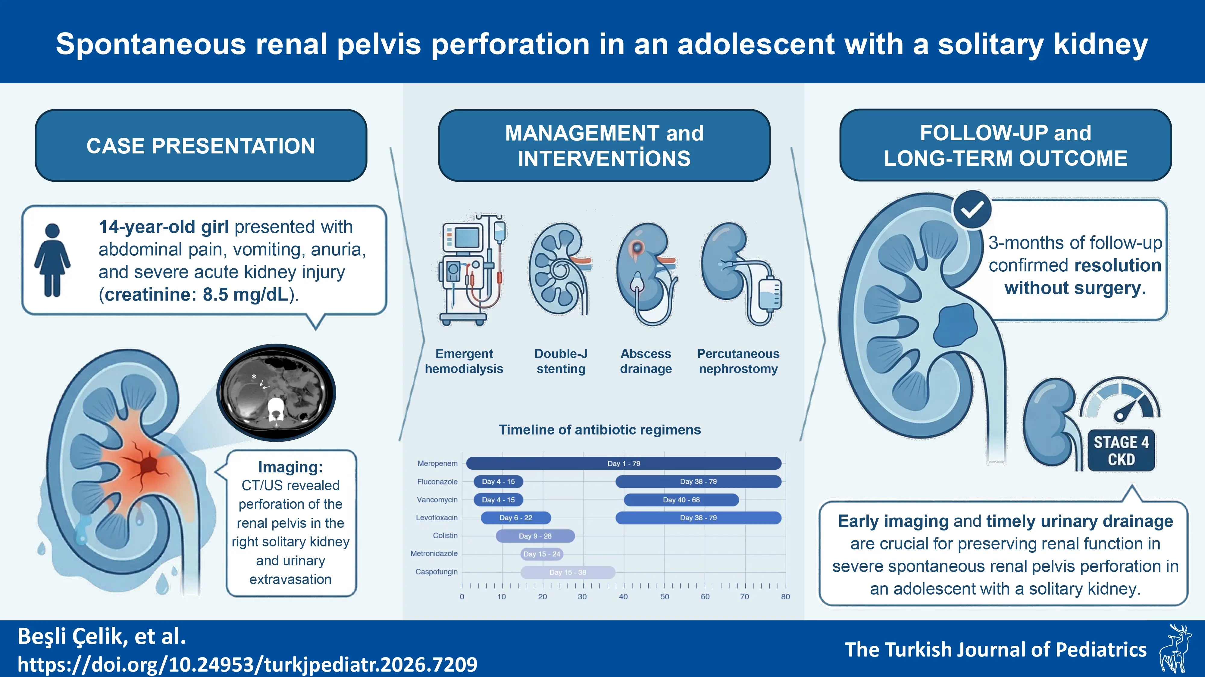
Background. Spontaneous renal pelvis perforation is a rare but serious condition, often associated with urinary obstruction, infection, or increased intrapelvic pressure. Here, we present a pediatric patient with a solitary kidney who developed this rare condition.
Case presentation. We report the case of a 14-year-old girl with a solitary kidney who presented with abdominal pain, vomiting, and anuria. Laboratory findings revealed acute kidney injury, requiring emergent hemodialysis. Imaging studies demonstrated a spontaneous perforation in the anteromedial renal pelvis. The patient was initially managed with antibiotics and a double-J (DJ) ureteral stent. However, persistent fever and worsening retroperitoneal fluid collection necessitated percutaneous abscess drainage. Despite initial clinical improvement, she later developed recurrent urine leakage due to DJ stent occlusion, which required percutaneous nephrostomy placement. At the three-month follow-up, spontaneous resolution of the perforation was confirmed, and the patient was diagnosed with stage 4 chronic kidney disease.
Conclusions. Spontaneous renal pelvis perforation remains a rare entity in the pediatric population, with limited cases documented in the literature. This case emphasizes the importance of early recognition, timely imaging, and appropriate urinary drainage to prevent long-term renal impairment. In pediatric patients with a solitary kidney, close monitoring and a multidisciplinary approach are crucial to optimizing outcomes.
Keywords: acute kidney injury, children, solitary kidney, spontaneous renal pelvis perforation, urinary extravasation
Introduction
Spontaneous perforation of the renal pelvis is an exceptionally uncommon condition in the pediatric population, characterized by the extravasation of urine into the perinephric and retroperitoneal spaces. It is also rarely seen in the adult population and is often associated with underlying congenital anomalies of the kidneys and urinary tract and is typically caused by infection or urolithiasis, often in the setting of urinary obstruction.1-4 Although the exact pathophysiology of perforation is not fully understood, increased intrapelvic pressure resulting from obstruction or infection is considered the primary mechanism.3,5
The clinical presentation is often nonspecific and can include symptoms such as abdominal pain, vomiting, oliguria, or anuria, which may delay diagnosis and increase the risk of serious complications such as sepsis, perirenal abscess formation, or acute kidney injury (AKI).6,7
Renal pelvis rupture is typically diagnosed through abdominopelvic computed tomography (CT) or retrograde pyelography.2-4,6 Management strategies generally focus on ensuring adequate urinary drainage through ureteral stenting or percutaneous nephrostomy, while addressing any underlying pathology. In some cases, spontaneous healing of the perforation site may occur without the need for surgical intervention.1-3,5,7
Here, we present a rare case of spontaneous renal pelvis perforation in a pediatric patient with a solitary kidney, complicated by retroperitoneal abscess formation and AKI, which required interventional management.
Case Presentation
A 14-year-old girl was admitted to a local hospital with complaints of abdominal pain and vomiting for three days, along with anuria for one day. Her antenatal ultrasonography was reported as normal. She had no history of urinary tract infections or urinary incontinence. Additionally, there was no known history of chronic disease or prior trauma. She was referred to our hospital due to AKI, with laboratory findings showing elevated creatinine levels (8 mg/dL) and hyperkalemia (7 mEq/L). On admission, her vital signs, including temperature, pulse rate, and blood pressure, were within normal limits. Physical examination revealed a weak and pale appearance, right-sided costovertebral angle tenderness, and growth retardation, with a weight of 35 kg (SDS: -3.75) and a height of 132 cm (SDS: -5.02).
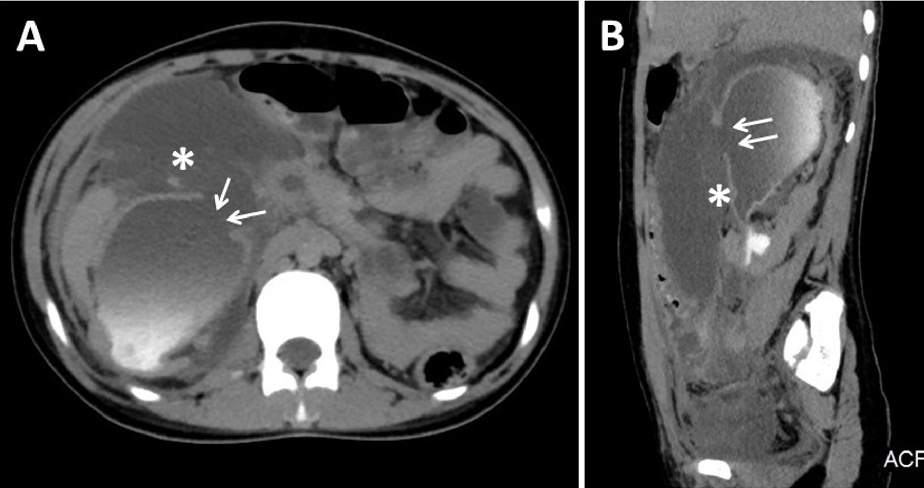
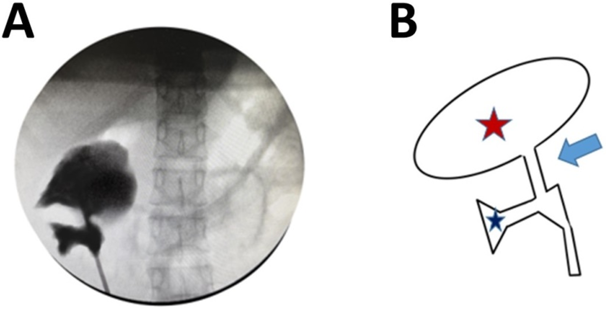
Laboratory tests showed: hemoglobin 9.3 g/dL, mean corpuscular volume 75 fL, leukocyte count 2,070 × 106/L, blood urea nitrogen 195 mg/dL, creatinine 8.5 mg/dL, potassium 7 mmol/L, and C-reactive protein levels exceeding 350 mg/L (0–5 mg/L), with arterial blood gas analysis revealing a pH of 7.40 and a bicarbonate level of 23.1 mEq/L. Emergent hemodialysis was initiated due to AKI and severe hyperkalemia. Abdominal ultrasonography and CT demonstrated a kidney size of 160×73×72 mm, grade 4 hydronephrosis, renal parenchymal thinning, and a perforation in the anteromedial renal pelvis of the right solitary kidney, accompanied by thickening of the proximal ureteral and bladder walls. A dense collection, appearing to be purulent and hemorrhagic, was observed in the perirenal and pararenal regions, extending into the pelvis along the retroperitoneal area (Fig. 1). The patient was started on intravenous meropenem (10 mg/kg per dose every 24 hours, adjusted for renal function) and was referred to pediatric urology. A DJ ureteral stent was inserted, and retrograde pyelography demonstrated a normal-appearing ureteropelvic junction, a narrow calyceal neck, and a markedly dilated upper calyceal system (Fig. 2). The patient started urinating immediately after the procedure. Urinalysis showed pyuria; however, urine culture was negative. Her kidney function gradually improved, allowing discontinuation of hemodialysis. Follow-up abdominopelvic magnetic resonance imaging (MRI) after DJ catheter insertion and medical treatment demonstrated the beginning resolution of the fluid collection.
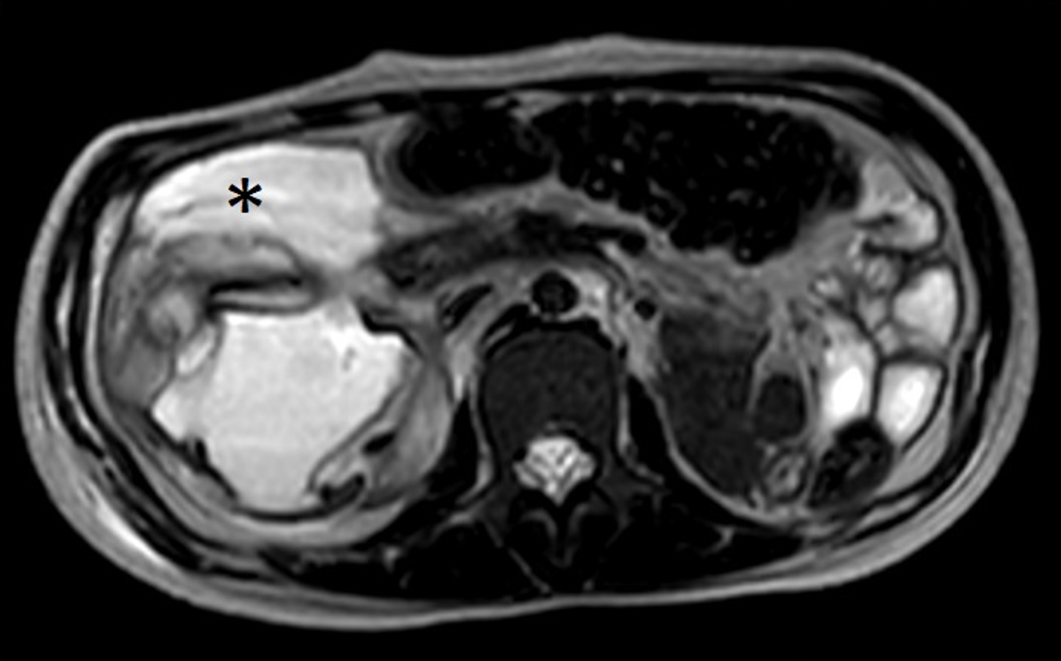
One week after the insertion of the DJ ureteral stent, the patient developed persistent fever, severe abdominal tenderness, and recurrent vomiting. A follow-up abdominopelvic MRI scan revealed a significant increase in the retroperitoneal fluid collection. In addition, the right renal parenchyma appeared thinned, particularly in the upper pole, with a heterogeneous parenchymal pattern. The ureteral wall surrounding the DJ stent was diffusely thickened and edematous along its entire course, and the bladder wall was also thickened (Fig. 3). Therefore, retroperitoneal abscess drainage was performed, and a drain was placed. Cultures for tuberculosis and other possible pathogens were negative. Following antibiotic revision and abscess drainage, the patient’s fever subsided, her overall condition improved, and she was able to resume oral intake. The antibiotic regimens administered to the patient and their respective durations are shown in Fig. 4. Over the course of one month, her creatinine levels gradually decreased to 1.6 mg/dL. Given the presence of elevated parathyroid hormone levels, growth retardation and according to imaging findings, AKI was considered to have occurred on a background of chronic kidney disease (CKD). Due to occlusion of the DJ stent, the patient underwent DJ stent revision two more times. Seven weeks after the initial intervention, a percutaneous nephrostomy was placed due to recurrent occlusion of the DJ stent and persistent urine leakage from the perforated renal pelvis. After successful nephrostomy placement, hydronephrosis resolved, and the patient was discharged at the end of 2.5 months of follow-up, with a serum creatinine level of 1.95 mg/dL and estimated glomerular filtration rate (eGFR) of 29.2 mL/min/1.73 m2.
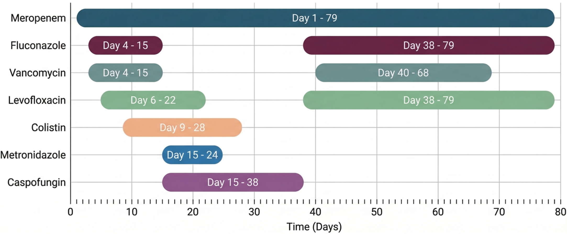
At the three-month follow-up, the nephrostomy catheter spontaneously dislodged. Urinary ultrasonography (US) did not reveal any evidence of a persistent perforation, and no further urine extravasation into the retroperitoneal area was observed. The patient has been under follow-up in our clinic for 17 months with a diagnosis of stage 4 CKD, with a current eGFR of 32.9 mL/min/1.73 m2 and a serum creatinine level of 1.73 mg/dL.
Discussion
Spontaneous renal pelvis rupture is characterized by extravasation of urine into the perinephric, paranephric, or retroperitoneal spaces without a history of trauma or recent surgery. The underlying pathophysiology is primarily linked to increased intraluminal pressure, which may result from urinary obstruction, urolithiasis, or infection. However, cases without a clear underlying etiology have also been reported.1-4 Gulati et al.2 reported three adult cases associated with conditions such as emphysematous pyelonephritis, ureteral stones, and malignancy. Tylski et al.7 described a case of idiopathic renal pelvis rupture in a 73-year-old patient with a solitary functioning kidney, requiring hemodialysis—a presentation similar to that of our patient.
Spontaneous renal pelvis perforation is a rare but clinically significant condition that can lead to severe complications, including retroperitoneal abscess formation and AKI. Given its nonspecific clinical presentation, which often includes abdominal or flank pain, vomiting, and costovertebral angle tenderness, the condition closely mimics renal colic. Although rare, it should be considered in the differential diagnosis, particularly in patients exhibiting signs of peritoneal irritation.1,5,8
Spontaneous renal pelvis perforation is more frequently observed in adults, whereas pediatric cases remain extremely rare. Taşkınlar et al.9 reported a case of an 18-month-old girl who developed spontaneous rupture of the renal pelvis due to an extruded kidney stone, a phenomenon rarely documented in the pediatric population. Similarly, Jiang et al.10 described a case of spontaneous kidney rupture in a 9-month-old boy, associated with urinary obstruction and infection, further highlighting the rarity of this condition in children. These cases underscore the importance of considering spontaneous renal pelvis perforation in the differential diagnosis of pediatric patients presenting with acute abdominal pain and urinary abnormalities, particularly in those with predisposing factors such as urinary obstruction or congenital anomalies.
In the present case, a 14-year-old girl with a solitary functioning kidney developed spontaneous renal pelvis perforation, complicated by AKI and a retroperitoneal abscess. Although the exact type of anomaly could not be clearly identified, the solitary kidney was considered to be dysplastic. It is hypothesized that the renal pelvis perforation occurred secondary to infection in this structurally abnormal kidney. Notably, despite the absence of an apparent obstructive etiology, the patient presented with severe renal dysfunction requiring hemodialysis. An increase in the retroperitoneal fluid collection, which initially regressed with stent placement, eventually required percutaneous drainage. This case highlights the importance of close monitoring and early interventional management in pediatric patients with renal pelvis perforation, particularly in those with a solitary kidney, where renal function is already compromised.
Management of renal pelvis perforation is primarily aimed at relieving urinary obstruction and promoting drainage. In most cases, DJ ureteral stent placement is the first-line intervention, facilitating spontaneous healing of the perforation.1,2,8 However, in cases with persistent urine leakage, recurrent stent occlusion, or large fluid collections, percutaneous nephrostomy may be necessary, as demonstrated in our case. While conservative treatment is often effective, surgical intervention may be warranted in cases of extensive rupture or failed drainage.1,8 However, it should be emphasized that renoprotective approaches are of great importance, especially in patients with a solitary kidney, as in our case.
Conclusion
Spontaneous renal pelvis perforation is a rare but clinically significant condition, particularly in pediatric patients. Given the limited number of cases reported in the literature, the optimal management strategy remains unclear. To our knowledge, none of the previous publications have reported spontaneous renal pelvis perforation in an adolescent with a solitary kidney. Early imaging, timely urinary drainage, and close follow-up are essential to prevent long-term renal impairment. This case highlights the importance of individualized management approaches and underscores the potential for healing in pediatric patients, even in the presence of severe complications.
Ethical approval
Informed consent was obtained from the patient’s parents for the publication.
Source of funding
The authors declare the study received no funding.
Conflict of interest
The authors declare that there is no conflict of interest.
References
- Porfyris O, Apostolidi E, Mpampali A, Kalomoiris P. Spontaneous rupture of renal pelvis as a rare complication of ureteral lithiasis. Turk J Urol 2016; 42: 37-40. https://doi.org/10.5152/tud.2015.92979
- Gulati A, Prakash M, Bhatia A, Mavuduru R, Khandelwal N. Spontaneous rupture of renal pelvis. Am J Emerg Med 2013; 31: 762.e1-762.e3. https://doi.org/10.1016/j.ajem.2012.12.021
- Assaker R, El Hasbani G, Thomas G, Sapire J, Kaye A. Spontaneous rupture of the renal calyx secondary to a vesicoureteral junction calculus. Clin Imaging 2020; 60: 169-171. https://doi.org/10.1016/j.clinimag.2019.10.021
- Weber T, DeSanto M, Ricchiuti D. An unusual case of spontaneous rupture of the renal pelvis. Urol Case Rep 2022; 43: 102060. https://doi.org/10.1016/j.eucr.2022.102060
- Yanaral F, Ozkan A, Cilesiz NC, Nuhoglu B. Spontaneous rupture of the renal pelvis due to obstruction of pelviureteric junction by renal stone: a case report and review of the literature. Urol Ann 2017; 9: 293-295. https://doi.org/10.4103/UA.UA_24_17
- Huri E, Ayyildiz A, Nuhoğlu B, Germiyanoğlu C. Spontaneous rupture and emergency repairment of the renal pelvis. Int Urol Nephrol 2007; 39: 413-415. https://doi.org/10.1007/s11255-006-9020-x
- Tylski M, Muras-Szwedziak K, Nowicki M. Idiopathic spontaneous rupture of renal pelvis in a single functioning kidney. Case Rep Nephrol Dial 2021; 11: 221-226. https://doi.org/10.1159/000512588
- Tas T, Cakıroglu B, Aksoy SH. Spontaneous renal pelvis rupture: unexpected complication of urolithiasis expected to passage with observation therapy. Case Rep Urol 2013; 932529. https://doi.org/10.1155/2013/932529
- Taşkınlar H, Yiğit D, Avlan D, Naycı A. Unusual complication of a urinary stone in a child: spontaneous rupture of the renal pelvis with the migration of calculus into the retroperitoneum. Turk J Urol 2016; 42: 48-50. https://doi.org/10.5152/tud.2015.94467
- Jiang H, Zhang Y, Zheng J, Li F. Surgical management of spontaneous kidney rupture in the setting of urinary obstruction in children. Asian J Surg 2022; 45: 1331-1332. https://doi.org/10.1016/j.asjsur.2022.02.006
Copyright and license
Copyright © 2026 The Author(s). This is an open access article distributed under the Creative Commons Attribution License (CC BY), which permits unrestricted use, distribution, and reproduction in any medium or format, provided the original work is properly cited.