Graphical Abstract

Abstract
Background. Studies on gastroptosis and gastroparesis in children are limited. This prospective study aimed to evaluate the frequency of gastroptosis and gastroparesis and their association with duodenogastric reflux (DGR) and treatment-resistant dyspeptic symptoms in children.
Methods. The study evaluated pediatric patients aged 2-18 years with dyspepsia who underwent upper gastrointestinal endoscopy between March 2020 and June 2022. Fluoroscopy and gastric emptying scintigraphy were performed in 88 patients (50 with DGR and 38 with treatment-resistant dyspepsia).
Results. The mean age of the patients was 13.7±3.6 years, and 62 (70.5%) were girls. Gastroptosis was detected in 22 (25%) and gastroparesis in 24 (27%) of all 88 patients. Gastroptosis was present in 10 (20%) and gastroparesis in 20 (40%) of the 50 patients with DGR. Gastroptosis was present in 12 (32%) and gastroparesis in 4 (11%) of the 38 patients with treatment-resistant dyspepsia. Gastroparesis was more common in those with DGR (40%) than in those with treatment-resistant dyspepsia (11%) (p=0.09). Gastroptosis was also detected in 14 (58.3%) patients with gastroparesis. The median body mass index (BMI) z-score of those with gastroptosis was significantly lower than those without gastroptosis (p=0.002).
Conclusions. Gastroptosis and gastroparesis were frequently identified in children with treatment-resistant dyspeptic symptoms or DGR. These conditions should be considered and investigated in this patient population.
Keywords: dyspepsia, duodenogastric reflux, fluoroscopy, gastroparesis, gastroptosis, scintigraphy
Introduction
Functional dyspepsia is a common gastrointestinal symptom with an overall prevalence of 7.6% in children.1 According to the Rome IV criteria, functional dyspepsia is characterized by one or more bothersome symptoms, including postprandial fullness, early satiation, or epigastric pain or burning not associated with defecation. These symptoms should be present at least once a week, with symptom onset at least 6 months before diagnosis and the diagnostic criteria fulfilled during the last 3 months, after appropriate evaluation excludes another medical condition.2 Pathophysiological factors causing dyspeptic symptoms include abnormal gastric accommodation, delayed gastric emptying, visceral hypersensitivity, gastrointestinal infections, Helicobacter pylori infection, psychological factors, duodenal increased sensitivity to acids and lipids, and duodenojejunal motility disorder.3
Gastroptosis is characterized by the abnormal downward displacement of the stomach, which can lead to constipation, tenesmus, dyspepsia, belching, loss of appetite, nausea and vomiting.4,5 Other than case reports, two studies have investigated the relationship between gastroptosis and dyspepsia. One study conducted in adults reported that dyspeptic symptoms were significantly less common in patients with gastroptosis.6 In contrast, a pediatric study published by Tangul et al. demonstrated that alkaline gastroesophageal reflux is more frequently observed in gastroptosis cases and that its incidence increases with the severity of gastroptosis.7 Gastroparesis is a gastric motility disorder characterized by postprandial epigastric pain, halitosis, bloating, postprandial fullness, weight loss, and vomiting.8 In childhood, gastroparesis is extremely rare and mainly occurs in premature infants, when the gastrointestinal tract is immature or due to an allergy to cow’s milk protein. The majority of gastroparesis cases are idiopathic or postviral. Delayed gastric emptying may be associated with diabetes mellitus, hypothyroidism, eosinophilic gastroenteropathy, celiac disease, cystic fibrosis, muscular dystrophy, or vagotomy.9 Gastroparesis and gastroptosis can be easily misdiagnosed. In the presence of persistent dyspepsia in children, especially in girls, patients should be examined in detail for the possibility of gastroparesis. Delayed or misdiagnosed gastroparesis may lead to secondary gastroptosis.4
Duodenogastric reflux (DGR) is the reverse flow of duodenal fluid composed of bile acid, pancreatic secretions, and intestinal secretions into the stomach due to impaired antroduodenal motility and pyloric function. Inflammation and ulceration of the gastric mucosa and intestinal metaplasia in the stomach may result from DGR,10 which causes not only reflux gastritis but also mucosal damage that can be related to esophagitis, gastric ulcers, and gastric and esophageal carcinoma.11
The aim of this study was to evaluate the frequency of gastroptosis and gastroparesis and their association with DGR and treatment-resistant dyspeptic symptoms in children with dyspepsia who had either DGR or persistent symptoms despite treatment.
Materials and Methods
Ethical approval was obtained from the Clinical Research Ethics Committee of Gülhane Training and Research Hospital (Decision No. 2020/111, dated March 17, 2020), and the study adhered to the principles of the Declaration of Helsinki. A total of 920 children aged 2 -18 years with complaints of dyspepsia (patients with epigastric pain, bloating, postprandial fullness, early satiety, nausea, vomiting, burping, and epigastric burning) were evaluated at the Gülhane Training and Research Hospital Pediatric Gastroenterology Outpatient Clinic between March 2020 and June 2022. Gastroptosis and gastroparesis were investigated in 88 of 920 patients who underwent endoscopy (DGR = 50, treatment-resistant dyspepsia = 38). Patients were categorized according to their clinical presentation as either DGR or treatment-resistant dyspepsia, and gastroptosis and gastroparesis were evaluated as diagnostic outcomes within these groups.
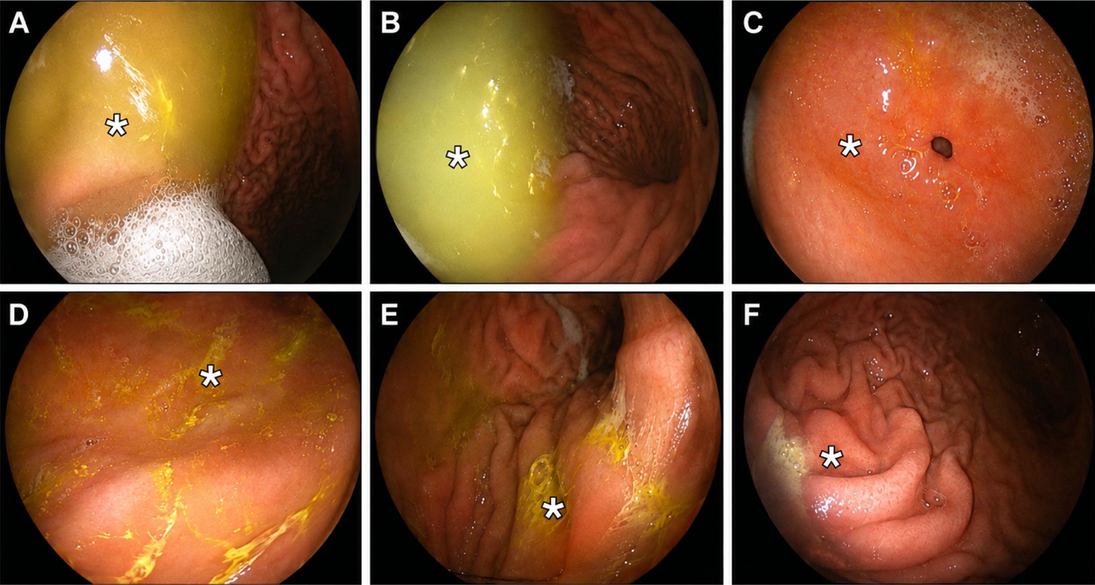
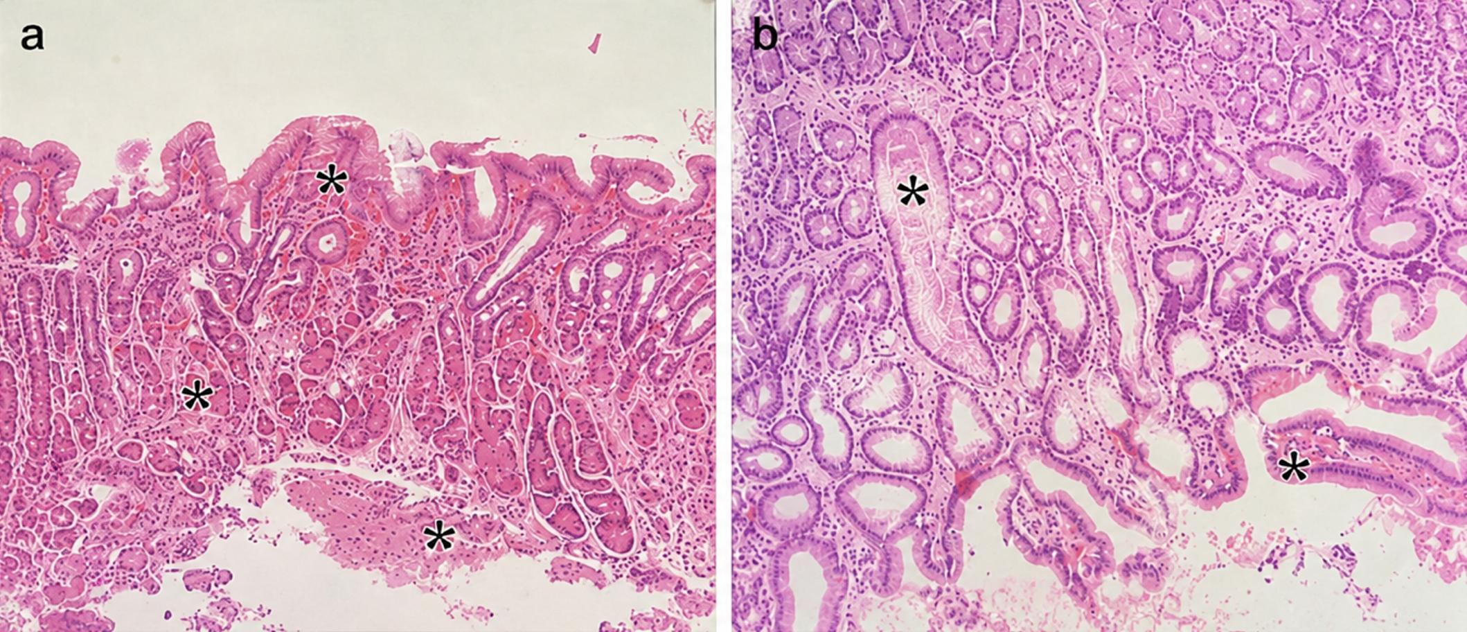
Under deep sedation induced by an anesthetist, an upper gastrointestinal (UGI) tract endoscopy was performed by 2 pediatric gastroenterologists using an Olympus X260 scope (Olympus Optical Corporation, Japan). Biopsy samples from the esophagus, stomach, and duodenum were delivered to pathology in a formalin fixation solution. Biopsy specimens were stained with hematoxylin-eosin and evaluated by light microscopy. Given the lack of access to advanced diagnostic tools such as Bilitec 2000, the diagnosis of DGR was based on a combination of endoscopic, histopathological, and clinical findings. During endoscopy, patients with hyperemia, fragility, edema, bile acid pool, and solid bile wastes in the gastric mucosa (Fig. 1), together with histopathological findings including inflammation and foveolar hyperplasia in gastric biopsies (Fig. 2), were evaluated as having DGR.12 Histopathological findings were not considered diagnostic alone but were interpreted together with endoscopic bile-related findings and clinical presentation to support the diagnosis of DGR. Because a fasting period (8 hours) before the endoscopy can also lead to bile pooling in the stomach, the mere detection of a bile pool was considered insufficient for the definition of DGR; the presence of solid bile wastes adherent to the stomach wall, and endoscopic and histopathological evidence of bile-related inflammation in the mucosa were also required.
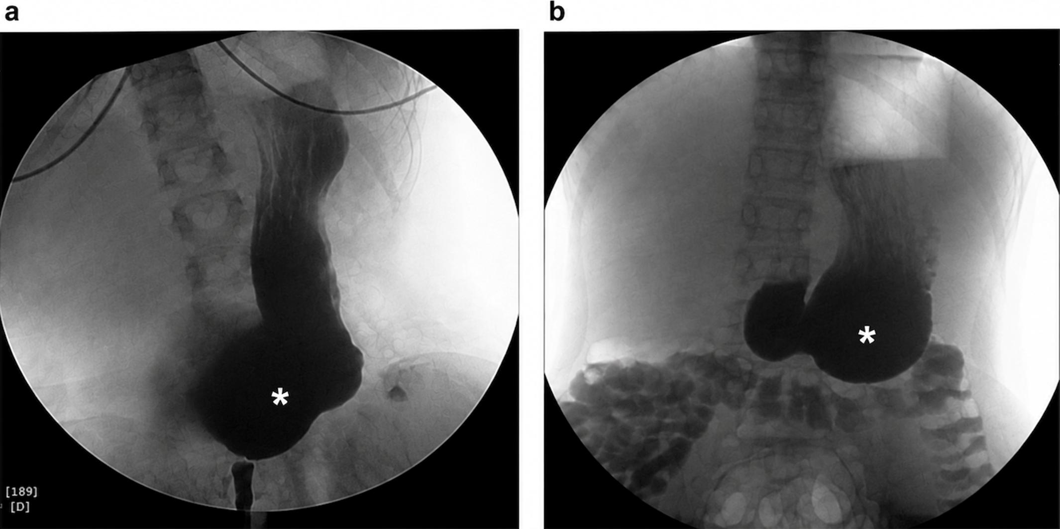
For the purposes of this study, treatment-resistant dyspepsia was defined as persistence of dyspeptic symptoms despite at least 8 weeks of proton pump inhibitor (PPI) therapy. To detect underlying gastroptosis and gastroparesis, UGI fluoroscopy examinations and gastric emptying scintigraphy were performed in patients with treatment-resistant dyspepsia or in patients with DGR. Gastroptosis was diagnosed by UGI fluoroscopy either with barium or water-soluble iodinated contrast according to the evaluation of the radiologists (Fig. 3). In the UGI fluoroscopy examination, elongation of the stomach’s greater curve down to the level of the iliac crests was considered gastroptosis.13 Gastroparesis was diagnosed by gastric emptying scintigraphy performed using a standardized solid-phase radiolabeled meal consisting of technetium-99m sulfur colloid–labeled eggs (equivalent to two eggs), accompanied by bread and water, in accordance with established gastric emptying scintigraphy protocols. Gastric emptying time was considered delayed if there was retention > 90% 1 hour after the meal, > 60% 2 hours, and > 10% 4 hours after.14
The anthropometric measurements, including body mass index (BMI) z-scores, were obtained from the patients’ medical records. BMI z-scores were calculated using the World Health Organization (WHO) growth assessment software.15 Patients who underwent endoscopy for dyspepsia and were found to have gastric inflammation were treated with PPIs. Those diagnosed with DGR received a combination of PPI and ursodeoxycholic acid. Prokinetic treatment (domperidone) was added in selected patients with gastroptosis and/or gastroparesis who had persistent dyspeptic symptoms or DGR. However, treatment response was not assessed using a standardized symptom severity scoring system, which is acknowledged as a limitation of the study.
Statistical analysis
Statistical data were evaluated using the Statistical Package for the Social Sciences (SPSS) for Windows version 20.0. Descriptive statistics were presented as numbers and percentages for categorical variables. Continuous variables were presented as mean ± standard deviation if normally distributed and as median (Q1–Q3) if the distribution was non-normal. The normality of numerical variables was assessed using the Kolmogorov–Smirnov test. Comparisons of normally distributed continuous variables were performed using the Student’s t-test, whereas non-normally distributed variables were compared using the Mann–Whitney U test. Categorical variables were compared using the chi-square test or Fisher’s exact test, as appropriate. A p value <0.05 was considered statistically significant.
Results
Gastroptosis and gastroparesis were investigated in 88 of 920 patients who underwent endoscopy with dyspepsia (DGR = 50, treatment-resistant dyspepsia = 38). The mean age of the patients was 13.7 ± 3.6 years, and 62 (70.5%) were girls. Gastroptosis was detected in 22 (25%) and delayed gastric emptying (gastroparesis) in 24 (27%) of all 88 patients. Gastroptosis was present in 10 (20%) and gastroparesis in 20 (40%) of the 50 patients with DGR. Gastroptosis was present in 12 (32%) and gastroparesis in 4 (11%) of the 38 patients with treatment-resistant dyspepsia. Gastroparesis was more common in those with DGR (40%) than in those with treatment-resistant dyspepsia (11%), (p = 0.09). (Table I). A notable overlap was observed between gastroptosis and gastroparesis, with gastroptosis present in 14 (58.3%) patients with gastroparesis.
| Chi-square test or Fisher's exact test, as appropriate. | |||
| Table I. The frequency of gastroptosis and gastroparesis among patient groups. | |||
| Variables |
|
|
|
| Upper gastrointestinal fluoroscopy |
|
||
| Normal |
|
|
|
| Gastroptosis |
|
|
|
| Gastric emptying scintigraphy |
|
||
| Normal |
|
|
|
| Gastroparesis |
|
|
|
The median BMI z-score of those with gastroptosis was significantly lower than those without (p = 0.002). There was no statistically significant difference in median BMI z- scores according to the presence of gastroparesis (p = 0.92) (Table II). While 12% of those with DGR were underweight according to their BMI z- score, there were no patients in the underweight category among those with treatment-resistant dyspepsia (p = 0.03). There was no significant difference between the mean ages according to the presence of gastroptosis (14.45 ± 3.05 vs. 13.52 ± 3.80 years, p = 0.38); however, the mean age of those with gastroparesis was significantly lower than those without (13.13 ± 1.95 vs. 14.48 ± 3.77 years, p = 0.03) and the mean age of those with DGR was significantly higher than those without (14.68 ± 2.64 vs. 12.53 ± 4.38 years, p = 0.02). There was no statistically significant difference in the frequency of gastroptosis between girls and boys (16/62 [25.8%] vs. 6/26 [23.1%], p = 0.787). Similarly, the frequency of gastroparesis did not differ significantly between girls and boys (18/62 [29.0%] vs. 6/26 [23.1%], p = 0.567).
|
Mann–Whitney U test. BMI z-scores given as median (Q1, Q3) BMI: body mass index, UGI: upper gastrointestinal |
||
| Table II. Comparison of BMI z- scores of patients with gastroptosis or gastroparesis with those without. | ||
|
|
|
|
|
|
|
|
|
|
|
|
|
|
|
|
Discussion
Gastroptosis is infrequently reported in the literature, particularly in pediatric populations, and its true prevalence remains unclear.16 Sukimo et al. reported that gastroptosis was seen in 12% of men and 43% of women.17 In a study in which Kusano et al. screened approximately 12,000 people in 2 years, gastroptosis was found in 500 women and 167 men.6 In our study, there was no significant difference between the frequency of gastroptosis according to age and sex, but similar to the literature, girls were the majority of patients with gastroptosis (16 girls, 6 boys).
It is believed that gastroptosis is due to the weakening and relaxation of the abdominal wall muscles, reduction of abdominal fat (especially the lesser omentum), and relaxation of the gastrohepatic and gastrocolic ligaments. The most important theory regarding its etiology is asthenic body structure and weak development of muscle and connective tissues (undifferentiated connective tissue dysplasia).18,19 Consistent with this theory, our study found that children with gastroptosis had significantly lower BMI z-scores than those without, supporting the hypothesis that reduced nutritional status or body mass may play a role in the development or worsening of gastroptosis.
In an adult study, Kusano et al. investigated the relationship between gastroptosis and dyspepsia in the Japanese population and found that dyspeptic symptoms were significantly less common in patients with gastroptosis.6 They hypothesized that since there is a close relationship between high BMI and gastroesophageal reflux disease, and patients with gastroptosis have lower BMIs than controls, subjects with gastroptosis monitor their weight and diet more carefully.6 In contrast, our findings showed gastroptosis in 32% of children with treatment-resistant dyspeptic complaints, with significantly lower BMI z-scores compared to those without gastroptosis. This indicates that dietary modification alone may be insufficient in pediatric dyspepsia, particularly when gastroptosis coexists. Moreover, a recent pediatric study demonstrated that alkaline reflux was more prevalent in children with gastroptosis, and its frequency increased with the severity of gastroptosis.7 In our cohort, gastroptosis was present in 20% of patients with DGR, supporting a potential association between gastric displacement and alkaline reflux.
Gastroparesis is a gastric motility disorder characterized by delayed gastric emptying in the absence of mechanical obstruction, for which the gold standard for diagnosis is gastric emptying scintigraphy.20 Large pediatric cohorts report a roughly equal sex distribution,21 consistent with our findings. While gastroparesis and functional dyspepsia are noticed more frequently in adults, the overall prevalence in children is unknown.22 The clinical presentations of patients with gastroparesis and functional dyspepsia are significantly similar.23 Gastric motility disorders, including delayed gastric emptying and reflux of duodenal contents into the stomach, are known to cause significant clinical problems.24 In our study, gastroparesis was identified in 11% of children with treatment-resistant dyspeptic symptoms. Notably, it was more prevalent in patients with DGR, being detected in 40% of these cases. These findings suggest that underlying gastric motility disturbances may contribute to the pathophysiology of DGR and persistent dyspeptic symptoms in children.
It has been suggested that delayed or misdiagnosed gastroparesis may contribute to the development of gastroptosis. The coexistence of gastroptosis and gastroparesis has been shown in only one case in the literature.4 In our study, gastroptosis was detected in 58.3% of patients with gastroparesis. This high rate of coexistence suggests a potential pathophysiological link between the two conditions, which has not previously been demonstrated in a pediatric population.
Nowadays, surgical treatment of gastroptosis is limited to a small number of complicated cases. Current treatment includes prokinetic drugs and adequate nutrition.18 Management of gastroparesis includes dietary modifications (small volume and frequent meals with low content in fat and non-digestible fibers), medical treatments (prokinetic drugs, antiemetics, PPIs, intrapyloric injection of botulinum toxin) and surgical interventions.20 In our study, prokinetic treatment with domperidone was used in selected patients with gastroptosis and/or gastroparesis who had persistent dyspeptic symptoms or DGR. These findings indicate that identifying underlying gastroptosis or gastroparesis may have practical implications for the clinical management in children with persistent symptoms. However, treatment response was assessed clinically during follow-up visits without the use of a standardized symptom severity scoring system, which represents an important limitation of the study.
The most reliable of the currently available tools for excessive bile reflux detection is the recent Bilitec 2000 method (Synectics Medical) consisting of a fiberoptic probe connected to a portable bilimeter which allows 24 h continuous endoluminal monitoring of bilirubin exposure based on its spectrophotochemical properties.25 The other limitation of our study is that this method could not be used to define DGR in our study, and the endoscopic and histopathologic findings may not fully reflect the 24-hour variability of DGR.
In conclusion, gastroptosis and gastroparesis were frequently identified among children with treatment-resistant dyspeptic complaints or DGR. Evaluating these conditions in this patient population may improve diagnostic accuracy, facilitate appropriate management, and help reduce complications related to delayed diagnosis.
Ethical approval
The study was approved by Clinical Research Ethics Committee of Gülhane Training and Research Hospital (date: March 17, 2020, number: 2020/111).
Source of funding
The authors declare the study received no funding.
Conflict of interest
The authors declare that there is no conflict of interest.
References
- Robin SG, Keller C, Zwiener R, et al. Prevalence of pediatric functional gastrointestinal disorders utilizing the Rome IV Criteria. J Pediatr 2018; 195: 134-139. https://doi.org/10.1016/j.jpeds.2017.12.012
- Rajindrajith S, Zeevenhooven J, Devanarayana NM, Perera BJC, Benninga MA. Functional abdominal pain disorders in children. Expert Rev Gastroenterol Hepatol 2018; 12: 369-390. https://doi.org/10.1080/17474124.2018.1438188
- Shava U, Srivastava A, Mathias A, et al. Functional dyspepsia in children: a study of pathophysiological factors. J Gastroenterol Hepatol 2021; 36: 680-686. https://doi.org/10.1111/jgh.15193
- Christianakis E, Bouchra K, Koliatou A, Paschalidis N, Filippou D. Gastroparesis associated with gastroptosis presenting as a lower abdominal bulking mass in a child: a case report. Cases J 2009; 2: 184. https://doi.org/10.1186/1757-1626-2-184
- Sullivan SN. Gastroptosis: a cause of postprandial abdominal bloating. Can J Gastroenterol Hepatol. 1991; 5: 3. https://doi.org/10.1155/1991/412406
- Kusano M, Moki F, Hosaka H, et al. Gastroptosis is associated with less dyspepsia, rather than a cause of dyspepsia, in Japanese persons. Intern Med 2011; 50: 667-671. https://doi.org/10.2169/internalmedicine.50.4582
- Tangul SU, Senayli A. Evaluation of the relationship between gastroptosis and reflux in pediatric patients. Front Med (Lausanne) 2025; 12: 1543297. https://doi.org/10.3389/fmed.2025.1543297
- Venkatasubramani N, Sood MR. Motility disorders of the gastrointestinal tract. Indian J Pediatr 2006; 73: 927-930. https://doi.org/10.1007/BF02859288
- Vandenplas Y, Hauser B, Salvatore S. Current pharmacological treatment of gastroparesis. Expert Opin Pharmacother 2004; 5: 2251-2254. https://doi.org/10.1517/14656566.5.11.2251
- Szarszewski A, Korzon M, Kamiñska B, Lass P. Duodenogastric reflux: clinical and therapeutic aspects. Arch Dis Child 1999; 81: 16-20. https://doi.org/10.1136/adc.81.1.16
- Pazzi P, Scalia S, Stabellini G, Trevisani L, Alvisi V, Guarneri M. Bile reflux gastritis in patients without prior gastric surgery: therapeutic effects of ursodeoxycholic acid. Current Therapeutic Research 1989; 45: 476-487.
- Türker SN, Barış Z, Şeker NS, Aydemir Y. Histopathological differences in pediatric duodenogastric reflux: a comparative study. Eur J Pediatr 2025; 184: 343. https://doi.org/10.1007/s00431-025-06163-z
- Van Welie AJM, Klein WM, Draaisma JMT. The clinical or radiographic diagnosis of gastroptosis: still relevant? Gastro Open J. 2017; 2: 14-19. https://doi.org/10.17140/GOJ-2-126
- Chogle A, Saps M. Gastroparesis in children: the benefit of conducting 4-hour scintigraphic gastric-emptying studies. J Pediatr Gastroenterol Nutr 2013; 56: 439-442. https://doi.org/10.1097/MPG.0b013e31827a789c
- World Health Organization (WHO). WHO Anthro and Macros. Version 3.2.2. [S. l.]. WHO; 2011. Available at: https://www.who.int/tools/child-growth-standards/software. (Accessed on March 1, 2019).
- Bestari MB, Chandra M, Joewono IR, et al. Gastroptosis due to gastric outlet obstruction secondary to duodenal tumor: Glenbard’s disease revisited. Case Rep Gastroenterol 2022; 16: 89-93. https://doi.org/10.1159/000521977
- Sukimo S, Kazutaka H. The forms of the stomach in healthy individuals: with special reference to analysis of relationships between the degree of gastroptosis and sex, age, comparative weight ratio or equivocal complaints. Kitasato Med 1986; 16: 211-226.
- Shulhai OM, Kabakova AB, Klym LA, Shulhai AMA, Glushko KT. The problem of gastroptosis as a manifestation of undifferentiated connective tissue dysplasia in the clinical practice of pediatric gastroenterologist. Child’s Health 2021; 13: 112-117. https://doi.org/10.22141/2224-0551.13.0.2018.131191
- Staszewska A, Jarzumbek A, Saran A, Gierak-Firszt S, Kwiecien J. Postprandial abdominal pain caused by gastroptosis-a case report. children (Basel) 2023; 10: 116. https://doi.org/10.3390/children10010116
- Saliakellis E, Fotoulaki M. Gastroparesis in children. Ann Gastroenterol 2013; 26: 204-211.
- Waseem S, Islam S, Kahn G, Moshiree B, Talley NJ. Spectrum of gastroparesis in children. J Pediatr Gastroenterol Nutr 2012; 55: 166-172. https://doi.org/10.1097/MPG.0b013e31824cf06e
- Islam S. Gastroparesis in children. Curr Opin Pediatr 2015; 27: 377-382. https://doi.org/10.1097/MOP.0000000000000216
- Stanghellini V, Tack J. Gastroparesis: separate entity or just a part of dyspepsia? Gut 2014; 63: 1972-1978. https://doi.org/10.1136/gutjnl-2013-306084
- Ramkumar D, Schulze KS. The pylorus. Neurogastroenterol Motil 2005; 17(Suppl 1): 22-30. https://doi.org/10.1111/j.1365-2982.2005.00664.x
- Hermans D, Sokal EM, Collard JM, Romagnoli R, Buts JP. Primary duodenogastric reflux in children and adolescents. Eur J Pediatr 2003; 162: 598-602. https://doi.org/10.1007/s00431-003-1259-y
Copyright and license
Copyright © 2026 The Author(s). This is an open access article distributed under the Creative Commons Attribution License (CC BY), which permits unrestricted use, distribution, and reproduction in any medium or format, provided the original work is properly cited.