Graphical Abstract
Abstract
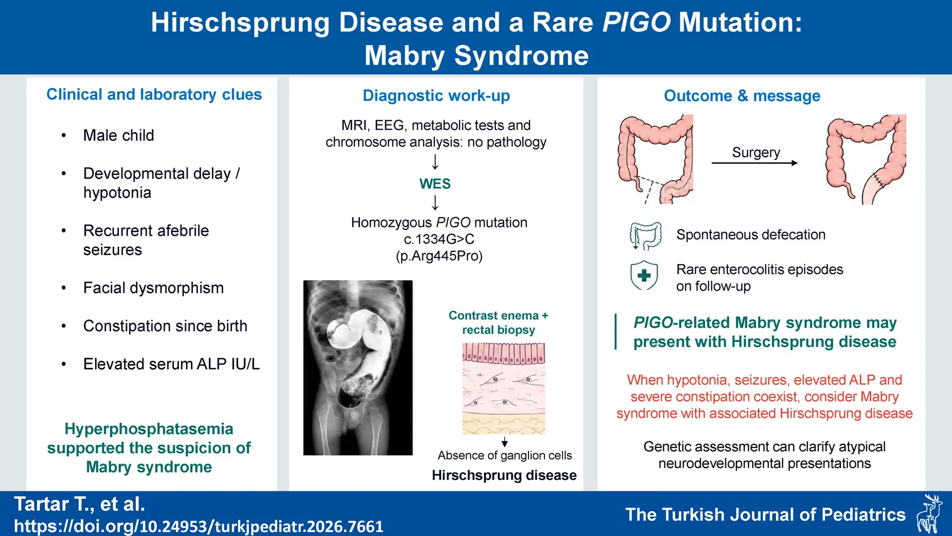
Background. Mabry syndrome is a very rare condition characterized by cognitive impairment, distinctive facial features, hypotonia, elevated alkaline phosphatase (ALP) levels in the blood (hyperphosphatasia), and recurrent generalized tonic-clonic seizures. It can be accompanied by gastrointestinal anomalies such as Hirschsprung’s disease (HD). In this study, we aim to present a case of hyperphosphatasia with impaired intellectual development syndrome 2 (HPMRS2) from Türkiye, featuring a novel homozygous p.Arg445Pro variant in the PIGO gene, occurring in association with HD.
Case Presentation. A two-year-old male patient was referred to the pediatric neurology clinic with decreased responsiveness to the environment, lack of trunk control, and failure to respond to his name. ALP value was 613 IU/L. No pathology was detected in metabolic tests, vitamin D levels, or cranial magnetic resonance imaging, and electroencephalography (EEG). At 11 months of age, he had a generalized tonic-clonic seizure without fever. Since no abnormalities were detected in chromosome analysis and array analysis, whole exome sequencing (WES) was performed. A homozygous mutation was detected in the PIGO gene (NM_032634.4): c.1334G>C (p.Arg445Pro). Sanger analysis revealed heterozygous carrier status in both parents. The patient had constipation that was unresponsive to medical treatment since birth. Physical examination revealed abdominal distension. Rectal examination revealed an explosive discharge of gas and stool. A rectal biopsy was performed. The pathology result was ganglion-negative, and the patient was diagnosed with HD. After definitive surgery, spontaneous defecation has been achieved during follow-up, except for rare episodes of enterocolitis.
Conclusions. It should be remembered that Mabry syndrome can be accompanied by gastrointestinal anomalies, primarily HD. This case highlights the increased phenotypic variability associated with PIGO mutations and demonstrates the importance of genetic assessment in atypical neurodevelopmental conditions.
Keywords: seizures, constipation, Hirschsprung’s disease, Mabry syndrome, child
Introduction
Mabry syndrome, which is a very rare condition, is also known as hyperphosphatasia with impaired intellectual development syndrome-2 (HPMRS2). Its metabolic pathophysiology is based on glycosylphosphatidylinositol (GPI) deficiency. The clinical presentation often includes intellectual developmental delay, marked hypotonia, delayed walking, hypertelorism, and facial dysmorphism characterized by long palpebral fissures. Seizures are frequently recurring generalized tonic-clonic seizures accompanied by loss of consciousness, and are often unresponsive to single medical therapy. Rarely, cardiac and gastrointestinal (anal atresia, Hirschsprung’s disease) anomalies may accompany. Serum alkaline phosphatase (ALP) levels are significantly elevated in most patients, and hyperphosphatasemia is the main biochemical marker of Mabry syndrome.1-3
In this study, we aim to report a rare case of HPMRS2 in a Turkish patient, identified with a novel homozygous p.Arg445Pro variant in the PIGO gene. The clinical phenotype is characterized by hypotonia, refractory epilepsy, and Hirschsprung’s disease (HD). This report seeks to expand the known clinical phenotypic diversity associated with this syndrome.
Case Presentation
A nine-month-old male patient was admitted to the pediatric neurology clinic with complaints of delayed head control, lack of body control without support, failure to respond to his name, decreased eye tracking, and chronic constipation. His prenatal and natal history was unremarkable, and the baby was born at term with a birth weight of 2900 grams. From the neonatal period, the patient had severe constipation and required rectal stimulation for defecation. Spontaneous defecation was not observed, and progressive abdominal distension was recorded during infancy. Physical examination revealed facial dysmorphism (micrognathia, wide and flat nose, long philtrum, and low-set ears) and generalized hypotonia. No organomegaly was detected. At one year of age, the patient’s measurements were as follows: length 70 cm (10th–25th percentile), weight 7.6 kg (3rd–10th percentile), and head circumference 44 cm (10th percentile).
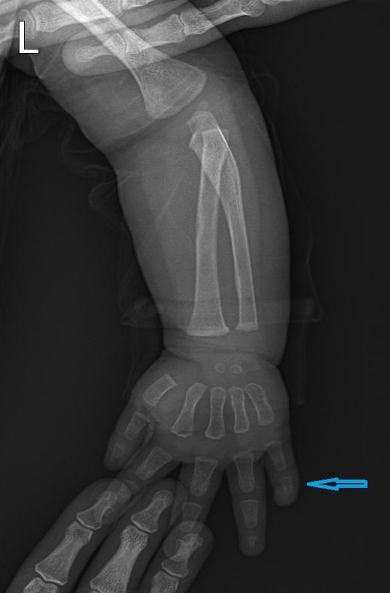
At 11 months of age, he had an afebrile generalized tonic-clonic seizure. No abnormalities were observed on electroencephalography (EEG). Levetiracetam was initiated after seizure recurrence during drug-free follow-up. However, seizures persisted, and the medication dose was adjusted to 60 mg/kg/d. Serum ALP levels were found to be elevated at 613 IU/L (reference range 0–240 IU/L). There was no vitamin D deficiency, which could cause elevated ALP levels. Metabolic tests, including those for liver disease and increased bone metabolism, were also normal. The patient’s forearm radiographs showed no abnormalities. The hand radiograph revealed hypoplasia in the distal phalanx of the 5th finger (Fig. 1). Cranial magnetic resonance imaging revealed no pathology. Since no abnormalities were detected in chromosome analysis and array analysis, whole exome sequencing (WES) was performed. A homozygous mutation was detected in the PIGO gene (NM_032634.4): c.1334G>C (p.Arg445Pro). Sanger sequencing revealed heterozygous carrier status in both parents.
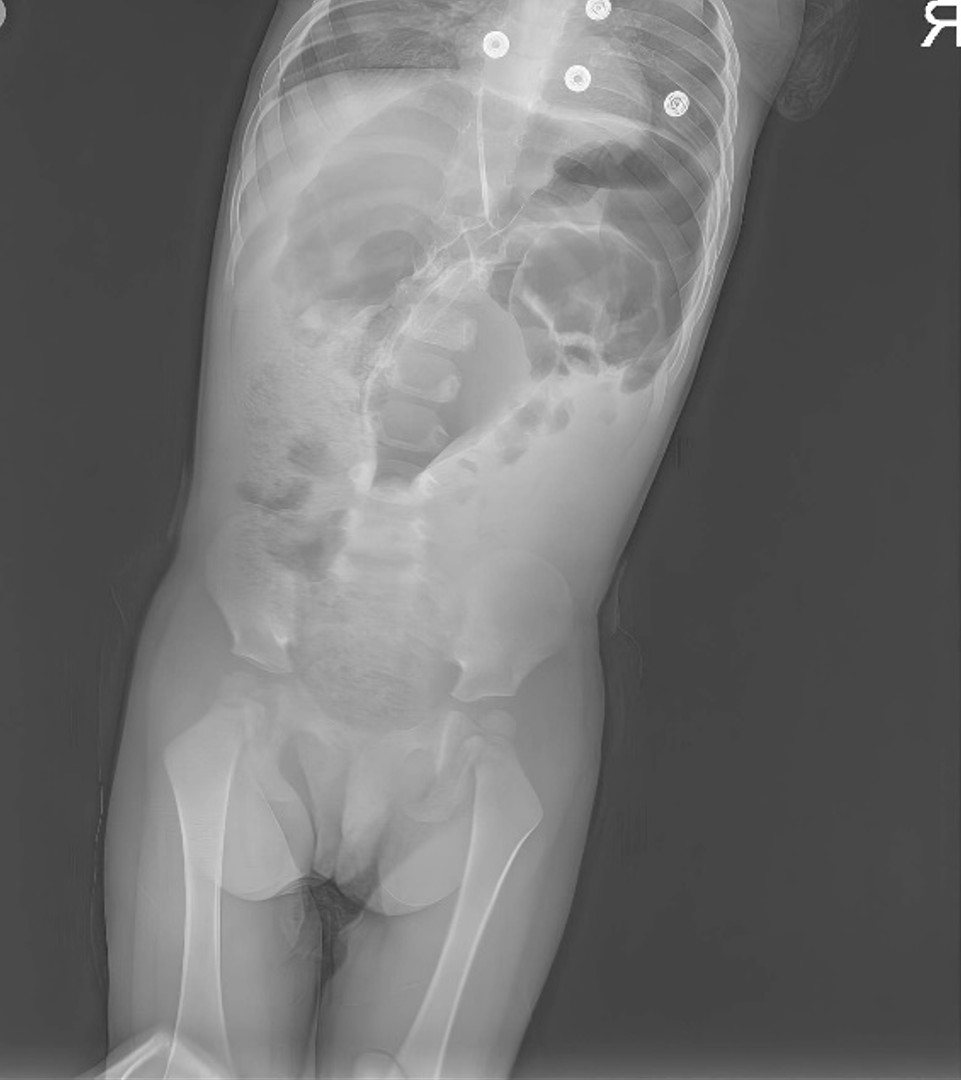
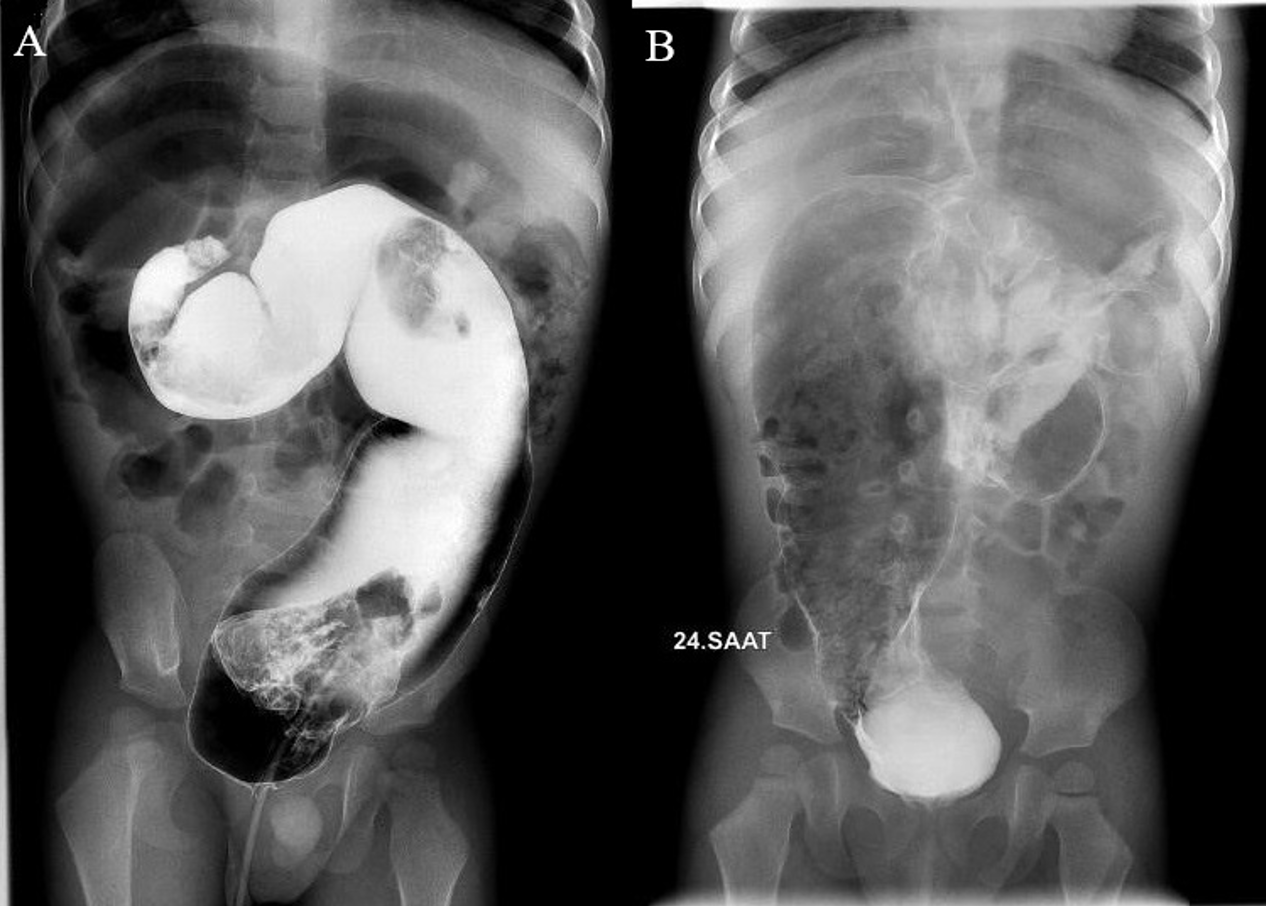
The Ankara Developmental Screening Inventory (AGTE) was administered at 18 months of age. Developmental age equivalents were 12 months in the Language–Cognitive domain, 14 months in the Fine Motor domain, 14 months in the Gross Motor domain, and 12 months in the Social–Self-Care domain. The patient was referred to the pediatric surgery clinic with complaints of constipation unresponsive to medical treatment. Physical examination revealed abdominal distension, but the abdomen was soft. Rectal examination revealed an explosive discharge of gas and stool. An upright abdominal X-ray showed that the pelvis contained almost no intestinal gas, but there were dilated bowel loops (Fig. 2). Contrast-enhanced colonography demonstrated a transition zone suggestive of HD (Fig. 3). A rectal biopsy confirmed the absence of ganglion cells in the submucosal plexus, and the patient was diagnosed with HD (Fig. 4). The aganglionic segment was limited to the distal sigmoid colon. The patient subsequently underwent transanal endorectal pull-through surgery. Histopathological examination of the resected specimen confirmed the aganglionic distal segment and normal ganglion cells in the proximal bowel. During follow-up, the patient achieved spontaneous defecation, except for rare episodes of enterocolitis.
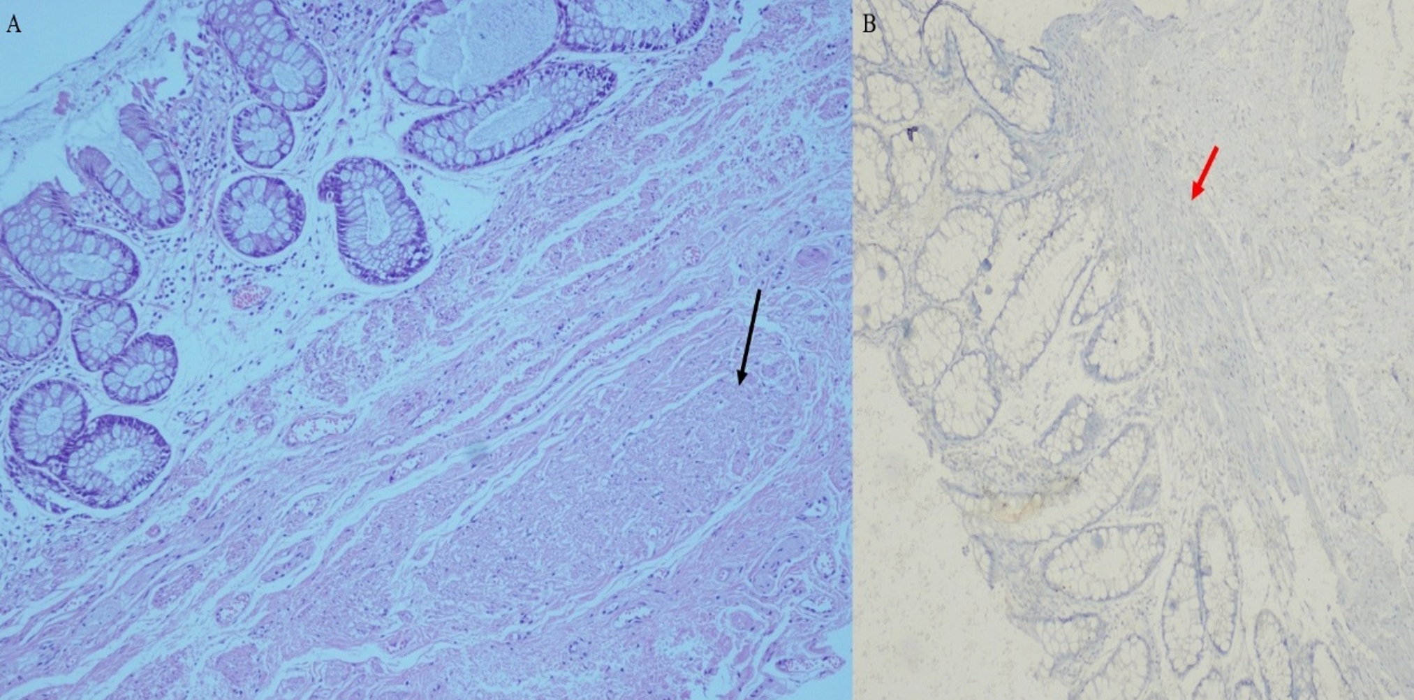
A) The illustration shows aganglionic rectal mucosa and hypertrophic nerve fibers (black arrow) (H&E, original magnification x20). B) Negative calretinin immunstaining of rectal mucosa (red arrow) (Calretinin IHC, original magnification x 20).
Informed consent for the publication of this case report was obtained from the patient’s parents. However, clinical photographs could not be included because parental consent for the publication of facial images could not be obtained.
Discussion
Mabry syndrome is a rare, autosomal recessive disease characterized by moderate/severe psychomotor developmental delay, refractory seizures, facial dysmorphism, brachytelephalangy, and hyperphosphatasemia.1,2 It develops as a result of mutations in genes that are involved in the biosynthesis of GPI anchors. The biosynthesis, protein binding, maturation, and intracellular transport of GPI anchors involve a complex process involving more than 30 genes.
Hirschsprung’s disease is a congenital aganglionosis characterized by the absence of ganglion cells in the distal intestine due to a failure in the migration, proliferation, differentiation, or survival of vagal and sacral neural crest cells, which form the precursor cells of the enteric nervous system (ENS) during the embryonic period. GPI-anchored proteins regulate signaling pathways that are important in the migration and differentiation of neural crest cells. Therefore, GPI biosynthesis deficiencies may be associated with HD because they can disrupt the normal development of the ENS.4,5
GPI-anchored proteins play a critical role in cell surface adhesion, signal transduction, and neurodevelopmental processes. The PIGO gene encodes one of the ethanolamine phosphate transferase enzymes responsible for the addition of ethanolamine phosphate to the GPI anchor, and mutations can lead to multisystem involvement.6,7 Few patients with PIGO mutations have been described in the literature, and the clinical spectrum is quite broad. In our case, a homozygous mutation of c.1334G>C (p.Arg445Pro) was detected in the PIGO gene (NM_032634.4), which has not been previously identified in Türkiye. Based on the assessment conducted in accordance with the ACMG guidelines, the identified variant was classified as a variant of uncertain significance. Nevertheless, the clinical constellation of hyperphosphatasia, hypotonia, intellectual disability, epilepsy, and HD observed in our patient exhibits a high degree of phenotypic specificity characteristic of HPMRS2 associated with PIGO mutations. In this context, the fulfillment of the PP4 criterion significantly bolsters the likelihood of pathogenicity and reinforces the correlation between the molecular findings and the clinical presentation. Comprehensive literature and database queries, including PubMed and ClinVar, confirmed that this variant has not been previously reported. Consequently, this study represents an original case report that contributes to the existing literature and provides critical diagnostic data for the future identification of similar cases.
According to the population data criterion (PM2), the variant is exceedingly rare and, to the best of our knowledge, is being reported for the first time in the literature. Although data regarding the clinical effects of the p.Arg445Pro variant are limited, no functional study directly evaluating this specific amino acid substitution in PIGO has yet been reported. Nevertheless, when considered together with previously published functional and structural data on PIGO variants, as well as the well-established helix-disrupting biochemical property of proline, the arginine-to-proline substitution is biologically highly likely to exert a deleterious effect on protein stability and/or conformation, and consequently on PIGO function.8 The present case is also thought to expand the clinical spectrum of Mabry syndrome, particularly with respect to the presence of refractory seizures, hypotonia, intellectual disability, hyperphosphatasemia, and its association with HD.
Holtz et al., in their study summarizing a literature review, evaluated 18 cases with PIGO mutations in detail, and observed developmental delay and hyperphosphatasemia in all cases. The most common dysmorphic features were distal digital hypoplasia (77.8%), seizures that usually began before the age of 2 years (66.7%), and nail anomalies (55.6%). Although variable, the most common facial dysmorphisms were ear anomalies (44.4%), and tent-shaped upper lip (33.3%). Hearing loss was also reported in 50% of cases. ALP levels exceeded 1000 U/L in 55.6% of the cases.1 Another study reported that GPI deficiencies were seen in a very wide clinical spectrum.2 In the presented case, digital anomalies and facial dysmorphism were present, consistent with the literature, but hearing loss was not detected and the serum ALP level was 613 IU/L.
In their study, Holtz et al. found gastrointestinal anomalies in 55.6% of the cases. The anomalies detected were anal atresia (33.3%), HD (33.3%), and esophageal atresia (11.1%).1 The significant number of gastrointestinal anomalies in Mabry syndrome suggests that PIGO mutations may have an impact on neuroenteric development. In the presented case, HD was identified, and definitive surgery was performed. In this respect, it is one of the first cases of HD caused by the PIGO mutation reported from Türkiye.
In conclusion, Mabry syndrome should be considered in cases of hypotonia, refractory seizures beginning before the age of 2, and unexplained hyperphosphatemia. It should be kept in mind that serious gastrointestinal abnormalities, particularly HD, may be present. This case highlights the increased phenotypic variability associated with PIGO mutations and demonstrates the importance of genetic assessment in atypical neurodevelopmental conditions.
Ethical approval
Informed consent for the publication of this case report was obtained from the patient’s parents.
Source of funding
The authors declare the study received no funding.
Conflict of interest
The authors declare that there is no conflict of interest.
References
- Holtz AM, Harrington AW, McNamara ER, et al. Expanding the phenotypic spectrum of Mabry Syndrome with novel PIGO gene variants associated with hyperphosphatasia, intractable epilepsy, and complex gastrointestinal and urogenital malformations. Eur J Med Genet 2020; 63: 103802. https://doi.org/10.1016/j.ejmg.2019.103802
- Bellai-Dussault K, Nguyen TTM, Baratang NV, Jimenez-Cruz DA, Campeau PM. Clinical variability in inherited glycosylphosphatidylinositol deficiency disorders. Clin Genet 2019; 95: 112-121. https://doi.org/10.1111/cge.13425
- Fleming L, Lemmon M, Beck N, et al. Genotype-phenotype correlation of congenital anomalies in multiple congenital anomalies hypotonia seizures syndrome (MCAHS1)/PIGN-related epilepsy. Am J Med Genet A 2016; 171: 77-86. https://doi.org/10.1002/ajmg.a.37369
- McKeown SJ, Stamp L, Hao MM, Young HM. Hirschsprung disease: a developmental disorder of the enteric nervous system. Wiley Interdiscip Rev Dev Biol 2013; 2: 113-129. https://doi.org/10.1002/wdev.57
- Lefeber DJ, Freeze HH, Steet R, Kinoshita T. Congenital disorders of glycosylation. In: Varki A, Cummings RD, Esko JD, et al., editors. Essentials of Glycobiology. 4th ed. Cold Spring Harbor (NY): Cold Spring Harbor Laboratory Press; 2022.
- Fujihara Y, Ikawa M. GPI-AP release in cellular, developmental, and reproductive biology. J Lipid Res 2016; 57: 538-545. https://doi.org/10.1194/jlr.R063032
- Horn D, Wieczorek D, Metcalfe K, et al. Delineation of PIGV mutation spectrum and associated phenotypes in hyperphosphatasia with mental retardation syndrome. Eur J Hum Genet 2014; 22: 762-767. https://doi.org/10.1038/ejhg.2013.241
- Susgun S, Ben-Mahmoud A, Rüschendorf F, et al. Macrocephaly and digital anomalies expand the phenotypic spectrum of PGAP2 variants in hyperphosphatasia with impaired intellectual development syndrome 3 (HPMRS3). Hum Mutat 2024; 5518289. https://doi.org/10.1155/2024/5518289
Copyright and license
Copyright © 2026 The Author(s). This is an open access article distributed under the Creative Commons Attribution License (CC BY), which permits unrestricted use, distribution, and reproduction in any medium or format, provided the original work is properly cited.