Abstract
Background. Congenital malformations of the external iliac arteries (EIAs) are rare and may be associated with other congenital anomalies, such as vertebral, cardiac, or limb anomalies, anal atresia, and tracheoesophageal fistula (VACTERL). These anomalies can be classified into three categories: anomalies of origin or course, hypoplasia or atresia with a persistent sciatic artery, and isolated hypoplasia or atresia. We report a term neonate with left leg cyanosis within the first 48 hours of life due to an isolated congenital EIA malformation.
Case Presentation. A male term neonate was delivered by emergency cesarean section at 40 weeks’ gestation for suspected asphyxia (birth weight 4040 g, length 52 cm, Apgar scores 10, 10). The pregnancy was uncomplicated except for gestational diabetes. The neonate showed no signs of distress or cyanosis at birth. Asymptomatic hypoglycemia was corrected with oral glucose. On the second day of life, the neonate developed cyanosis of the left lower limb, accompanied by delayed capillary refill (6 seconds), weak distal pulses, and reduced oxygen saturation (60–70%) as measured by pulse oximetry. Echocardiography excluded cardiac defects. Doppler ultrasound revealed high-grade stenosis of the left EIA, which was confirmed by magnetic resonance angiography, showing collateral circulation and no persistent sciatic artery. Coagulation tests were normal, and the neonate remained asymptomatic. At the 1-year follow-up, he demonstrated normal growth and neurodevelopment, with clinically normal development of the affected lower limb.
Conclusions. Neonatal limb cyanosis should prompt further evaluation after excluding other, more common conditions. Persistent cyanosis, especially when accompanied by weakness, atrophy, or length discrepancy, should raise suspicion for EIA malformations. To the best of our knowledge, this is the third reported case of EIA malformation and the first case of congenital segmental stenosis in a neonate. Despite their rarity, these malformations require consideration and early non-invasive imaging in unilateral limb cyanosis.
Keywords: external iliac artery, external iliac artery malformation, limb cyanosis, neonate
Introduction
Congenital malformations of the external iliac arteries (EIAs) are rare and have been described only a few times in the literature.1 The first reported case was published in 1957.2 The precise incidence remains undetermined.3 These arterial malformations have been classified into three categories by Tamisier et al: 1) anomalies in the origin or course of the artery, 2) hypoplasia or atresia compensated by a persistent sciatic artery (PSA), and 3) isolated hypoplasia or atresia.4 Unlike the first group, which is usually discovered at autopsy, the remaining categories are more likely to cause chronic ischemia of the leg, especially the third group, due to thrombotic tendency or intrinsic arterial wall abnormalities associated with obstructive lesions.4,5 Chronic leg ischemia due to hypoplasia or atresia usually occurs in childhood or young adulthood, however two cases of congenital malformation of the EIAs in neonates (aged 1 to 6 days) have been described in the literature.6,7 We report a case of a term neonate who developed cyanosis of the left leg within the first 48 hours of life due to an isolated congenital malformation of EIA, documented with corresponding clinical and radiographic images.
Case presentation
A male term neonate was delivered by emergency cesarean section at 40 weeks’ gestation due to suspected asphyxia to a 36-year-old mother. Birth weight was 4040 g (83rd centile), length 52 cm (65th centile), with Apgar scores of 10 and 10 at the 1st and 5th minutes, respectively. The neonate was in good clinical condition at birth, displaying no signs of distress or cyanosis, with normal cardiorespiratory function. As part of routine screening for neonates of mothers with gestational diabetes, asymptomatic hypoglycemia was detected, and successfully corrected with oral glucose, with subsequent blood glucose measurements remaining within normal range.
On the second day of life, the neonate developed cyanosis of the left lower limb, accompanied by delayed capillary refill (6 seconds), weak distal pulses, and reduced oxygen saturation (60–70%) as measured by pulse oximetry. The other extremities demonstrated normal saturation levels. Both legs were symmetrical in length and circumference, with normal spontaneous movements and no tenderness. Femoral pulses were palpable on both legs. The neonate was admitted to the neonatal intensive care unit (NICU) for further evaluation (Fig. 1).

Due to elevated inflammatory markers (C-reactive protein [CRP] 63.7 mg/L), a seven-day course of antibiotics was administered. Blood cultures remained negative. Acute onset of unilateral limb cyanosis warrants consideration of thromboembolism, catheter-related thrombosis, and traumatic vascular injury in the differential diagnosis. There was no history of umbilical or femoral arterial catheterization or traumatic procedures, and physical examination did not reveal signs of soft tissue injury or hematoma. The coagulation profile was within normal limits. Cranial, renal, and urinary tract ultrasound findings were normal, and echocardiography revealed no structural cardiac defects or intracardiac thrombi. Doppler ultrasound was performed, revealing high-grade stenosis of the left external iliac artery. Peak systolic velocity reached 200 cm/s at the stenotic segment with reduced post-stenotic flow, consistent with hemodynamically significant stenosis. Doppler findings localized to the external iliac artery without evidence of embolic occlusion in more distal vessels made an acquired thromboembolic event less likely and favored an underlying congenital arterial malformation. Magnetic resonance angiography (MRA) of the abdominal aorta and pelvic arteries on the third day of life showed a segmental absence of signal in the left external iliac artery, while the pelvic arteries on the right side displayed normal continuity. Collateral circulation was observed via circumflex branches of the iliac artery. No evidence of a PSA or other vascular anomalies was seen (Fig. 2). This lesion can be classified as an isolated EIA malformation (group 3) according to Tamisier’s classification4, representing a congenital segmental stenosis.
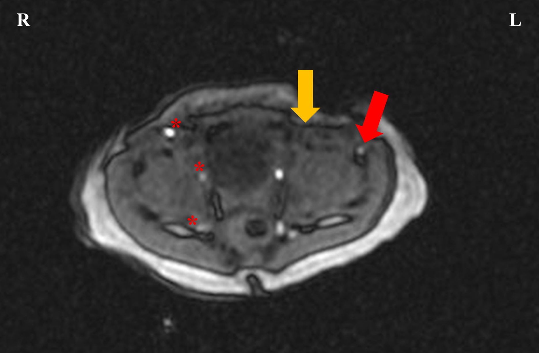
The neonate remained stable during hospitalization. No further episodes of cyanosis were observed, and the neonate maintained satisfactory oxygenation levels in the affected limb prior to discharge. Given the absence of acute complications, progressive ischemia or neurologic deficit, presence of collateral circulation, and spontaneous improvement in limb perfusion, a conservative management approach was chosen in agreement with pediatric surgery and vascular teams. Surgical/endovascular intervention was deferred, because the risk of procedural complications in a clinically stable neonate was considered to outweigh the potential benefit. Close clinical and outpatient follow-up were planned to monitor limb growth, function, and perfusion over time. At subsequent evaluations and pediatric surgical follow-ups, the infant remained symptom-free and demonstrated normal growth and development at 1 year of age. A follow-up MRA is planned at 2 years of age.
Written informed consent was obtained from the patient’s legal guardians for publication of this case report.
Discussion
The development of the arterial system of the lower extremities starts in a 6 mm human embryo (approximately 35-37 days of gestation). When the umbilical artery reaches the dorsal aspect of the Wolffian duct, it develops a branch called the axial artery which is the main embryonic lower extremity artery.8 Its proximal portion is referred to as the sciatic artery. EIA develops shortly thereafter, proximal to the origin of the axial (sciatic) artery, and then gives rise to the femoral artery, which forms an anastomosis with the sciatic artery by 42 to 49 days of gestation.7 As the femoral vessels form, the sciatic artery usually regresses. Therefore, in cases of inadequate development of the femoral or external iliac arteries, the sciatic artery may persist.9 The definitive arterial anatomy is usually completed by the third month of gestation. There have been speculations that hyperglycemia and/or hyperinsulinemia may contribute to the development of arterial malformations. However, no studies have confirmed this association to date.7
The average age of patients with EIA anomalies at diagnosis is 28 years, but ages range from 1 day to 73 years old with male predominance. Laterality of EIA anomalies have been reported as 42.9% on the right side and 57.1% on the left side.1 The PSA occurs with an estimated incidence of 0.03-0.06%.1,10
Growth of the lower extremities appears to be unaffected in utero despite arterial malformations, owing to the rich supply from the primary capillary plexus and the development of adequate collateral circulation.11 However, chronic limb ischemia in later life may lead to limb atrophy, leg length discrepancy, general weakness, and consequently motor delay.7
The presence of PSA mandates close monitoring, as almost 50% of patients develop an aneurysm of this remnant artery, which often manifests as sciatic neuropathy due to nerve compression. The lack of arterial elastic tissue in the PSA can contribute to aneurysm formation and thrombosis, leading to lower limb ischemia.12 Therefore, early surgical or endovascular intervention may be required when indicated.7
Some of these cases have been associated with other organ anomalies such as vertebral defects, anal atresia, cardiac defects, tracheoesophageal fistulas, renal anomalies and limb abnormalities (VACTERL association).1 The associated cardiac defects (3.8%) include type B1 distal interruption of the aortic arch, aortopulmonary window, patent ductus arteriosus and persistent foramen ovale.6 In our case, echocardiography excluded cardiac defects.
The molecular control of vascular development is not well understood and variations in regulation, genetic predisposition, and environmental factors (hypoxemia) can lead to variations in vessel growth and patterning.13
In our case, the diagnosis of external iliac artery malformation was established using duplex doppler ultrasound and MRA. Doppler ultrasound allows non-invasive, real-time assessment of arterial flow and hemodynamic significance of stenoses, while MRA provides detailed visualization of the aorto-iliac and femoropopliteal segments, including collateral pathways, and avoids ionizing radiation or iodinated contrast in neonates. Although three-dimensional reconstructions are sometimes used, only limited-quality cross-sectional images could be obtained in our case due to the use of a 2-Tesla system, the inability to perform MRA under anesthesia, and motion artefacts. Despite these limitations, the combination of doppler ultrasound and MRA was sufficient to confirm an isolated external iliac artery malformation and to guide conservative management. Consistent with our approach, previous reports have emphasized the role of doppler ultrasound and cross-sectional angiographic techniques (computered tomography or MRA) in visualizing external iliac or iliac artery malformations in neonates and children.14,15
In conclusion, limb cyanosis in infants should prompt further diagnostic evaluation after exclusion of more common neonatal conditions, such as infection, respiratory distress syndrome, cardiac anomalies, or hypothermia. Persistent cyanosis, particularly when accompanied by leg weakness, atrophy or length discrepancy, should raise suspicion for EIA anomalies. Although rare, these malformations may have significant long-term consequences if not recognized. To the best of our knowledge, this represents only the third reported neonatal case of EIA malformation and the first case of congenital segmental stenosis, emphasizing its exceptional rarity and adding to the limited existing literature.6,7
Ethical approval
Written informed consent was obtained from the patient’s legal guardians for the publication of this case report.
Source of funding
The authors declare the study received no funding.
Conflict of interest
The authors declare that there is no conflict of interest.
References
- Ezzone A, Al-Embideen S, Nazzal M, Osman M. A case report of congenital atresia of the right external iliac artery associated with congenital cardiac defect. Ann Vasc Surg Brief Rep Innov 2023; 3: 100163. https://doi.org/10.1016/j.avsurg.2023.100163
- Howard JM, Goudelock WJ, Couves CM. Congenital atresia of the external iliac artery. AMA Arch Surg 1957; 75: 296-299. https://doi.org/10.1001/archsurg.1957.01280140134025
- Xu Y, Yuan J, Li C. Unveiling the uncommon: hypoplasia of external iliac artery-a case report and literature review. J Cardiothorac Surg 2025; 20: 7. https://doi.org/10.1186/s13019-024-03202-y
- Tamisier D, Melki JP, Cormier JM. Congenital anomalies of the external iliac artery: case report and review of the literature. Ann Vasc Surg 1990; 4: 510-514. https://doi.org/10.1016/S0890-5096(07)60081-8
- Koyama T, Kawada T, Kitanaka Y, et al. Congenital anomaly of the external iliac artery: a case report. J Vasc Surg 2003; 37: 683-685. https://doi.org/10.1067/mva.2003.102
- Tůma S, Hruda J, First T, Vĕtrovská L. Agenesis of the external iliac artery and congenital heart defects. Case report of multiple vascular and organ anomalies. Cesk Pediatr 1989; 44: 675-677.
- Patel M, Chonat S, Olomu I, Arrington S, Kadrofske M. Absent left common and left external iliac artery presenting in a neonate. J Perinatol 2013; 33: 407-409. https://doi.org/10.1038/jp.2012.132
- Senior HD. The development of the arteries of the human lower extremity. Am J Anat 1919; 25: 54-95. https://doi.org/10.1002/aja.1000250105
- Link DP, Garza AS, Monsky WL. Congenital single, pelvic iliac artery: a case report. J Vasc Interv Radiol 2009; 20: 1231-1234. https://doi.org/10.1016/j.jvir.2009.05.042
- van Hooft IM, Zeebregts CJ, van Sterkenburg SM, de Vries WR, Reijnen MM. The persistent sciatic artery. Eur J Vasc Endovasc Surg 2009; 37: 585-591. https://doi.org/10.1016/j.ejvs.2009.01.014
- Lippert H, Pabst R. Arterial variations in man: classification and frequency. München: Springer; 1985: 54-61.
- Fung HS, Lau S, Chan MK, Tang KW, Cheung YL, Chan SC. Persistent sciatic artery complicated by aneurysm formation and thrombosis. Hong Kong Med J 2008; 14: 492-494.
- Qazi E, Wilting J, Patel NR, et al. Arteries of the lower limb-embryology, variations, and clinical significance. Can Assoc Radiol J 2022; 73: 259-270. https://doi.org/10.1177/08465371211003860
- Tekgündüz KŞ, Ceviz N, Kantarcı M, et al. Rare cause of absence of femoral arterial pulse: bilateral common iliac artery hypoplasia. Pediatr Int 2014; 56: 909-910. https://doi.org/10.1111/ped.12343
- Zhao S. Congenital atresia of external iliac artery: diagnosing a rare vascular anomaly cause of unilateral lower extremity claudication. 2024. Available at: https://msrads.web.unc.edu/wp-content/uploads/sites/15695/2024/09/RADY-External-Iliac-Artery-Atresia.pdf (Accessed on April 1, 2026).
Copyright and license
Copyright © 2026 The Author(s). This is an open access article distributed under the Creative Commons Attribution License (CC BY), which permits unrestricted use, distribution, and reproduction in any medium or format, provided the original work is properly cited.