Abstract
Background. Stuve–Wiedemann syndrome is a rare autosomal recessive bent-bone dysplasia characterized by dysautonomia and distinctive skeletal abnormalities, most commonly caused by biallelic LIFR variants.
Methods. Eleven patients from ten unrelated families with molecularly confirmed Stuve–Wiedemann syndrome were evaluated. Detailed demographic, perinatal, clinical, radiographic, and molecular data were collected. All patients underwent Sanger sequencing of LIFR.
Results. Respiratory problems and episodic hyperthermia were present in all patients, while feeding difficulties, hypotonia, growth failure, developmental delay, craniofacial dysmorphism, oral abnormalities, and ocular involvement were also common. All patients demonstrated long-bone bowing, cortical thickening, and flared metaphyses. A biallelic LIFR variant was identified in all patients, occurring in the homozygous state in nine patients and in the compound heterozygous state in two patients. The recurrent c.2074C>T; p.(Arg692Ter) variant was homozygous in seven patients and compound heterozygous in one patient. Three novel LIFR variants were also identified.
Conclusion. Stuve–Wiedemann syndrome is characterized by a high mortality rate during the first two years of life due to severe dysautonomia, and orthopedic complications worsen progressively with age; therefore, early diagnosis and lifelong multidisciplinary follow-up are essential. Furthermore, the high rate of consanguinity in Türkiye, together with the recurrent p.(Arg692Ter) variant in LIFR identified in multiple unrelated families, supports the possibility of a founder effect and suggests that the frequency of Stuve–Wiedemann syndrome in our population may be higher than expected. Our findings further characterize the previously reported clinical manifestations of Stuve–Wiedemann syndrome while expanding the molecular spectrum through the identification of three novel LIFR variants.
Keywords: Stuve-Wiedemann syndrome, LIFR, dysautonomia, skeletal dysplasia
Introduction
Stuve–Wiedemann syndrome (SWS; OMIM #601559), also referred to as neonatal Schwartz–Jampel syndrome type 2 (SJS2), is a rare autosomal recessive disorder characterized by dysautonomia, episodic hyperthermia, respiratory insufficiency, feeding difficulties, and distinctive skeletal abnormalities. The disorder was first described in 1971 by Stüve and Wiedemann in two female siblings and a male cousin who exhibited congenital bowing of the long bones, hand anomalies, craniofacial abnormalities, and respiratory distress, suggesting autosomal recessive inheritance.1
SJS, also known as chondrodystrophic myotonia, is primarily defined by myotonia and characteristic skeletal abnormalities. A distinct subgroup presenting at birth with prominent skeletal features, including fetal hypokinesia, congenital contractures, shortening and bowing of the long bones, undertubulation of the metaphyses, and osteoporosis, has been described as SJS type 2 (a neonatal-onset form with Pyle disease–like bone dysplasia).2 Notably, beyond its clinical characteristics, this subgroup has been shown to lack genetic linkage to the classical SJS locus on 1p36–34.2 Subsequent studies showed that SJS type 2 and SWS share essentially the same clinical spectrum with identical skeletal features and differ only by the presence of myotonia on electromyography in SJS, leading to the conclusion that these two conditions represent a single entity.3
In 2004, a study involving 19 affected families demonstrated that null mutations in the leukemia inhibitory factor receptor (LIFR) gene (OMIM *151443) located on 5p13.1 are responsible for the disorder, thereby providing molecular confirmation that the two entities are indeed the same condition.4 Furthermore, SWS patients negative for LIFR mutations have been identified, and in this clinically and molecularly heterogeneous disorder, biallelic loss-of-function variants in IL6ST (OMIM *600694) have also been shown to cause the phenotype, leading to the designation of SWS type 2 (OMIM #619751).
It has been reported that while the risk of death due to dysautonomia is high during infancy, this risk decreases in childhood, during which orthopedic complications begin to predominate in the clinical course. Early recognition of SWS is crucial, as timely diagnosis enables appropriate management of dysautonomia and prevention of progressive orthopedic complications.
To date, more than 90 patients with SWS have been reported in the literature.5-20 Here, we report 11 patients from 10 unrelated families diagnosed with SWS, providing detailed clinical, radiographic, and molecular characterization to further delineate the disorder.
Methods
A total of 11 patients with a molecularly confirmed diagnosis of SWS who were followed at a single tertiary referral center were included in this study. Detailed demographic and clinical data, including perinatal features, respiratory problems, feeding difficulties, dysautonomic episodes, growth and developmental outcomes, skeletal abnormalities, and other associated comorbidities, were collected.
Peripheral blood samples were obtained from all patients and available parents. Genomic DNA was extracted using the standard salt precipitation method after written informed consent had been obtained. All patients underwent Sanger sequencing of the LIFR gene (NM_001127671.2). Specific primers were used to amplify all coding exons and exon–intron boundaries by polymerase chain reaction (PCR). PCR products were subjected to capillary electrophoresis on an ABI 3500 Genetic Analyzer (Thermo Fisher Scientific, Waltham, Massachusetts, USA). Sequence chromatograms were evaluated using standard sequencing analysis software, and genotypes were determined accordingly. Primer pairs are available upon request.
Variants were classified according to the ACMG guidelines using available databases including ClinVar, gnomAD, HGMD, and in silico prediction tools such as MutationTaster, SIFT, and PolyPhen.21 Segregation analysis was performed whenever parental samples were available.
Statistical analysis
Given the descriptive nature of the study, only descriptive statistical analyses were performed. Continuous variables are presented as median (range), whereas categorical variables are presented as numbers and percentages.
The study was approved by Hacettepe University Ethics Committee (SBA 25/1013, date: 17/12/2025).
Results
Demographic and clinical findings
A total of 11 patients from 10 families, including eight females and three males, were included in the study. The demographic, clinical, and molecular characteristics of the patients are summarized in Table I. Consanguinity was present in 6 of 10 families (60%), and two additional families originated from the same village. A history of sibling death was reported in five (50%) families. The median age at diagnosis was 12 months, ranging from postnatal day 1 to 4 years. Patient 4 died at 7 months of age due to sudden infant death, whereas Patient 6 died suddenly at 4 years of age, with a presumed cerebrovascular event as the cause of death. Prenatal abnormalities were documented in six patients, most commonly limb shortening/bowing and decreased fetal movements.
| ASD: Atrial septal defect, AR: Aortic regurgitation, F: Female, M: Male, MR: Mitral regurgitation, MVP: Mitral valve prolapse, n.a.:Not available, PAH: Pulmonary arterial hypertension, PEV: Pes equinovarus, PFO: Patent foramen ovale, TR: Tricuspid regurgitation, VUS: Variant of Uncertain Significance. The novel variants are indicated in bold. | ||||||||||||
| Table I. Demographic, clinical, and molecular characteristics of patients with Stuve-Wiedemann syndrome. | ||||||||||||
| Family | F1 | F2 | F3 | F4 | F5 | F6 | F7 | F8 | F9 | F10 | ||
| Patient ID | P1 | P2 | P3 | P4 | P5 | P6 | P7 | P8 | P9 | P10 | P11 | |
| Demographic featu res | Sex | F | M | F | F | F | F | F | M | F | M | F |
| Consanguinity | + | + | + | - |
- (Same village) |
+ | + | + | + | - |
- (Same village) |
|
| History of sibling death | - | - | + | + | + | - | + | - | - | + | - | |
| Age at diagnosis | 12 months | 3 years | 1.5 years | Day 1 | 4 years | 6 months | 3 months | 3 years | 4 months | 4 months | 2 years | |
| Survival status | Alive | Alive | Alive | Died at 7 months | Alive | Died at 4 years | Alive | Alive | Alive | n.a. | Alive | |
| Clinical findings | Perinatal features | Increased nuchal translucency, and lower limb bowing and shortening | Limb shortening, and decreased fetal movements | Oligohydramnios, limb shortening, and decreased fetal movements | Limb bowing | - (No prenatal care) | Femoral shortening, and decreased fetal movements | Oligohydramnios, short femoral neck, and decreased fetal movements | - | - (No prenatal care) | - (No prenatal care) | - |
| Hypotonia | + | + | + | + | + | + | + | + | - | n.a. | + | |
| Respiratory problems | + | + | + | + | + | + | + | + | + | + | + | |
| Feeding difficulty | + | + | + | + | + | + | + | - | + | + | + | |
| Episodic hyperthermia | + | + | + | + | + | + | + | + | + | + | + | |
| Growth failure | + | + | + | + | + | + | + | - | + | n.a. | + | |
| Developmental delay | - | Motor delay | Motor delay | Motor delay | - | Global delay | Global delay | Speech delay | Motor delay | n.a. | Motor delay | |
| Facial dysmorphism | Bitemporal narrowing, depressed nasal bridge, microretrognathia, and short neck | Depressed nasal bridge and microretrognathia | Bitemporal narrowing, depressed nasal bridge, and short neck | - | Narrow forehead with frontal hypertrichosis | Microretrognathia, prominent metopic suture, frontal bossing, depressed and broad nasal bridge, everted lower lip, high-arched palate | Mild frontal bossing, midfacial hypoplasia, incomplete eyelid closure, bulbous nasal tip, prognathism, short neck | Frontal bossing, hypertrichosis, synophrys, deep-set eyes, bulbous nasal tip, high and narrow palate | Micrognathia | Dolichocephaly | Blepharophimosis, microstomia, micrognathia | |
| Pursed lips | + | + | + | + | + | + | + | + | + | n.a. | + | |
| Oral/dental findings | Oligodontia, early dental caries, and atrophy of the lingual papillae | Oligodontia, premature tooth loss, dry mouth, and atrophy of the lingual papillae | Oligodontia, and atrophy of the lingual papillae | n.a. | Premature tooth loss, orthodontic problems and atrophy of the lingual papillae | Malocclusion, atrophy of the lingual papillae | Premature tooth loss, malocclusion, dry mouth, and atrophy of the lingual papillae |
Multiple dental caries, dry mouth and atrophy of the lingual papillae | Multiple dental caries, premature tooth loss, and dry mouth | n.a. | Multiple dental caries and premature tooth loss | |
| Ocular findings | Keratitis, corneal opacity and dry eye | Keratitis, corneal opacity and dry eye | Hypermetropia, keratitis, corneal opacity, dry eye, and strabismus | Dry eye | - | Keratitis, corneal opacity and dry eye | Myopia, keratitis, corneal opacity and dry eye | Keratitis, corneal opacity, and strabismus | Keratitis, corneal opacity, and dry eye | n.a. | Keratitis, corneal opacity, and dry eye | |
| Skeletal features | Camptodactyly | + | + | - | + | + | - | + | - | + | + | + |
| Joint contracture | + | + | + | + | + | - | + | - | + | + | + | |
| Scoliosis | + | + | + | - | + | + | + | - | + | n.a. | + | |
| Osteoporosis/ Bone fracture | +/+ | n.a. / + | + / - | n.a. / n.a. | +/ + | + / + | + / + | + / - | + / - | n.a. / n.a. | +/ + | |
| Bowing of the long bones | + | + | + | + | + | + | + | + | + | + | + | |
| Cortical thickening of the long bones | + | + | + | + | + | + | + | + | + | + | + | |
| Wide, flared metaphyses | + | + | + | + | + | + | + | + | + | + | + | |
| Anesthetic complications | - | - | - | - | - | Respiratory distress after anesthesia | - | - | - | n.a. | - | |
| Other clinical findings | PEV | Morgagni hernia | Moderate TR, papular skin lesions | - | Papular skin lesions | Microcephaly, mildly thin corpus callosum, PAH, MVP, mild-to-moderate MR, PFO and PEV | Hearing loss, minimal AR, bicuspid aortic valve, and aortic root dilatation | Microcephaly, small ASD, PFO | - | Seizures | - | |
| Molecular findings |
LIFR variant NM_001127671.2 |
c.2074C>T, p.(Arg692Ter) | c.2074C>T, p.(Arg692Ter) | c.823G>T, p.(Glu275Ter) | c.274C>T, p.(Gln92Ter) / c.1429delC, p.(Gln477LysfsTer11) | c.2074C>T, p.(Arg692Ter) | c.2074C>T, p.(Arg692Ter) | c.2074C>T, p.(Arg692Ter) / c.1105_1106insT, p.(Tyr369LeufsTer5) | c.890T>A, p.(Val297Asp) | c.2074C>T, p.(Arg692Ter) | c.2074C>T, p.(Arg692Ter) | c.2074C>T, p.(Arg692Ter) |
| Zygosity | Homozygous | Homozygous | Homozygous | Compound heterozygous | Homozygous | Homozygous | Compound heterozygous | Homozygous | Homozygous | Homozygous | Homozygous | |
| ACMG Classification | Pathogenic | Pathogenic | Likely pathogenic | Likely pathogenic /Likely pathogenic | Pathogenic | Pathogenic | Pathogenic / Likely pathogenic | VUS | Pathogenic | Pathogenic | Pathogenic | |
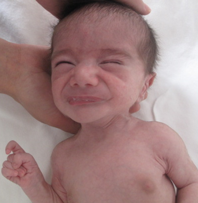
Respiratory problems and episodic hyperthermia were observed in all patients, while feeding difficulties were present in 10 of 11 patients (90.9%), hypotonia in 9 of 10 patients (90%), and growth failure in 9 of 10 patients (90%). Developmental delay was present in the majority of patients (80%), manifesting as isolated speech delay, motor delay, or global developmental delay. Characteristic craniofacial findings were variably observed and pursed lips were noted in all evaluated patients (10/10, 100%) (Fig. 1). Oral/dental abnormalities were observed in all evaluated patients (9/9, 100%), including oligodontia, multiple dental caries, dry mouth, and atrophy of the lingual papillae. Ocular involvement was frequent (9/10, 90%) and included keratitis, corneal opacity, dry eye, refractive errors, and strabismus.
Radiological and skeletal findings
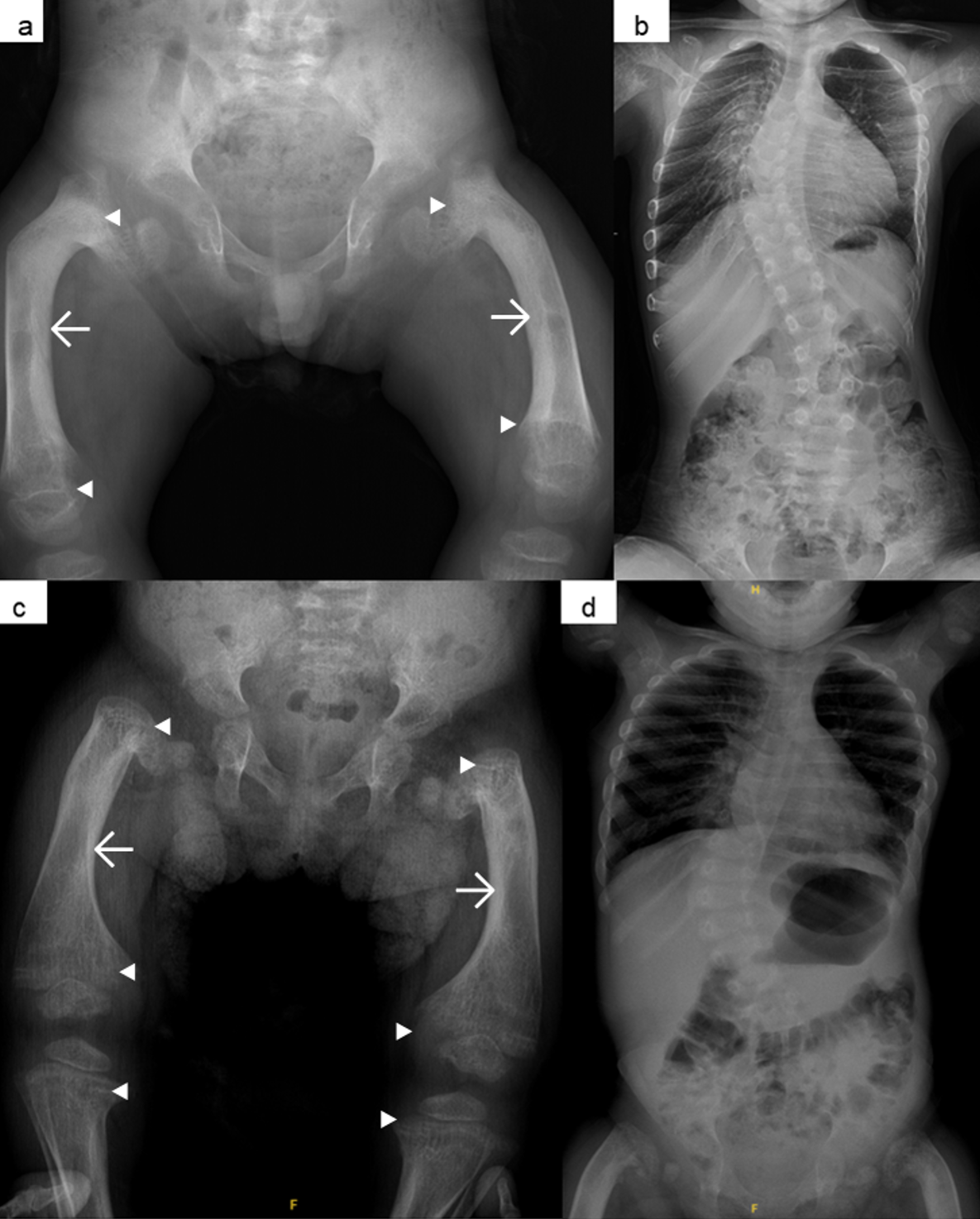
Among the skeletal findings, bowing of the long bones, cortical thickening, and flared metaphyses were present in all patients, whereas camptodactyly was observed in 8 of 11 patients (72.7%), joint contractures in 9 of 11 (81.8%), scoliosis in 8 of 10 (80%), and bone fractures in 6 of 9 (66.6%) (Fig. 1 and Fig. 2).
Molecular findings
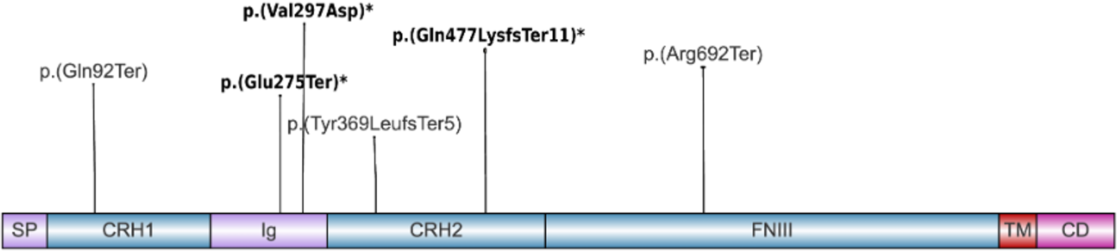
Molecular analysis identified biallelic LIFR variants in all patients. The recurrent pathogenic variant c.2074C>T; p.(Arg692Ter) was identified in the homozygous state in patients from seven families, whereas in one patient it was detected in the compound heterozygous state in combination with an additional variant. Overall, the c.2074C>T; p.(Arg692Ter) variant accounted for 15 of 22 disease-associated alleles (68.2%) in the cohort. Moreover, three previously unreported variants in the LIFR gene were identified, comprising one nonsense variant, one missense variant, and one frameshift variant. Among the novel variants detected in our cohort, c.890T>A, p.(Val297Asp), was classified as a variant of uncertain significance (VUS) based on ACMG guidelines; however, several lines of evidence raised the possibility of pathogenicity. This variant is not present in the gnomAD database (PM2_Supporting), and in silico prediction tools suggest a deleterious impact on protein function, as supported by AlphaMissense (score: 0.754; PP3_Supporting) and PolyPhen-2, which classifies it as probably damaging (score: 1.000). In addition, segregation analysis supported an autosomal recessive inheritance pattern, and the patient’s phenotype was strongly consistent with SWS, with no alternative molecular diagnosis identified. The novel variants were submitted to the ClinVar database (SUB16043420, SUB16045977, SUB16046006), and all identified variants were marked on the schematic representation of the LIFR protein (Fig. 3).
Discussion
SWS is a rare autosomal recessive disorder characterized by skeletal abnormalities and severe dysautonomic manifestations, particularly during early childhood, whereas orthopedic complications become more prominent with age.5,9 All patients in our cohort demonstrated the characteristic skeletal phenotype of SWS, including universal bowing of the long bones and frequent camptodactyly, accompanied by varying degrees of autonomic dysfunction (Table I).
Short and bowed limbs have been reported in all patients with SWS, and camptodactyly has been described in approximately 80% of cases.5 Consistent with these observations, bowing of the long bones was present in all of our patients, while camptodactyly was observed in 72.7% of the cohort. Characteristic radiological findings, including bowed long bones with cortical thickening, enlarged metaphyses, and an abnormal trabecular pattern, as well as scoliosis and osteoporosis-related fractures observed in our patients were consistent with the previously reported clinical spectrum and natural course of the disorder.5
Prenatal warning signs include growth restriction, oligohydramnios, reduced fetal movements, and characteristic skeletal findings such as short and bowed long bones, pronounced tibial bowing (greater than femoral involvement), relative sparing of the fibula and upper limb bones, camptodactyly, and talipes.8,22 It has been emphasized that, in the prenatal differentiation of SWS from other bent-bone disorders, the presence of camptodactyly together with a normal scapula is an important distinguishing feature.22 In our cohort, six prenatally monitored patients showed findings such as increased nuchal translucency, limb shortening, decreased fetal movements, and oligohydramnios. However, none of the patients received a molecularly confirmed diagnosis of SWS during the prenatal period. This may be attributed to the relatively nonspecific prenatal skeletal findings of SWS, the absence of severe abnormalities suggestive of a lethal skeletal dysplasia leading to limited prenatal recognition, restricted access to prenatal genetic testing, and the lack of a previously diagnosed affected sibling in the family, which may have reduced the likelihood of recommending molecular analysis.
A narrow thorax, one of the main prenatal indicators of lethality in skeletal dysplasias, is typically absent in SWS. Nevertheless, infant mortality remains high because of dysautonomic complications.8 The disorder is considered potentially fatal before the age of 2 years, primarily because of respiratory distress and episodic hyperthermia. Sudden death and anesthesia-related malignant hyperthermia have also been reported. Mortality is notably high, estimated at approximately 46% overall, reaching 42% in patients younger than 2 years and 10% in older individuals.5 In our cohort, two patients died, one following a presumed cerebrovascular event and one because of sudden death. In addition, Patient 4 had two siblings who had previously died with similar clinical features. Anesthetic complications were also documented in one of our patients who subsequently died (Table I, Patient 6). Additionally, pulmonary arterial hypertension, which has been suggested to be associated with poor prognosis, was present in one of our patients, who died at 4 years of age.5
Additional features associated with the disorder include corneal involvement, oral and dental abnormalities, and dysmorphic facial features, particularly the characteristic pursed-mouth appearance, as well as neurological manifestations such as hypotonia, seizures, and delayed motor development.5 Ectodermal findings such as milia, multiple eruptive vellus hair cysts, and dystrophic nails have been reported, and similarly, papular skin lesions were noted in two of our patients.7 One of our patients was found to have a Morgagni hernia; to the best of our knowledge, this association has not previously been reported and may represent a coincidental finding.
Although the disorder has been reported more frequently in Arab populations, with an estimated prevalence of 0.52 per 10,000 births, recent reports suggest that it may not be as rare in Türkiye as previously assumed.23,24 This distribution may be related to the high rate of consanguinity in these populations, which increases the prevalence of autosomal recessive disorders. A systematic review published in 2022 reported consanguinity in 65% of families with SWS.5 In our cohort, the corresponding rate was 60%, further supporting the contribution of consanguinity to disease occurrence. In addition, a founder variant in the Turkish population has been proposed in the LIFR gene, and this variant, c.2074C>T; p.(Arg692Ter), was homozygous in seven of our patients, while one patient carried it in trans with another pathogenic LIFR variant, consistent with a compound heterozygous genotype.23 Notably, the fact that this variant accounted for the majority of disease-associated alleles in our cohort and was identified in apparently unrelated families further supports the possibility of a founder effect in the Turkish population. Although this variant has also been reported in other populations, it has been predominantly described in Turkish patients.19,23,25,26 Moreover, a previous study demonstrated a shared haplotype spanning the LIFR locus among affected individuals, supporting a possible founder effect.23 However, future haplotype analyses would be required to confirm a shared ancestral origin.
Null mutations in LIFR disrupt the JAK/STAT3 signaling pathway, and studies in Lifr−/− mice have demonstrated impaired bone remodeling, reduced astrocyte numbers, and perinatal lethality.4,27 Similar to CRLF1-related cold-induced sweating syndrome 1 (Crisponi syndrome; OMIM #272430), SWS belongs to the spectrum of ciliary neurotrophic factor receptor (CNTFR) pathway–related disorders characterized by autonomic nervous system dysfunction.11,27,28 In the differential diagnosis of SWS, Crisponi syndrome is an important consideration, as it shares multiple clinical features, such as episodic hyperthermia, feeding difficulties, respiratory problems, facial trismus, and camptodactyly. However, bowing of the long bones is highly characteristic of SWS and serves as a key distinguishing feature. Owing to this hallmark feature, SWS is classified within Group 20, the bent-bones dysplasia group, in the current Nosology of genetic skeletal disorders.29 Nevertheless, the presence of dysautonomic features distinguishes SWS from other entities within this group.
This study has several limitations. First, the sample size was small, which is expected for a rare disorder but limits the generalizability of our findings. Second, the retrospective design resulted in incomplete data for some variables, as reflected by the variable denominators across clinical features. Third, the cohort was drawn from a population with a high rate of consanguinity, which may have influenced both the frequency of homozygous variants and the apparent predominance of the recurrent p.(Arg692Ter) allele. Finally, although three previously unreported LIFR variants were identified, no functional studies were performed; therefore, their pathogenicity relies on ACMG-based interpretation, segregation data, and phenotype correlation.
In conclusion, SWS should be considered in children with dysautonomic manifestations accompanied by long-bone bowing, camptodactyly, and characteristic craniofacial features. Early recognition is essential for appropriate orthopedic surveillance, timely physical therapy, and surgical interventions when necessary. Early calcium and vitamin D supplementation may also help prevent osteoporosis-related fractures. Equally important is the careful assessment of dysautonomia-related symptoms and the prevention of anesthesia-associated complications, including malignant hyperthermia, which may be life-threatening. Our cohort further delineates the phenotypic and molecular spectrum of the disorder and underscores the recurrent p.(Arg692Ter) LIFR variant in the Turkish population.
Ethical approval
The study was approved by Hacettepe University Ethics Committee (date: December 17, 2025, number: SBA 25/1013).
Source of funding
The authors declare the study received no funding.
Conflict of interest
The authors declare that there is no conflict of interest.
References
- Stüve A, Wiedemann HR. Congenital bowing of the long bones in two sisters. Lancet 1971; 2: 495. https://doi.org/10.1016/s0140-6736(71)92666-3
- Giedion A, Boltshauser E, Briner J, et al. Heterogeneity in Schwartz-Jampel chondrodystrophic myotonia. Eur J Pediatr 1997; 156: 214-223. https://doi.org/10.1007/s004310050587
- Superti-Furga A, Tenconi R, Clementi M, et al. Schwartz-Jampel syndrome type 2 and Stüve-Wiedemann syndrome: a case for “lumping”. Am J Med Genet 1998; 78: 150-154. https://doi.org/bfm7zs
- Dagoneau N, Scheffer D, Huber C, et al. Null leukemia inhibitory factor receptor (LIFR) mutations in Stuve-Wiedemann/Schwartz-Jampel type 2 syndrome. Am J Hum Genet 2004; 74: 298-305. https://doi.org/10.1086/381715
- Warnier H, Barrea C, Bethlen S, Schrouff I, Harvengt J. Clinical overview and outcome of the Stuve-Wiedemann syndrome: a systematic review. Orphanet J Rare Dis 2022; 17: 174. https://doi.org/10.1186/s13023-022-02323-8
- Alallah J, Alamoudi LO, Makki RM, Shawli A, AlHarbi AT. Stüve-Wiedemann syndrome with a novel mutation in a Saudi infant. Int J Pediatr Adolesc Med 2022; 9: 143-146. https://doi.org/10.1016/j.ijpam.2021.10.002
- Almuhanna N, Alogayell L, Alkhezzi S, et al. A rare presentation of multiple eruptive vellus hair cysts and dystrophic nails in a pediatric patient with Stüve-Wiedemann syndrome. JAAD Case Rep 2023; 38: 89-91. https://doi.org/10.1016/j.jdcr.2023.05.042
- Begam MA, Hasan M, Chedid F, Mirghani H. The fetus points to the diagnosis of rare skeletal dysplasia: Stuve-wiedemann syndrome: retrospective case series and prenatal review. J Med Ultrasound 2024; 33: 142-147. https://doi.org/10.4103/jmu.jmu_177_23
- Bhalla D, Sati S, Basel D, Karody V. A novel termination site in a case of Stüve-Wiedemann syndrome: case report and review of literature. Front Pediatr 2024; 12: 1341841. https://doi.org/10.3389/fped.2024.1341841
- Braun D, Amylidi-Mohr S, Ahrens O, Trippel M, Schiller R, Zweier C. A fetal case of Stüve-Wiedemann syndrome due to a novel homozygous truncating variant in IL6ST. Eur J Med Genet 2025; 77: 105040. https://doi.org/10.1016/j.ejmg.2025.105040
- Chen Y, Liu Y, He S, Gui J, Wang Y, Zheng M. Heat stroke associated with novel leukaemia inhibitory factor receptor gene variant in a Chinese infant. Open Life Sci 2025; 20: 20251195. https://doi.org/10.1515/biol-2025-1195
- Cores ML, de Los Bueis AB. Implications of neuropathy and management of the corneal surface in a patient with Stuve-Wiedemann syndrome. Rom J Ophthalmol 2023; 67: 412-5. https://doi.org/10.22336/rjo.2023.66
- Hamasharef KH, Najmadden ZB, AKhailany R, Rashid MJ, Hamakarim K. Novel genetic findings in Stuve-Wiedemann syndrome: a case report and review of literature. Clin Case Rep 2026; 14: e71709. https://doi.org/10.1002/ccr3.71709
- Hernández-García S, Valdivia HG, Bartomeu JP, Molina JS. Neuroparalytic keratopathy in Stüve-Wiedemann syndrome treated with tarsoconjunctival flap. Indian J Ophthalmol 2023; 71: 1651-1653. https://doi.org/10.4103/IJO.IJO_3260_22
- Jin J, Rothämel P, Büchel J, et al. Case report: Stüve-Wiedemann syndrome-a rare cause of persistent pulmonary hypertension of the newborn. Front Pediatr 2024; 11: 1329404. https://doi.org/10.3389/fped.2023.1329404
- Kandari A, Alshamali W, ALnakkas D, Alfadhli H, Alibraheem I. A case report on the oral manifestations of Stüve-Wiedemann syndrome. J Oral Sci 2026; 68: 48-50. https://doi.org/10.2334/josnusd.25-0252
- Kelly C, Morand M, Ray JW. Adult case of Stüve-Wiedemann syndrome. Am J Med Genet A 2026. https://doi.org/10.1002/ajmg.a.70214
- McDermott H, Simmonds J, Thyagarajan M, et al. Paediatric survivors beyond infancy with Stüve-Wiedemann syndrome - a case series from the West Midlands, UK. Eur J Med Genet 2023; 66: 104788. https://doi.org/10.1016/j.ejmg.2023.104788
- Melnik E, Sharova M, Kenis V, Morgul A, Zabnenkova V, Markova T. Case report: new phenotype of late-onset Stüve-Wiedemann syndrome due to a C-terminal variant in the LIFR gene. Front Pediatr 2024; 12: 1442624. https://doi.org/10.3389/fped.2024.1442624
- Wiebe JE, Rowe LW, Boente CS, Borschel GH. Single-stage bilateral corneal neurotization for neurotrophic keratopathy in Stüve-Wiedemann syndrome: a case report and literature review. J Pediatr Ophthalmol Strabismus 2024; 61: e54-e58. https://doi.org/10.3928/01913913-20240807-04
- Richards S, Aziz N, Bale S, et al. Standards and guidelines for the interpretation of sequence variants: a joint consensus recommendation of the American College of Medical Genetics and Genomics and the Association for Molecular Pathology. Genet Med 2015; 17: 405-424. https://doi.org/10.1038/gim.2015.30
- Begam MA, Alsafi W, Bekdache GN, Chedid F, Al-Gazali L, Mirghani HM. Stuve-Wiedemann syndrome: a skeletal dysplasia characterized by bowed long bones. Ultrasound Obstet Gynecol 2011; 38: 553-558. https://doi.org/10.1002/uog.8967
- Yeşil G, Lebre AS, Santos SD, et al. Stuve-Wiedemann syndrome: is it underrecognized? Am J Med Genet A 2014; 164A: 2200-2205. https://doi.org/10.1002/ajmg.a.36626
- Al-Gazali LI, Bakir M, Hamid Z, et al. Birth prevalence and pattern of osteochondrodysplasias in an inbred high risk population. Birth Defects Res A Clin Mol Teratol 2003; 67: 125-132. https://doi.org/10.1002/bdra.10009
- Guran T, Guran O, Paketci C, et al. Effects of leukemia inhibitory receptor gene mutations on human hypothalamo-pituitary-adrenal function. Pituitary 2015; 18: 456-460. https://doi.org/10.1007/s11102-014-0594-5
- Romeo Bertola D, Honjo RS, Baratela WAR. Stüve-Wiedemann syndrome: update on clinical and genetic aspects. Mol Syndromol 2016; 7: 12-18. https://doi.org/10.1159/000444729
- Mikelonis D, Jorcyk CL, Tawara K, Oxford JT. Stüve-Wiedemann syndrome: LIFR and associated cytokines in clinical course and etiology. Orphanet J Rare Dis 2014; 9: 34. https://doi.org/10.1186/1750-1172-9-34
- Akawi NA, Ali BR, Al-Gazali L. Stüve-Wiedemann syndrome and related bent bone dysplasias. Clin Genet 2012; 82: 12-21. https://doi.org/10.1111/j.1399-0004.2012.01852.x
- Unger S, Ferreira CR, Mortier GR, et al. Nosology of genetic skeletal disorders: 2023 revision. Am J Med Genet A 2023; 191: 1164-1209. https://doi.org/10.1002/ajmg.a.63132
Copyright and license
Copyright © 2026 The Author(s). This is an open access article distributed under the Creative Commons Attribution License (CC BY), which permits unrestricted use, distribution, and reproduction in any medium or format, provided the original work is properly cited.