Abstract
Background. Congenital disorders of glycosylation (CDG) represent a group of inherited metabolic diseases characterized by multisystem involvement. SLC35A2-CDG (OMIM #300896) is a rare X-linked subtype resulting from pathogenic variants in the SLC35A2 gene, which encodes the UDP-galactose transporter. Approximately 70 cases have been documented, predominantly presenting with early-onset epilepsy, developmental delay, hypotonia, and dysmorphic features. Phenotypic variability remains incompletely characterized, and novel features continue to expand the clinical spectrum.
Case Presentation. We report a 6-year-old girl with SLC35A2-CDG harboring a novel de novo heterozygous frameshift variant (NM_005660.3: c.105_106insCG, p.K36Rfs*6). The patient presented with epileptic spasms, severe global developmental delay, hypotonia, and distinctive dysmorphic features including strabismus and inverted nipples. Notably, she exhibited bilateral patellar luxation and perineal dysmorphism with a shortened anogenital distance, features that have not been previously associated with SLC35A2-CDG. Additional findings included a thin corpus callosum, a pineal cyst, right-sided aortic arch, bilateral vesicoureteral reflux, and central hypothyroidism. Oral D-galactose supplementation was initiated and resulted in subjective improvements in language comprehension, pain responsiveness, and motor function, although no improvement in seizures was observed.
Conclusions. This casa may expand the phenotypic spectrum of SLC35A2-CDG by describing bilateral patellar luxation and perineal dysmorphism as potentially novel clinical features, along with a previously unreported pathogenic variant. Recognition of such unique features is crucial for improving our clinical understanding of this rare disorder, guiding differential diagnosis, and contributing to a more refined characterization of genotype–phenotype correlations.
Keywords: SLC35A2-CDG, congenital disorders of glycosylation, epileptic spasms, perineal dysmorphism, patellar luxation, CDG-IIm
Introduction
Congenital disorders of glycosylation (CDG) are inherited metabolic diseases characterized by defects in glycan attachment or processing of glycoproteins and glycolipids.1 The phenotypic spectrum has expanded rapidly since the condition was first described by Jaeken in 1980.2 In 2009, Jaeken et al.3 proposed a revised nomenclature using only the official gene symbol followed by ‘-CDG.’ By 2024, the number of recognized subtypes had exceeded 200.4 CDG can present with symptoms affecting various organs, primarily the nervous system, including hypotonia, developmental delay, and seizures.5 More than half of patients exhibit endocrine abnormalities, as glycoproteins regulated many endocrine functions, with abnormalities reported in the thyroid, adrenal, and growth axes, sexual development, and metabolism.6
Among more than 200 known subtypes, SLC35A2-CDG (OMIM #300896) is a rare X-linked dominant disorder caused by pathogenic variants in the SLC35A2 gene on Xp11.23,7 with approximately 70 documented cases.8 SLC35A2 encodes the UDP-galactose transporter (UGT), which transports UDP-galactose from the cytoplasm into the Golgi apparatus for incorporation of galactose into the oligosaccharide chains. Mutations can disrupt glycosylation pathways, leading to incomplete oligosaccharides.9
SLC35A2-CDG primarily affects females, with most cases occurring in female heterozygotes with de novo mutations. In males, a mutation in a single gene copy is typically lethal, though some survive through somatic mosaicism.10 Clinically, the disorder manifests in early infancy with refractory seizures, infantile spasms and West syndrome, severe hypotonia, and developmental delay. Dysmorphic features include coarse facial features, broad nasal bridge, thick lips, and strabismus. Patients may also show ocular abnormalities, hepatic dysfunction, and brain MRI findings including cerebral atrophy and a thin corpus callosum.10,11
We report the case of a 6-year-old girl with confirmed SLC35A2-CDG presenting with epileptic encephalopathy, developmental delay, hypotonia, dysmorphic features, and systemic involvement. Our patient also exhibited perineal dysmorphism and bilateral patellar luxation, which have not been previously reported in SLC35A2-CDG and may represent a potential expansion of the phenotypic spectrum, although a causal relationship between these findings and the disorder remains uncertain.
Case presentation
A 6-year-old girl was initially referred to our clinic at 4 months of age because of new-onset seizures. These seizures commenced 2–3 days after routine vaccination and were classified as epileptic spasms. Electroencephalography (EEG) demonstrated a hypsarrhythmic pattern. The patient was admitted with a diagnosis of West syndrome. At 38 weeks and 5 days of gestation, she was delivered via cesarean section due to cephalopelvic disproportion, with a birth weight of 2880 g (−0.93 SDS) and a length of 46 cm (−1.88 SDS). Her head circumference at birth was not documented. Written informed consent was obtained from the patient’s parents for the publication of this case report and any accompanying images and videos.
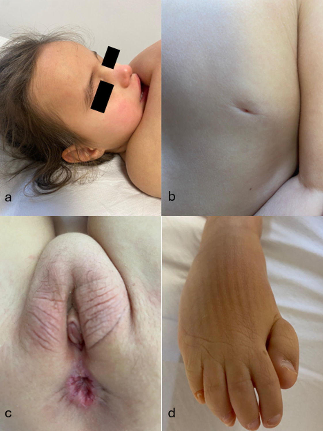
At the age of 6 years, anthropometric measurements showed a weight of 17.3 kg (−1.2 SDS), height of 106 cm (−1.93 SDS) which was consistent with the target height (158.8 cm, −0.73 SDS), and a head circumference of 49.3 cm (−1.26 SDS). The sitting height/height ratio was 0.57, indicating a proportionally large trunk. Hair was sparse in both anterior and posterior regions, particularly at the vertex. The forehead was wide and prominent. Joint laxity and hypermobility were observed. Bilateral patellar luxation was evident, with notable mobility of the right patella; the patient had undergone surgery for left patellar luxation (Video 1). Inverted nipples were present. The toes were proximally positioned, and the distal phalanx of the right index finger exhibited radial deviation. Organomegaly was not observed. The anogenital distance (AGD), measured from the posterior fourchette to the center of the anus as described by Andreasen et al.12 was 17 mm. The dysmorphic features of the patient are shown in Fig. 1.
During the neonatal period, she experienced significant constipation, necessitating the use of enemas until the age of one year. Spontaneous defecation was subsequently possible without an enema. Recurrent urinary tract infections were observed until the age of two. The patient achieved head control at 2 years of age and independent sitting at 3.5 years. She exhibited persistent food-seeking behavior suggestive of hyperorality, along with repetitive biting episodes involving self-injury. Diminished pain perception was evident based on reduced responses to noxious stimuli. No ocular deviation was observed before the onset of seizures; however, the patient subsequently developed strabismus.
Considering the diagnosis of West syndrome and the presence of multiple dysmorphic features, a thorough evaluation was conducted. Cranial magnetic resonance imaging (MRI) revealed a thin corpus callosum and a 4 mm pineal cyst. Echocardiography revealed a right-sided aortic arch. Imaging of the urinary system confirmed bilateral grade 3 vesicoureteral reflux (VUR). Endocrine examination at 7.5 months revealed labial hypoplasia with anterior displacement of the anus. The external genital score (EGS) was 0, confirming female sex, with marked labial rugae. Follicle stimulating hormone, luteinizing hormone, and estradiol levels were consistent with minipuberty, with normal growth and adrenal axes. Thyroid function tests suggested central hypothyroidism (thyroid stimulating hormone 3.92 mIU/L, free thyroxine 0.61 ng/dL). Mild hypoglycemia (50 mg/dL) occurred during infancy. Abdominal/pelvic MRI confirmed the presence of the uterus and ovaries. During the follow-up period, serum alanine aminotransferase (ALT) and aspartate aminotransferase (AST) levels generally remained within the normal range, although occasional mild elevations were observed.
Whole-exome sequencing revealed a heterozygous pathogenic variant of SLC35A2 (NM_005660.3: c.105_106insCG, p.K36Rfs*6), confirming the diagnosis of SLC35A2-CDG. This variant was absent in both parents, indicating a de novo origin, and was not detected in existing databases (ClinVar, gnomAD, dbSNP), confirming its classification as a novel variant. A transferrin isoelectric focusing study performed prior to the initiation of treatment had been reported as normal, although the corresponding numerical values were no longer available for review. Following genetic confirmation, oral D-galactose therapy was initiated. The most recent transferrin isoelectric focusing panel during therapy revealed the following values: disialotransferrin 0.87% (reference range, 2–2.5%), trisialotransferrin 7.51% (reference range, 4.5–9%), tetrasialotransferrin 76.76% (reference range, 64–80%), and pentasialotransferrin 14.86% (reference range, 12–18%).
Discussion
The clinical presentation of our patient closely corresponded with the established spectrum of SLC35A2-CDG.11 She presented with early infantile-onset epilepsy characterized by epileptic spasms. In alignment with most documented cases, she demonstrated significant global developmental delay, marked by the absence of age-appropriate speech and motor milestones, hypotonia, and intellectual disability.11 Her dysmorphic features were consistent with those described in glycosylation disorders, including strabismus and inverted nipples.11,13-15 Brain MRI revealed thinning of the corpus callosum, a recurrent observation in SLC35A2-CDG.11,13 In addition, a 4 mm pineal cyst was identified, which, although likely incidental, represents a previously unreported neuroimaging feature of this subtype.
Hepatic involvement in CDG can range from mild to severe transaminase elevation and coagulopathy.16 In our case, ALT and AST levels largely remained within normal ranges, with only occasional mild elevations. These mild fluctuations were attributed to the long-term use of multiple antiseizure medications rather than primary hepatic dysfunction. In a review, liver involvement, defined as mild transaminase elevation, was observed in 12 of 30 cases (40%).11
Although there was considerable overlap with the established phenotype, our case also presented features atypical of SLC35A2-CDG. The patient had VUR and a right-sided aortic arch, which are not commonly reported in this disorder. In a series of 30 cases, cardiac anomalies were observed in 8 out of 30 cases (27%) and renal anomalies in 1 out of 30 cases (3%).11 While congenital cardiac and renal/urinary tract anomalies are not characteristic hallmarks of CDG, their presence in this patient suggests that SLC35A2-CDG may have broader systemic manifestations than previously appreciated.
It is well established that glycosylation plays a crucial role in the development of the urogenital system.17 Notably, the patient exhibited a short anogenital distance (17 mm) with a normal external genital score. To the best of our knowledge, reduced AGD has not been previously reported in SLC35A2-CDG. This raises the possibility that glycosylation defects in SLC35A2 may subtly influence the embryological development of the caudal midline or genital structures, although the precise pathophysiological link remains unclear. Glycosylation is integral to numerous developmental signaling pathways; thus, impairment of glycan assembly may contribute to abnormal morphogenesis of the perineum. Conversely, it is conceivable that this perineal finding represents an unrelated congenital variant unique to our patient. Given the rarity of SLC35A2-CDG, even isolated anomalies may be significant, and documenting them is valuable because it may enable other clinicians to recognize similar features in future cases.
Given the known association between glycosylation defects and endocrine abnormalities, it has been recommended that all endocrine axes be assessed, together with bone mineralization and glucose metabolism from birth through puberty. The patient’s history of constipation may be attributable to central hypothyroidism and/or the abnormal AGD, both of which were present.
A 2021 review by Lipiński et al.18 systematically examined skeletal involvement in patients with CDG and identified a broad spectrum of abnormalities, including joint laxity, brachydactyly, scoliosis, vertebral malformations, and hip dislocation. However, to date, patellar luxation has not been reported as a clinical feature of any CDG subtype including SLC35A2-CDG. Therefore, the presence of bilateral patellar luxation in our patient may represent a novel skeletal manifestation associated with CDG, expanding the known phenotypic spectrum of SLC35A2-related disorders.
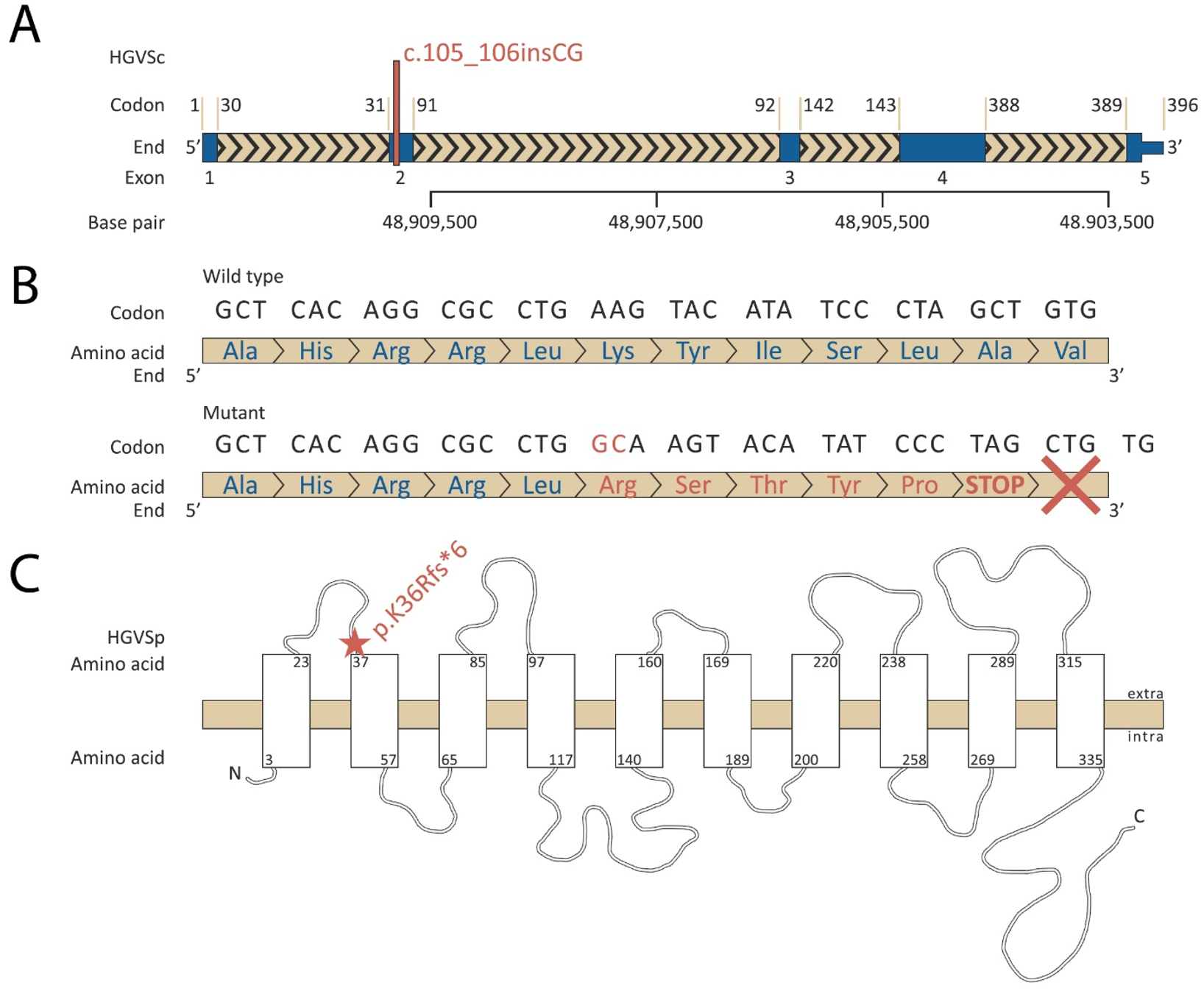
Genetic analysis revealed a heterozygous NM_005660.3: c.105_106insCG (p.K36Rfs*6) variant. At the DNA level, the insertion of CG between nucleotides 105 and 106, located within exon 2 of SLC35A2, leads to a frameshift mutation (Fig. 2). The mutant protein is predicted to be severely truncated and likely non-functional or subject to nonsense-mediated mRNA decay. Given its nature as a frameshift variant leading to a premature stop codon in a gene for which loss-of-function is a known disease mechanism, this variant fulfills the PVS1 criterion under the ACMG guidelines.19 Parental testing confirmed its de novo origin (PS2), the variant was absent from population databases (PM2), and the patient’s phenotype was highly consistent with SLC35A2-CDG (PP4). Therefore, the variant was classified as pathogenic. Importantly, this specific variant has not been previously reported in major variant databases and is thus considered a novel mutation.
A broad spectrum of pathogenic SLC35A2 variants has been reported to underlie this disorder, encompassing missense, nonsense, frameshift, and splice-site variants as well as small in-frame insertions and deletions; the majority arise de novo and are predicted to result in loss of function of the UDP-galactose transporter.7,9,11 In addition to germline variants responsible for the multisystem congenital phenotype, postzygotic somatic mosaic SLC35A2 variants have been increasingly recognized as a recurrent cause of mild malformation of cortical development with oligodendroglial hyperplasia in epilepsy (MOGHE).20 Hemizygous variants in affected males are generally considered lethal, with survival reported mainly in the setting of somatic mosaicism.10
Oral galactose supplementation has been investigated to enhance UDP-galactose availability for glycoprotein synthesis in patients with SLC35A2-CDG. Witters et al.21 administered oral D-galactose to ten patients and demonstrated significant improvement on the Nijmegen Pediatric CDG Rating Scale (NPCRS), with advances in postural control, response to stimuli, and chewing function, as well as improvements in gastrointestinal symptoms. While our patient’s quantitative pretreatment biochemical results were unavailable, the family reported enhanced language comprehension, increased pain responsiveness, and the ability to bear weight and stand briefly with support. She was also able to articulate two or three words. However, no beneficial effects on seizures or EEG findings were observed.
In conclusion, we describe a 6-year-old female with SLC35A2-CDG who displays prototypical features of this rare glycosylation disorder along with two previously unreported anomalies: perineal dysmorphism and bilateral patellar luxation. The causative SLC35A2 variant (NM_005660.3: c.105_106insCG, p.K36Rfs*6) is a novel pathogenic mutation. This case emphasizes that patients with CDG may occasionally manifest unique anatomical variations. Recognition of such atypical features is important, as it can prompt more thorough clinical examinations and help clinicians develop a more complete picture of the manifestations of this disorder. Further reporting of similar cases is essential to determine whether features such as perineal dysmorphism or patellar luxation are recurrent findings associated with SLC35A2 variants or isolated anomalies. Ultimately, our report contributes a new piece to the puzzle of SLC35A2-CDG.
Supplementary materials
Supplementary materials for this article are available online at https://cdn.turkjpediatr.org/html-files/TJP-8349-Video-1.mp4
Ethical approval
Written informed consent was obtained from the patient’s parents for the publication of this case report and any accompanying images and videos. The study was conducted in accordance with the Declaration of Helsinki.
Source of funding
The authors declare the study received no funding.
Conflict of interest
The authors declare that there is no conflict of interest.
References
- Freeze HH, Eklund EA, Ng BG, Patterson MC. Neurology of inherited glycosylation disorders. Lancet Neurol 2012; 11: 453-466. https://doi.org/10.1016/S1474-4422(12)70040-6
- Jaeken J, Vanderschueren-Lodeweyckx M, Casaer P, et al. Familial psychomotor retardation with markedly fluctuating serum prolactin, FSH and GH levels, partial TBG-deficiency, increased serum arylsulphatase A and increased CSF protein: a new syndrome. Pediatr Res 1980; 14: 179. https://doi.org/10.1203/00006450-198002000-00117
- Jaeken J, Hennet T, Matthijs G, Freeze HH. CDG nomenclature: time for a change! Biochim Biophys Acta 2009; 1792: 825-826. https://doi.org/10.1016/j.bbadis.2009.08.005
- Ng BG, Freeze HH, Himmelreich N, Blau N, Ferreira CR. Clinical and biochemical footprints of congenital disorders of glycosylation: proposed nosology. Mol Genet Metab 2024; 142: 108476. https://doi.org/10.1016/j.ymgme.2024.108476
- Paprocka J, Jezela-Stanek A, Tylki-Szymańska A, Grunewald S. Congenital disorders of glycosylation from a neurological perspective. Brain Sci 2021; 11: 88. https://doi.org/10.3390/brainsci11010088
- Ünsal Y, Özön ZA. Endocrine implications of congenital disorders of glycosylation. J Clin Res Pediatr Endocrinol 2026; 18: 11-29. https://doi.org/10.4274/jcrpe.galenos.2025.2024-10-7
- Kodera H, Nakamura K, Osaka H, et al. De novo mutations in SLC35A2 encoding a UDP-galactose transporter cause early-onset epileptic encephalopathy. Hum Mutat 2013; 34: 1708-1714. https://doi.org/10.1002/humu.22446
- Abuduxikuer K, Wang JS. Four new cases of SLC35A2-CDG with novel mutations and clinical features. Front Genet 2021; 12: 658786. https://doi.org/10.3389/fgene.2021.658786
- Quelhas D, Correia J, Jaeken J, et al. SLC35A2-CDG: novel variant and review. Mol Genet Metab Rep 2021; 26: 100717. https://doi.org/10.1016/j.ymgmr.2021.100717
- National Library of Medicine (US). SLC35A2-congenital disorder of glycosylation. MedlinePlus. Available at: https://medlineplus.gov/genetics/condition/slc35a2-congenital-disorder-of-glycosylation/ (Accessed on Apr 15, 2025).
- Ng BG, Sosicka P, Agadi S, et al. SLC35A2-CDG: functional characterization, expanded molecular, clinical, and biochemical phenotypes of 30 unreported individuals. Hum Mutat 2019; 40: 908-925. https://doi.org/10.1002/humu.23731
- Andreasen SM, Gehrt L, Hagen CP, et al. Correlation of anogenital distance from childhood to age 9 years-a prospective population-based birth cohort-the Odense child cohort. Hum Reprod Open 2024; 2024: hoae050. https://doi.org/10.1093/hropen/hoae050
- Online Mendelian Inheritance in Man (OMIM). SLC35A2-CDG clinical synopsis. Available at: https://omim.org/clinicalSynopsis/300896 (Accessed on Apr 15, 2025).
- Kimizu T, Takahashi Y, Oboshi T, et al. A case of early onset epileptic encephalopathy with de novo mutation in SLC35A2: clinical features and treatment for epilepsy. Brain Dev 2017; 39: 256-260. https://doi.org/10.1016/j.braindev.2016.09.009
- Eklund EA, Freeze HH. The congenital disorders of glycosylation: a multifaceted group of syndromes. NeuroRx 2006; 3: 254-263. https://doi.org/10.1016/j.nurx.2006.01.012
- Lipiński P, Bogdańska A, Socha P, Tylki-Szymańska A. Liver involvement in congenital disorders of glycosylation and deglycosylation. Front Pediatr 2021; 9: 696918. https://doi.org/10.3389/fped.2021.696918
- Varki A, Freeze HH, Vacquier VD. Glycans in development and systemic physiology. In: Varki A, Cummings RD, Esko JD, et al., editors. Essentials of Glycobiology. 2nd ed. Cold Spring Harbor (NY): Cold Spring Harbor Laboratory Press; 2009.
- Lipiński P, Stępień KM, Ciara E, Tylki-Szymańska A, Jezela-Stanek A. Skeletal and bone mineral density features, genetic profile in congenital disorders of glycosylation: review. Diagnostics (Basel) 2021; 11: 1438. https://doi.org/10.3390/diagnostics11081438
- Richards S, Aziz N, Bale S, et al. Standards and guidelines for the interpretation of sequence variants: a joint consensus recommendation of the American College of Medical Genetics and Genomics and the Association for Molecular Pathology. Genet Med 2015; 17: 405-424. https://doi.org/10.1038/gim.2015.30
- Bonduelle T, Hartlieb T, Baldassari S, et al. Frequent SLC35A2 brain mosaicism in mild malformation of cortical development with oligodendroglial hyperplasia in epilepsy (MOGHE). Acta Neuropathol Commun 2021; 9: 3. https://doi.org/10.1186/s40478-020-01085-3
- Witters P, Tahata S, Barone R, et al. Clinical and biochemical improvement with galactose supplementation in SLC35A2-CDG. Genet Med 2020; 22: 1102-1107. https://doi.org/10.1038/s41436-020-0767-8
Copyright and license
Copyright © 2026 The Author(s). This is an open access article distributed under the Creative Commons Attribution License (CC BY), which permits unrestricted use, distribution, and reproduction in any medium or format, provided the original work is properly cited.