Abstract
Background. Primary hypoparathyroidism is a rare endocrine disorder in childhood; data on its clinical and etiological spectrum in pediatric populations remain limited. We aimed to characterize the clinical, biochemical, etiological, and therapeutic spectrum of pediatric primary hypoparathyroidism at a tertiary center in Southeastern Türkiye.
Methods. A retrospective, single-center observational cohort study was conducted between January 2016 and January 2026. Medical records of 27 children (0–18 years) with nonsurgical primary hypoparathyroidism were reviewed. The diagnosis required persistent hypocalcemia with hyperphosphatemia and inappropriately low or normal intact parathyroid hormone on at least two measurements, in the absence of secondary causes. Demographic, clinical, biochemical, imaging, genetic testing (fluorescence in situ hybridization/multiplex ligation-dependent probe amplification for 22q11.2; targeted sequencing for candidate genes), and treatment data were collected.
Results. Median age at diagnosis was 3 months (interquartile range: 1.6–111 months; range: 3 days–17 years); 9 patients (33.3%) were diagnosed in the neonatal period. Seizures were the most frequent presenting feature in 12 patients (44.4%). A specific etiological diagnosis was established in 11 patients (40.7%): DiGeorge syndrome in 5 (18.5%), Kenny–Caffey syndrome type 1 in 3 (11.1%), Sanjad–Sakati syndrome in 1 (3.7%), autoimmune polyglandular syndrome type 1 in 1 (3.7%), and autosomal dominant hypocalcemia type 1 due to an activating calcium-sensing receptor (CASR) mutation in 1 (3.7%). Twenty-four patients (88.9%) received combined oral calcium and calcitriol therapy. Two patients (7.4%) died from sepsis.
Conclusions. In this Southeastern Turkish pediatric cohort, DiGeorge syndrome and tubulin-specific chaperone E gene (TBCE) -related disorders were the most prominent identified etiologies; the enrichment of TBCE-related disorders likely reflects regional consanguinity and Arab-origin demographics. An integrated diagnostic approach combining clinical, biochemical, and genetic evaluation is essential in similar populations. Prospective multicenter studies with systematic genetic testing are warranted.
Keywords: primary hypoparathyroidism, hypocalcemia, childhood, genetic etiology, DiGeorge syndrome, tubulin-specific chaperone E gene
Introduction
Primary hypoparathyroidism in childhood is an uncommon endocrine disorder characterized by deficient production or secretion of parathyroid hormone (PTH) leading to persistent hypocalcemia accompanied by hyperphosphatemia. Hypoparathyroidism may result from intrinsic abnormalities of the parathyroid glands, including developmental defects, genetic disorders, and autoimmune destruction, or from acquired causes such as surgical removal or damage to the parathyroid glands. While postsurgical hypoparathyroidism represents the most common form overall, the present study focuses on nonsurgical primary hypoparathyroidism and its underlying intrinsic etiologies.1 Only a few population-based studies have evaluated the epidemiology of nonsurgical primary hypoparathyroidism. Reported prevalence estimates range from 0.7 per 100,000 in Japan to 5 per 100,000 in Rochester, USA, while a nationwide Danish study reported a prevalence of 2.3 per 100,000 inhabitants.2-4 Although rare, pediatric hypoparathyroidism carries significant clinical consequences, ranging from subtle biochemical abnormalities to life-threatening neuromuscular and neurodevelopmental complications such as seizures and tetany; early recognition is therefore essential to prevent acute morbidity and long-term sequelae.5 The variable clinical spectrum and potential for atypical presentations complicate timely diagnosis and management in children.
The etiological landscape of pediatric nonsurgical primary hypoparathyroidism is heterogeneous, encompassing isolated genetic defects, syndromic conditions, and developmental anomalies of the parathyroid glands.6 Among syndromic causes, 22q11.2 deletion syndrome (DiGeorge syndrome) represents one of the most frequently identified etiologies in pediatric cohorts.7 In regions with high rates of consanguineous marriage, autosomal recessive conditions such as Kenny–Caffey syndrome type 1 (KCS1) and Sanjad–Sakati syndrome (SSS), both caused by pathogenic variants in the tubulin-specific chaperone E (TBCE) gene, are disproportionately represented.8,9 Genetic mechanisms implicated include defects in calcium-sensing receptor (CaSR) signaling, abnormalities of parathyroid development, and perturbations in transcriptional regulators of parathyroid function (e.g., GCM2, GATA3, and PTH), as well as autoimmune polyendocrine syndrome type 1 (APS-1) caused by pathogenic variants in the autoimmune regulator (AIRE) gene.7,10,11
Despite the clinical importance of pediatric hypoparathyroidism, published data from the Eastern Mediterranean and Middle Eastern regions remain sparse. Southeastern Türkiye is characterized by elevated rates of consanguineous marriage and a noteworthy Arab-origin population, demographic factors that may substantially influence the etiological spectrum of rare recessive disorders.12,13 The aim of this study was to characterize the clinical, biochemical, etiological, and therapeutic profile of children with nonsurgical primary hypoparathyroidism, with particular emphasis on intrinsic genetic and syndromic etiologies who were followed at a tertiary pediatric endocrinology center in this region.
Materials and Methods
This retrospective, single-center observational cohort study was conducted in the pediatric endocrinology department of a tertiary care center in Southeastern Türkiye. Medical records of children diagnosed with nonsurgical primary hypoparathyroidism and followed between January 2016 and January 2026 were reviewed. Patients aged 0–18 years with a diagnosis of primary hypoparathyroidism, defined as persistent hypocalcemia with hyperphosphatemia and inappropriately low or normal PTH levels in the absence of secondary causes, were included. Patients with postsurgical hypoparathyroidism, vitamin D deficiency, renal insufficiency, transient neonatal hypocalcemia, malabsorption disorders, or hypomagnesemia-related syndromes were excluded.
Demographic data collected included age at diagnosis, sex, and parental consanguinity. Clinical data comprised presenting symptoms (e.g., seizures, tetany, lethargy), age at symptom onset, family history of hypoparathyroidism or unexplained infant deaths, comorbid conditions, and dysmorphic features. Laboratory data obtained from electronic medical records at diagnosis and during follow-up included total serum calcium, ionized calcium, serum phosphate, magnesium, alkaline phosphatase (ALP), intact PTH, 25-hydroxyvitamin D, creatinine, spot urine calcium-to-creatinine ratio, and 24-hour urinary calcium excretion when available. Clinical characteristics and biochemical parameters reported in this study were obtained at the time of diagnosis. All biochemical measurements were performed in the hospital laboratory using standard automated analyzers; assay-specific reference ranges and units recorded at the time of testing were noted in the dataset when available. Serum intact PTH concentrations were measured using an electrochemiluminescence immunoassay (ECLIA) on Cobas e402 and Cobas e801 analyzers (Roche Diagnostics, Mannheim, Germany). The laboratory reference range for intact PTH was 15–65 pg/mL. Age-adjusted phosphate reference ranges and urine Ca/Cr reference values were applied according to local laboratory standards.
Imaging data included renal ultrasonography to assess nephrocalcinosis and cranial computed tomography (CT) or magnetic resonance imaging (MRI) to detect basal ganglia calcifications when clinically indicated. Audiological and transthoracic echocardiographic evaluations were performed in patients with syndromic features or clinical suspicion, and available results were recorded. Treatment modalities, including oral calcium supplementation, active vitamin D analogues (calcitriol), magnesium replacement, and recombinant PTH when applicable, were documented. Treatment-related data, including elemental calcium and calcitriol doses, were recorded from the most recent follow-up visit available in the medical records. Treatment response was evaluated based on biochemical parameters, clinical improvement, and treatment-related complications such as hypercalciuria and nephrocalcinosis.
Genetic analysis
Available genetic test results were retrospectively reviewed. Genetic investigations had been performed during routine clinical care according to clinical suspicion and test availability. Targeted testing for 22q11.2 deletion was performed using fluorescence in situ hybridization (FISH) and/or multiplex ligation-dependent probe amplification (MLPA). In selected patients with characteristic phenotypic findings, candidate genes including TBCE and CASR were analyzed by targeted Sanger sequencing. Comprehensive next-generation sequencing panels and whole-exome sequencing were not routinely available during the study period. Accordingly, genetic confirmation could not be obtained for all patients with clinically suspected syndromic diagnoses.
Statistical analysis
Descriptive statistics were used for all analyses. Continuous variables were summarized according to their distribution characteristics. Variables with skewed distributions are presented as median (interquartile range, IQR: 25th–75th percentile), whereas normally distributed variables are presented as mean ± standard deviation (SD). Categorical variables are expressed as frequencies and percentages. Statistical analyses were performed using SPSS version 25.0 (IBM Corp., Armonk, NY, USA).
Ethical approval
The study protocol was reviewed and approved by the Ethics Committee of Adnan Menderes University (approval number: 2026/101), and the requirement for informed consent was waived given the retrospective design.
Results
Patient demographics
A total of 27 patients were included in the study, comprising 15 males (55.6%) and 12 females (44.4%). The median age at diagnosis was 3 months (IQR: 1.6–111 months; range: 3 days–17 years), and 9 patients (33.3%) were diagnosed during the neonatal period. Parental consanguinity was present in 23 patients (85.1%). The median duration of follow-up was 3.75 years (IQR: 2.2–6.5 years; range: 3 months–15 years).
Presentation and clinical features
Seizures were the most frequent presenting symptom in 12 patients (44.4%), followed by neonatal detection of hypocalcemia in 9 patients (33.3%) and muscle cramps in 4 patients (14.8%). One patient (3.7%) was diagnosed incidentally during treatment for pneumonia and one (3.7%) during routine follow-up for previously known as APS-1. Dysmorphic features were observed in 10 patients (37.0%). Cardiac anomalies were present in 8 patients (29.6%): ventricular septal defect in 2 patients (7.4%), pulmonary stenosis in one patient (3.7%), atrial septal defect in one patient (3.7%), patent foramen ovale in 3 patients (11.1%), and left ventricular dilatation in one patient (3.7%). Sensorineural hearing loss occurred in 4 patients (14.8%) and renal anomalies in 2 patients (7.4%). Cranial imaging was performed in only 3 patients (11.1%); basal ganglia calcification was detected in 1 of these 3 (33.3%) patients, which likely resulted in an underestimation of its true prevalence.
Biochemical findings at diagnosis
All patients fulfilled the diagnostic criteria for primary hypoparathyroidism and therefore presented with hypocalcemia and hyperphosphatemia at diagnosis. Biochemical findings are summarized in Table I. Two patients (7.4%) had PTH levels within the normal reference range despite documented hypocalcemia, whereas one patient (3.7%) had a slightly elevated PTH level. These patients were subsequently diagnosed with an activating CASR mutation, DiGeorge syndrome, and APS-1, respectively.
|
Data are given as median (IQR) or n (%). IQR: interquartile range, PTH: parathyroid hormone. |
|
| Table I. Baseline characteristics of study patients (n = 27). | |
| Characteristic |
|
| Number of patients |
|
| Female/Male |
|
| Median age at diagnosis (months) |
|
| Neonatal presentation |
|
| Parental consanguinity |
|
| Presenting symptoms | |
| Seizures |
|
| Neonatal hypocalcemia |
|
| Muscle cramps |
|
| Incidental diagnosis |
|
| Biochemical data at diagnosis | |
| Total serum calcium (mg/dL) |
|
| Serum phosphate (mg/dL) |
|
| Intact PTH (pg/mL) |
|
| 25-Hydroxyvitamin D (ng/mL) |
|
| Serum magnesium (mg/dL) |
|
| Urinary calcium/creatinine (mg/mg creatinine) |
|
| Median follow-up duration (years) |
|
Etiology of primary hypoparathyroidism
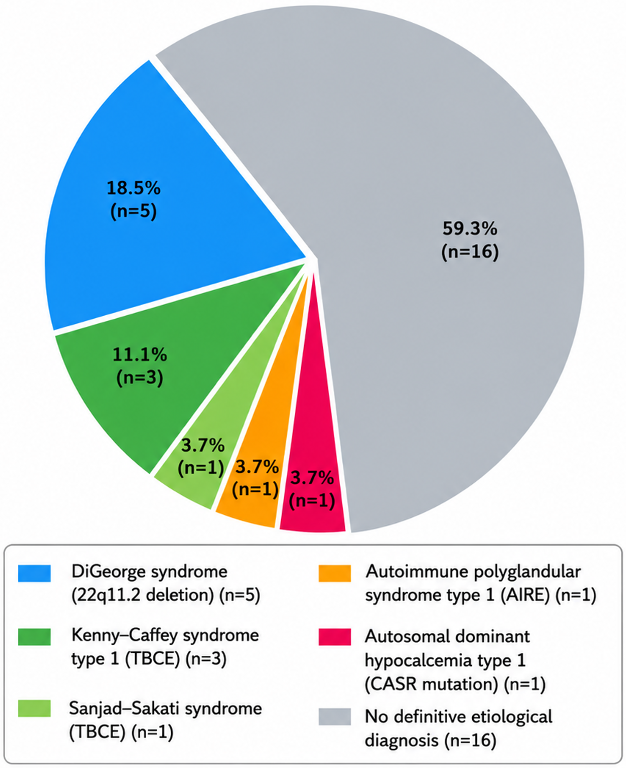
A specific etiological diagnosis was established in 11 patients (40.7%) based on clinical and/or genetic findings (Table II). Among these, molecular confirmation was available in 8 patients, whereas the remaining diagnoses were established on the basis of characteristic clinical features and associated findings. The identified etiologies included DiGeorge syndrome in 5 patients (18.5%), KCS1 in 3 patients (11.1%), SSS in one patient (3.7%), APS-1 in one patient (3.7%), and autosomal dominant hypocalcemia type 1 (ADH1, CASR mutation) in one patient (3.7%). All genetic variants identified in our cohort had been previously reported in the literature and were consistent with the corresponding clinical phenotypes.7,8,14 However, segregation analysis could not be performed because of the retrospective design of the study and the unavailability of parental genetic testing. The remaining 16 patients (59.3%) had no confirmed genetic or clinical diagnosis, commonly due to limited access to genetic testing during the study period. The etiological distribution is illustrated in Fig. 1. Among the patients without genetic testing or a definitive clinical diagnosis, 4 patients (25%) exhibited foramen ovale or mild cardiac abnormalities, and only one patient (6.2%) had sensorineural hearing loss. Most did not display characteristic syndromic features suggestive of a specific genetic syndrome. Nevertheless, because comprehensive molecular testing was not available for all patients, an underlying genetic etiology cannot be excluded. Detailed individual clinical and genetic characteristics of patients with established etiological diagnoses are presented in Table II.
| ADH1: autosomal dominant hypocalcemia type 1; APS: autoimmune polyglandular syndrome; ASD: atrial septal defect; BGC: basal ganglia calcification; CASR: calcium-sensing receptor gene; Dx: diagnosis; Echo: echocardiography; KCS: Kenny–Caffey syndrome; mo: months; NA: not available (genetic analysis could not be performed); NB: newborn; ND: not done (imaging not performed); PFO: patent foramen ovale; TBCE: tubulin-specific chaperone E gene; VSD: ventricular septal defect; yr: years. | ||||||||||||
| Table II. Clinical, laboratory, and genetic characteristics of patients with genetically or syndromically defined primary hypoparathyroidism. | ||||||||||||
| Pt | Diagnosis |
Dx |
|
Initial presentation |
features |
(mg/dL) |
(mg/dL) |
(pg/mL) |
Echo | Nephro calcinosis |
Genetic finding |
|
| 1 | DiGeorge syndrome |
|
|
Neonatal hypocalcemia |
|
|
|
|
Pulmonary stenosis | No | NA |
|
| 2 | DiGeorge syndrome |
|
|
Neonatal hypocalcemia |
|
|
|
|
Normal | No | 22q11.21 microdeletion |
|
| 3 | DiGeorge syndrome |
|
|
Neonatal hypocalcemia |
|
|
|
|
VSD | No | NA |
|
| 4 | DiGeorge syndrome |
|
|
Neonatal hypocalcemia |
|
|
|
|
VSD + PFO | No | 22q11.21 microdeletion |
|
| 5 | DiGeorge syndrome |
|
|
Neonatal hypocalcemia |
|
|
|
|
ASD | No | 22q11.21 microdeletion |
|
| 6 | KCS1 |
|
|
Convulsion |
|
|
|
|
Normal | No | TBCE: c.155_166del (p.Ser52_Gly55del), homozygous |
|
| 7 | KCS1 |
|
|
Convulsion |
|
|
|
|
Normal | No | TBCE: c.155_166del (p.Ser52_Gly55del), homozygous |
|
| 8 | KCS1 |
|
|
Convulsion |
|
|
|
|
Normal | No | TBCE: c.155_166del (p.Ser52_Gly55del), homozygous |
|
| 9 | Sanjad–Sakati syndrome |
|
|
Convulsion |
|
|
|
|
Normal | No | TBCE: c.155_166del (p.Ser52_Gly55del), homozygous |
|
| 10 | APS type 1 |
|
|
Follow-up exam |
|
|
|
|
Normal | No | NA |
|
| 11 | ADH1 |
|
|
Muscle cramps |
|
|
|
|
Normal | No | CASR: c.407C>T (p.Pro136Leu), heterozygous |
|
Treatment and outcomes
Three patients (11.1%) received calcitriol monotherapy, and 24 patients (88.9%) received combined oral calcium and calcitriol therapy. At the most recent follow-up visit, the median elemental calcium dose was 28 (IQR: 22–33) mg/kg/day and the median calcitriol dose was 18.75 (IQR: 13–25.75) ng/kg/day. The wide range in calcitriol dosing reflects age-related pharmacokinetic differences (neonates and infants typically require higher weight-adjusted doses), as well as dose adjustments made in response to treatment-related complications such as hypercalciuria, particularly in the patient who developed nephrocalcinosis and chronic kidney disease (CKD). Hypercalcemia and nephrocalcinosis occurred only in one patient with KCS1, who subsequently developed CKD; no patient progressed to end-stage renal disease. Overall mortality was 7.4% (2/27). One patient with 22q11.2 deletion syndrome died during the neonatal period due to sepsis. The second patient, who had KCS1, died from sepsis at 3 years of age.
Discussion
DiGeorge syndrome was the most frequent identifiable cause in our series, affecting 5 patients (18.5%), consistent with prior pediatric reports that show a wide prevalence range (approximately 10–59%) depending on cohort composition and the extent of genetic evaluation.15 All five affected children in our cohort presented during the neonatal period, consistent with the well-described early manifestation of 22q11.2 deletion-related hypoparathyroidism, although later presentations have been documented in other series. Genetic confirmation was achieved in only 3/5 patients due to technical and access limitations, representing an important constraint on etiological ascertainment. None of the patients with DiGeorge syndrome had a documented family history of 22q11.2 deletion syndrome, consistent with the predominantly de novo occurrence of this condition. We recommend routine consideration of 22q11.2 deletion testing in neonates with unexplained hypocalcemia, even in the absence of overt cardiac or immunological features.
KCS1 (n = 3) and SSS (n = 1) together accounted for 4 patients (14.8%), a proportion higher than typically reported in non-Middle Eastern pediatric series but consistent with reports from Saudi Arabia and Kuwait, where TBCE-related disorders are relatively common.16-18 Both conditions are autosomal recessive and result from pathogenic variants in the TBCE gene; they have been repeatedly associated with Arab ancestry and increased parental consanguinity, factors known to elevate the prevalence of recessive disorders.8,12,18 The demographic features of Southeastern Türkiye, including high rates of consanguineous marriage and a substantial Arab-origin population, likely underlie the observed enrichment of TBCE-related diagnoses in our cohort.12,13,19 Consanguinity is well established as a major driver of autosomal recessive pediatric endocrine disorders across the Eastern Mediterranean region.20 Intrafamilial variability is illustrated by two affected siblings with KCS1 who had divergent clinical courses, emphasizing variable expressivity even with the same genotype.16,18 Although KCS1 and SSS share a genetic basis, they differ phenotypically: SSS typically presents with more severe neurodevelopmental impairment, whereas KCS1 is notable for skeletal features such as cortical thickening and medullary stenosis of long bones.17,18 This genotype–phenotype complexity suggests that modifying genetic, epigenetic, or environmental factors should be considered during genetic counseling and prognostic discussions.17
Among our 27 patients, two had inappropriately normal PTH levels despite documented hypocalcemia and were diagnosed with an activating CASR mutation and DiGeorge syndrome, whereas one patient with APS-1 had a slightly elevated PTH level despite hypocalcemia. This finding illustrates that a “normal” PTH in level the setting of low calcium reflects an inadequate parathyroid response rather than true euparathyroidism. This phenomenon may result from partial gland dysfunction, impaired parathyroid reserve, altered CaSR signaling, or early autoimmune destruction.21,22 Clinically, PTH must always be interpreted alongside concurrent serum calcium levels to avoid false reassurance.
The patient with an activating CASR mutation did not develop nephrocalcinosis or hypercalciuria despite the established renal risk associated with such mutations;22,23 early diagnosis and maintenance of conservative serum calcium targets may have mitigated this risk. The APS-1 case demonstrated the classic sequential evolution of disease, consistent with natural history cohorts reporting hypoparathyroidism in approximately 79–84% of patients,24,25 underscoring the need for long-term multidisciplinary endocrine surveillance in suspected APS-1. Nephrocalcinosis developed in one patient with KCS1, who subsequently progressed to CKD, underscoring the renal vulnerability described in TBCE-related disorders.17,18 No patient progressed to end-stage renal disease during follow-up.
Basal ganglia calcification was identified in only one patient; however, cranial imaging had been performed in just 3 patients, a limitation that likely led to substantial underestimation of intracranial calcifications compared with reported prevalences of 40–70% in long-standing hypoparathyroidism.22,23 Routine neuroimaging, preferably cranial CT, in patients with chronic disease should be considered to detect subclinical calcifications and guide long-term management.
The majority of patients (24/27, 88.9%) were managed with combined oral calcium and calcitriol, consistent with standard-of-care recommendations.23 All patients included in the present study had persistent primary hypoparathyroidism by definition; therefore, treatment discontinuation due to transient hypoparathyroidism was not expected.
Two patients (7.4%) died from sepsis during follow-up; both had genetically confirmed syndromic diagnoses (22q11.2 deletion and KCS1), conditions associated with immune dysfunction and infection susceptibility. While mortality data in pediatric hypoparathyroidism series are limited, the available literature suggests that deaths are uncommon in isolated hypoparathyroidism but occur at higher rates in syndromic forms, particularly in DiGeorge syndrome, where T-cell deficiency predisposes to serious infections.7,15 Our mortality rate, while seemingly elevated for a pediatric endocrine cohort, must be interpreted in the context of the predominantly syndromic composition of our diagnosed subgroup and the limited access to specialized immunological care in the region during the study period. Despite generally favorable metabolic control, these observations suggest a potential increased susceptibility to infections in these subgroups, emphasizing the need for vigilant multidisciplinary supportive care.
The majority of patients in this cohort (16/27, 59.3%) remained without a confirmed genetic or clinical diagnosis. This likely reflects the limited availability of comprehensive next-generation sequencing during the study period rather than a true absence of genetic etiology. Candidate genes not fully evaluated in this cohort include GCM2 (the most common cause of isolated familial hypoparathyroidism), GATA3 (associated with HDR syndrome: hypoparathyroidism, deafness, and renal anomalies), SOX3 (X-linked hypoparathyroidism), and FAM111A (Kenny–Caffey syndrome type 2). Given the high consanguinity rate (85.1%), novel autosomal recessive variants are plausible in a substantial proportion of the undiagnosed group. Whole-exome or whole-genome sequencing would be the recommended next diagnostic step for these patients.
The present study adds to the limited literature on pediatric nonsurgical primary hypoparathyroidism by providing data from Southeastern Türkiye, a region characterized by high rates of parental consanguinity and a substantial burden of rare autosomal recessive disorders.12,13,20 Furthermore, the presence of patients with inappropriately normal PTH concentrations despite hypocalcemia underscores an important diagnostic challenge and emphasizes the need to interpret PTH values in relation to serum calcium concentrations rather than relying solely on laboratory reference intervals.1,7,21 However, the retrospective single-center design introduces potential selection bias and incomplete data ascertainment, and the small sample size (n = 27), while reflective of disease rarity, limits generalizability. Another major limitation of this study is the relatively low etiological diagnostic yield (40.7%). This likely reflects restricted access to comprehensive molecular testing during much of the study period rather than a true absence of identifiable genetic causes. Therefore, the reported frequencies should be interpreted as the distribution of confirmed etiologies within the cohort and may underestimate the actual prevalence of specific genetic disorders.
Conclusions
In this cohort of 27 children from Southeastern Türkiye, a specific etiological diagnosis was established in 40.7% of patients, with DiGeorge syndrome and TBCE-related disorders being the most prominent identified causes; the latter likely reflects the region’s high consanguinity rates and substantial Arab-origin population. A normal PTH concentration does not exclude hypoparathyroidism and must always be interpreted alongside simultaneous serum calcium measurements. 22q11.2 deletion testing should be considered in all neonates with unexplained hypocalcemia, and routine neuroimaging merits consideration given the likely underestimation of intracranial calcifications. Children with TBCE-related disorders require vigilant infection surveillance. An integrated diagnostic strategy combining clinical assessment, targeted biochemical evaluation, and systematic genetic testing is essential in populations with high consanguinity. Prospective multicenter studies with routine next-generation sequencing (including whole-exome and/or whole-genome sequencing) are needed to improve etiological yield and guide management in pediatric hypoparathyroidism.
Ethical approval
The study protocol was reviewed and approved by the Ethics Committee of Adnan Menderes University (approval date: 13.03.2026 number: 2026/101), and the requirement for informed consent was waived given the retrospective design.
Source of funding
The authors declare the study received no funding.
Conflict of interest
The authors declare that there is no conflict of interest.
References
- Shoback D. Clinical practice. Hypoparathyroidism. N Engl J Med 2008; 359: 391-403. https://doi.org/10.1056/NEJMcp0803050
- Nakamura Y, Matsumoto T, Tamakoshi A, et al. Prevalence of idiopathic hypoparathyroidism and pseudohypoparathyroidism in Japan. J Epidemiol 2000; 10: 29-33. https://doi.org/10.2188/jea.10.29
- Clarke B, Leibson C, Emerson J, Ransom J, Lagast H. Co-morbid medical conditions associated with prevalent hypoparathyroidism: a population-based study. J Bone Miner Res 2011; 26(Suppl 1): S182. https://doi.org/10.1002/jbmr.564
- Underbjerg L, Sikjaer T, Mosekilde L, Rejnmark L. The epidemiology of nonsurgical hypoparathyroidism in Denmark: a nationwide case finding study. J Bone Miner Res 2015; 30: 1738-1744. https://doi.org/10.1002/jbmr.2501
- Bilezikian JP, Khan A, Potts JT, et al. Hypoparathyroidism in the adult: epidemiology, diagnosis, pathophysiology, target-organ involvement, treatment, and challenges for future research. J Bone Miner Res 2011; 26: 2317-2337. https://doi.org/10.1002/jbmr.483
- Thakker RV. Genetic developments in hypoparathyroidism. Lancet 2001; 357: 974-976. https://doi.org/10.1016/s0140-6736(00)04254-9
- Mannstadt M, Bilezikian JP, Thakker RV, et al. Hypoparathyroidism. Nat Rev Dis Primers 2017; 3: 17055. https://doi.org/10.1038/nrdp.2017.55
- Parvari R, Hershkovitz E, Grossman N, et al. Mutation of TBCE causes hypoparathyroidism-retardation-dysmorphism and autosomal recessive Kenny-Caffey syndrome. Nat Genet 2002; 32: 448-452. https://doi.org/10.1038/ng1012
- Naguib KK, Gouda SA, Elshafey A, et al. Sanjad-Sakati syndrome/Kenny-Caffey syndrome type 1: a study of 21 cases in Kuwait. East Mediterr Health J 2009; 15: 345-352.
- Pearce SH, Williamson C, Kifor O, et al. A familial syndrome of hypocalcemia with hypercalciuria due to mutations in the calcium-sensing receptor. N Engl J Med 1996; 335: 1115-1122. https://doi.org/10.1056/NEJM199610103351505
- Hannan FM, Kallay E, Chang W, Brandi ML, Thakker RV. The calcium-sensing receptor in physiology and in calcitropic and noncalcitropic diseases. Nat Rev Endocrinol 2018; 15: 33-51. https://doi.org/10.1038/s41574-018-0115-0
- Koc I. Prevalence and sociodemographic correlates of consanguineous marriages in Turkey. J Biosoc Sci 2008; 40: 137-148. https://doi.org/10.1017/S002193200700226X
- Akbayram S, Sari N, Akgün C, et al. The frequency of consanguineous marriage in eastern Turkey. Genet Couns 2009; 20: 207-214.
- Kocabey Sütçü Z, Aytaç Kaplan EH, Önal H. Pediatric hypoparathyroidism: etiological and clinical evaluation in a tertiary center. Endocrine 2025; 87: 1235-1245. https://doi.org/10.1007/s12020-024-04110-w
- Kim JH, Shin YL, Yang S, et al. Diverse genetic aetiologies and clinical outcomes of paediatric hypoparathyroidism. Clin Endocrinol (Oxf) 2015; 83: 790-796. https://doi.org/10.1111/cen.12944
- Sanjad SA, Sakati NA, Abu-Osba YK, Kaddoura R, Milner RD. A new syndrome of congenital hypoparathyroidism, severe growth failure, and dysmorphic features. Arch Dis Child 1991; 66: 193-196. https://doi.org/10.1136/adc.66.2.193
- Parvari R, Hershkovitz E, Kanis A, et al. Homozygosity and linkage-disequilibrium mapping of the syndrome of congenital hypoparathyroidism, growth and mental retardation, and dysmorphism to a 1-cM interval on chromosome 1q42-43. Am J Hum Genet 1998; 63: 163-169. https://doi.org/10.1086/301915
- Al-Gazali LI, Dawodu A. The syndrome of hypoparathyroidism, severe growth failure, developmental delay and distinctive facies. Clin Dysmorphol 1997; 6: 233-237. https://doi.org/10.1097/00019605-199707000-00006
- Tunçbilek E. Clinical outcomes of consanguineous marriages in Turkey. Turk J Pediatr 2001; 43: 277-279. https://doi.org/10.24953/turkjpediatr.2001.3070
- Amaratunga SA, Tayeb TH, Dusatkova P, Pruhova S, Lebl J. Invaluable role of consanguinity in providing insight into paediatric endocrine conditions: lessons learnt from congenital hyperinsulinism, monogenic diabetes, and short stature. Horm Res Paediatr 2022; 95: 1-11. https://doi.org/10.1159/000521210
- Korkmaz HA, Ozkan B. Hypoparathyroidism in children and adolescents. Ann Pediatr Endocrinol Metab 2023; 28: 159-167. https://doi.org/10.6065/apem.2346096.048
- Shoback DM, Bilezikian JP, Costa AG, et al. Presentation of hypoparathyroidism: etiologies and clinical features. J Clin Endocrinol Metab 2016; 101: 2300-2312. https://doi.org/10.1210/jc.2015-3909
- Brandi ML, Bilezikian JP, Shoback D, et al. Management of hypoparathyroidism: summary statement and guidelines. J Clin Endocrinol Metab 2016; 101: 2273-2283. https://doi.org/10.1210/jc.2015-3907
- Sharifinejad N, Zaki-Dizaji M, Tebyanian S, et al. Clinical, immunological, and genetic features in 938 patients with autoimmune polyendocrinopathy candidiasis ectodermal dystrophy (APECED): a systematic review. Expert Rev Clin Immunol 2021; 17: 807-817. https://doi.org/10.1080/1744666X.2021.1925543
- Ahonen P, Myllärniemi S, Sipilä I, Perheentupa J. Clinical variation of autoimmune polyendocrinopathy-candidiasis-ectodermal dystrophy (APECED) in a series of 68 patients. N Engl J Med 1990; 322: 1829-1836. https://doi.org/10.1056/NEJM199006283222601
Copyright and license
Copyright © 2026 The Author(s). This is an open access article distributed under the Creative Commons Attribution License (CC BY), which permits unrestricted use, distribution, and reproduction in any medium or format, provided the original work is properly cited.