Abstract
Background. Familial Mediterranean Fever (FMF) is an autosomal recessive autoinflammatory disease caused by mutations in the Mediterranean Fever (MEFV) gene. Despite its monogenic basis, FMF shows marked clinical heterogeneity, suggesting additional regulatory mechanisms. This exploratory study aimed to investigate host gene–dependent mechanisms underlying altered miR-197-3p expression in FMF.
Materials and Methods. Peripheral blood mononuclear cells from homozygous FMF patients (n=6) and matched healthy controls (n=6) were analyzed. miR-197-3p expression was quantified using RT-qPCR. DNA methylation patterns in regulatory regions of the GNAI3 promoter and the MIR197 upstream candidate regulatory region were assessed by bisulfite sequencing.
Results. miR-197-3p expression was significantly reduced in FMF patients compared with controls, as expected. No evidence supporting regulation through the MIR197 upstream candidate regulatory region was observed. Differential methylation within the GNAI3 promoter was statistically significant (p = 0.0093), supporting biological relevance despite the limited cohort size.
Conclusion. These pilot findings provide preliminary molecular evidence supporting host gene–dependent regulation of miR-197-3p in FMF. Given the small cohort size, these findings should be considered exploratory and require validation in larger, independent cohorts.
Keywords: autoinflammation, DNA methylation, GNAI3, Familial Mediterranean Fever, microRNA, miR-197-3p
Introduction
Familial Mediterranean Fever (FMF) is an autoinflammatory disease characterized by recurrent episodes of fever and serosal inflammation and is caused by mutations in the Mediterranean Fever (MEFV) gene encoding the pyrin protein.1,2 Although FMF is inherited as an autosomal recessive disorder, patients carrying identical MEFV mutations frequently exhibit substantial variability in disease severity, attack frequency, and response to treatment, indicating that factors beyond genetic variation contribute to disease pathogenesis.3
Epigenetic regulation has emerged as an important modifier of inflammatory and rheumatic diseases, influencing gene expression without altering the underlying DNA sequence.4,5 Among epigenetic mechanisms, microRNAs (miRNAs) play a central role in the post-transcriptional regulation of genes involved in innate immune signaling, cytokine production, and inflammasome activation.4 Altered miRNA expression profiles have been reported in several rheumatic and autoinflammatory diseases, including rheumatoid arthritis, systemic lupus erythematosus, and periodic fever syndromes.6,7
miR-197-3p has been identified as an inflammation-associated miRNA with regulatory effects on cytokine signaling pathways. In our previous work, we demonstrated that miR-197-3p expression is significantly reduced in homozygous FMF patients compared with healthy controls.8,9 Functional analyses further showed that miR-197-3p has anti-inflammatory effects by modulating cytokine release, caspase-1 activation, and cell migration.8 Despite these findings, the molecular mechanisms responsible for the decreased expression of miR-197-3p in FMF have remained unclear.
miR-197-3p is encoded from the MIR197 gene within the G Protein Subunit Alpha I3 (GNAI3) gene, and to date, no independent promoter has been described for the MIR197 locus.10 This genomic organization suggests that miR-197-3p expression is likely regulated in coordination with transcriptional control of its host gene rather than through a separate promoter-driven mechanism. Epigenetic modifications affecting host gene transcription, such as DNA methylation, therefore may contribute to miR-197-3p dysregulation.11,12
In this context, investigating regulatory features of the GNAI3 locus may provide insight into miR-197-3p expression changes observed in FMF patients. Clarifying whether altered miR-197-3p levels are linked to host gene–dependent regulation is essential for understanding miRNA-mediated control of inflammation and may reveal novel regulatory pathways relevant to autoinflammatory disease pathogenesis.12,13
In the present study, we aimed to investigate the molecular basis of miR-197-3p dysregulation in FMF, focusing on transcriptional and epigenetic mechanisms associated with its host gene, GNAI3.
Materials and Methods
Study group
The study group consisted of six patients with FMF homozygous for the M694V variant and six healthy controls without MEFV mutations. The FMF and control groups each included three female and three male participants. The mean age was 11.35 ± 3.60 years in the FMF group and 12.25 ± 3.88 years in the healthy control group. Disease duration among FMF patients ranged from 2.9 to 12 years. All patients were receiving colchicine treatment at the time of sampling (1 mg/day, n=5; 2 mg/day, n=1) and had an Autoinflammatory Disease Activity Index (AIDAI) score of 0, indicating clinically inactive disease. No patient had clinical evidence of an acute FMF attack at the time of sampling. MEFV mutation analysis was performed using restriction enzyme digestion–based mutation analysis, and homozygosity for the M694V variant was confirmed in all FMF patients. Available hematological parameters and C-reactive protein levels were recorded at the time of sampling. All procedures involving human participants were approved by the Hacettepe University Non-Interventional Clinical Research Ethics Committee (date: 20.09.2022, number: 2022/14-35). Written informed consent was obtained from all participants or their legal guardians prior to enrollment, and the study was conducted in accordance with institutional ethical standards.
Peripheral blood mononuclear cell isolation and stimulation
Peripheral blood samples were collected into heparinized tubes. Peripheral blood mononuclear cells (PBMCs) were isolated by density gradient centrifugation using lympholyte-poly solution. Isolated PBMCs were washed and incubated in serum-free Roswell Park Memorial Institute (RPMI) (Gibco) 1640 medium for 30 minutes. Following incubation cells were harvested for RNA and DNA isolation.
DNA isolation and bisulfite treatment
Genomic DNA was isolated from PBMCs using phenol–chloroform–isoamyl alcohol extraction method. DNA concentration and purity were assessed by spectrophotometry. Bisulfite conversion of genomic DNA was performed using the EpiTect Bisulfite Kit (QIAGEN) according to the manufacturer’s instructions to assess DNA methylation patterns. Following conversion, bisulfite-treated DNA was purified and stored at −20°C until further analysis in Tris-EDTA (TE) buffer.
DNA methylation analysis
DNA methylation analysis was conducted using the EpiTect Bisulfite Kit (QIAGEN) according to the manufacturer’s instructions by bisulfite sequencing. Regions selected for analysis included regulatory sequences associated with the promoter of GNAI3 and the region upstream of the MIR197 gene. Bisulfite-specific primers were designed to amplify CpG-rich regions while avoiding CpG sites within primer sequences. PCR amplification of bisulfite-converted DNA was performed using DreamTaq DNA Polymerase (5 U/µL; Thermo Scientific) on a SimpliAmp Thermal Cycler (Applied Biosystems). Sanger sequencing was conducted using the BigDye Terminator v3.1 Cycle Sequencing Kit (Applied Biosystems), followed by capillary electrophoresis on an ABI 3130 Genetic Analyzer (Applied Biosystems). Methylation levels at individual CpG sites were determined by direct analysis of sequencing chromatograms.
Because no experimentally validated independent promoter has been established for MIR197, the upstream region was investigated as a candidate regulatory region rather than a confirmed promoter. The analyzed regions were selected to evaluate two alternative regulatory models: host gene–dependent regulation associated with the GNAI3 promoter and possible local regulation associated with the genomic region upstream of MIR197. Region selection was guided by genomic position, CpG density, CpG-associated features, and regulatory annotations within the GNAI3/MIR197 locus.
Primer sequences used for amplification of bisulfite-treated DNA targeting regulatory regions of the GNAI3 locus and regions upstream of MIR197 are listed in Table I. Primer design was based on the length, CpG density, and sequence characteristics of the target regions to ensure efficient amplification of bisulfite-converted DNA and accurate region-specific methylation assessment.
| Fw, forward; Rv, reverse. | ||
| Table I. Primer sequences used in this study. List of primers used for amplification of bisulfite-treated DNA targeting regulatory regions of the GNAI3 locus and regions upstream of the MIR197 gene. Primer sequences are provided in the 5′–3′ direction. | ||
| Gene | Primer | Sequences |
| MIR197 | 1 |
Fw: TTTTTTAGGTGATTTTGATGTAGTTAAT Rv: CCACTATACTCAACCTATATTAATCTTT |
| 2 |
Fw: GAGTGTTATGTTGTTATTTAAAAAGTTT Rv: CCACCATACCTAACTAATTTTTATATTTT |
|
| 3 |
Fw: GTTAATATGGTGAAATTTYGTTTTTATT Rv: CCCAACAATTTTCAATAATTTTCTTAAT |
|
| 4 |
Fw: ATTAAGAAAATTATTGAAAATTGTTGGG Rv: ACTAACACTAAACATACAAAAACTTTTA |
|
| 5 |
Fw: TATGTAAAAGTTTTTGTATGTTTAGTGT Rv: AAAACCTTTATCCTACATAAACAAAAAT |
|
| 6 |
Fw: TTGTTTTGTTTATTTTTGTTTATGTAGG Rv: ACGTTAAATACCTTTATTAATACTACCT |
|
| 7 |
Fw: AGGTAGTATTAATAAAGGTATTTAACGT Rv: TTCTATCTTTTTACCATAAAAAAACACT |
|
| GNAI3 | 1 |
Fw: TTTAAATTAGGTAGTCGAAAGGTAAATG Rv: TTTAAACCACTCTCCTCAAAAAAAA |
| 2 |
Fw: TTGATAGAATTTCGAAATTTTGTAGATT Rv: AAACTAAAACACCGTCTATTACTAAA |
|
| 3 |
Fw: GAGAGGGTTATCGTTTAGTAATAGA Rv: GAATAACAACAACTTCACTTCTTT |
|
| 4 |
Fw: GTTAAAGAAGTGAAGTTGTTGTTAT Rv: ATACTCCCAAATCTCTCTACTAAAC |
|
RNA isolation and reverse transcription quantitative polymerase chain reaction (RT-qPCR)
Total RNA was isolated from PBMCs using RNeasy Mini Kit (QIAGEN) according to the manufacturer’s instructions. RNA concentration and quality were assessed prior to reverse transcription. For analysis of miR-197-3p expression, complementary DNA (cDNA) synthesis was performed using miRNA-specific reverse transcription protocols with the TaqMan® MicroRNA Reverse Transcription Kit (Applied Biosystems). All reactions were carried out using MicroRNA TaqMan® hsa-miR-197-3p, 000497 and U6 snRNA, 001973 assays (Applied Biosystems) with TaqMan® Universal PCR Master Mix and no AmpErase® UNG (Applied Biosystems) kit. The reactions were conducted on the BioRad IQ5 (Biorad). Small nuclear RNA U6 were used as internal controls, as appropriate. Relative expression levels were calculated using the comparative Ct method (2−ΔCt).
Statistical Analysis
Statistical analyses were performed using GraphPad Prism 10.4.1 (GraphPad Software). Data distribution was assessed using the Shapiro–Wilk test, and no significant deviation from normality was detected (p > 0.05). Differences between patient and control groups were evaluated using an unpaired t-test with Welch’s correction, which does not assume equal variances between groups. Average CpG methylation values (ranging from 0 to 1) were derived from normalized C/(C+T) ratios and are presented as percentages for clarity. Direct bisulfite sequencing was chosen to assess regional methylation patterns rather than single-allele resolution, as the aim of the study was to compare overall methylation differences between FMF patients and healthy controls. Data are presented as mean ± standard deviation. A p value of ≤0.05 was considered statistically significant.
Results
Clinical characteristics of the study population
Six patients with FMF homozygous for the M694V variant and six healthy controls without MEFV mutations were included. The groups were balanced for sex, with three females and three males in each group, and had comparable age distributions. The mean age was 11.35 ± 3.60 years in the FMF group and 12.25 ± 3.88 years in the healthy control group. Disease duration among FMF patients ranged from 2.9 to 12 years. All FMF patients were receiving colchicine treatment and had an AIDAI score of 0 at the time of sampling, indicating clinically inactive disease. No patient had clinical evidence of an acute FMF attack at the time of sampling. Demographic, clinical, and available laboratory characteristics of the study population are summarized in Table II and Table III.
| AIDAI, Autoinflammatory Disease Activity Index; SD, standard deviation. All FMF patients were attack-free at the time of sampling (AIDAI score = 0) and were receiving colchicine treatment. Dates are presented in DD/MM/YYYY format. | |||||||||||
| Table II. Individual and summary of demographic and clinical characteristics of FMF patients and healthy controls. | |||||||||||
| Date of Birth | Age (years) | Age (years), mean ± SD | Sex | Sex, female/male | Date of diagnosis | Disease duration (years) | Disease duration (years), mean ± SD | Colchicine treatment status | Disease activity measures (AIDAI) | ||
| MEFVM694V/M694V patients | 1 | 15.08.2011 | 12.3 | 11.35 ± 3.60 | Male | 3/3 | 01.01.2012 | 12 | 8.15 ± 3.00 | 1 mg/day | 0 |
| 2 | 28.03.2014 | 9.7 | Female | 01.10.2015 | 8.2 | 1 mg/day | 0 | ||||
| 3 | 21.03.2014 | 9.7 | Male | 01.06.2016 | 7.5 | 1 mg/day | 0 | ||||
| 4 | 16.09.2017 | 6.2 | Female | 01.02.2021 | 2.9 | 1 mg/day | 0 | ||||
| 5 | 24.06.2007 | 16.5 | Female | 01.12.2014 | 9 | 2 mg/day | 0 | ||||
| 6 | 07.04.2010 | 13.7 | Male | 01.09.2014 | 9.3 | 1 mg/day | 0 | ||||
|
Healthy controls No MEFV mutation |
1 | 25.09.2012 | 11.2 | 12.25 ± 3.88 | Male | 3/3 | |||||
| 2 | 31.10.2017 | 6.1 | Female | ||||||||
| 3 | 29.01.2014 | 9.9 | Female | ||||||||
| 4 | 10.02.2008 | 15.9 | Female | ||||||||
| 5 | 03.10.2008 | 15.2 | Male | ||||||||
| 6 | 03.10.2008 | 15.2 | Male | ||||||||
|
RV, Reference Value. *The analytical instrument was changed; therefore, reference ranges and units were updated accordingly. |
|||||||||
| Table III. Individual laboratory characteristics of FMF patients and healthy controls at the time of sampling. | |||||||||
|
Leukocyte x103/µL |
Lymphocyte % |
Monocyte % |
Neutrophil % |
Eosinophil % |
Basophil % |
Thrombocyte x103/µL |
C-reactive protein (CRP) mg/dL |
||
| Reference Values |
|
|
|
|
|
|
|
|
|
| MEFVM694V/M694V patients | 1 |
|
|
|
|
|
|
|
|
| 2 |
|
|
|
|
|
|
|
|
|
| 3 |
|
|
|
|
|
|
|
|
|
| 4 |
|
|
|
|
|
|
|
|
|
| 5 |
|
|
|
|
|
|
|
|
|
| 6 |
|
|
|
|
|
|
|
|
|
|
Healthy controls No MEFV mutation |
1 |
|
|
|
|
|
|
|
|
| 2 |
|
|
|
|
|
|
|
|
|
| 3 |
|
|
|
|
|
|
|
|
|
| 4 |
|
|
|
|
|
|
|
|
|
| 5 |
|
|
|
|
|
|
|
|
|
| 6 |
|
|
|
|
|
|
|
|
|
DNA methylation analysis of the GNAI3 and MIR197 locus
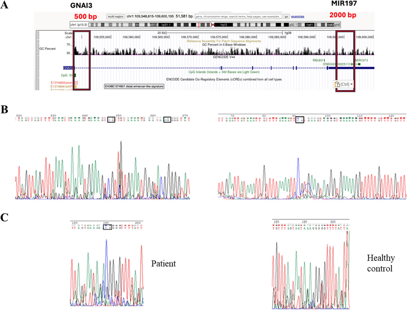
Bisulfite sequencing was used to evaluate DNA methylation patterns within regulatory regions including a ~500 bp region within the putative GNAI3 promoter and a ~2000 bp region upstream of the MIR197 gene (Fig. 1A). Representative bisulfite sequencing chromatograms of the selected regions confirm successful bisulfite conversion and enable discrimination between methylated and unmethylated CpG sites (Fig. 1B), while distinct CpG methylation patterns are observed between FMF patients and healthy controls within the GNAI3 promoter region (Fig. 1C).
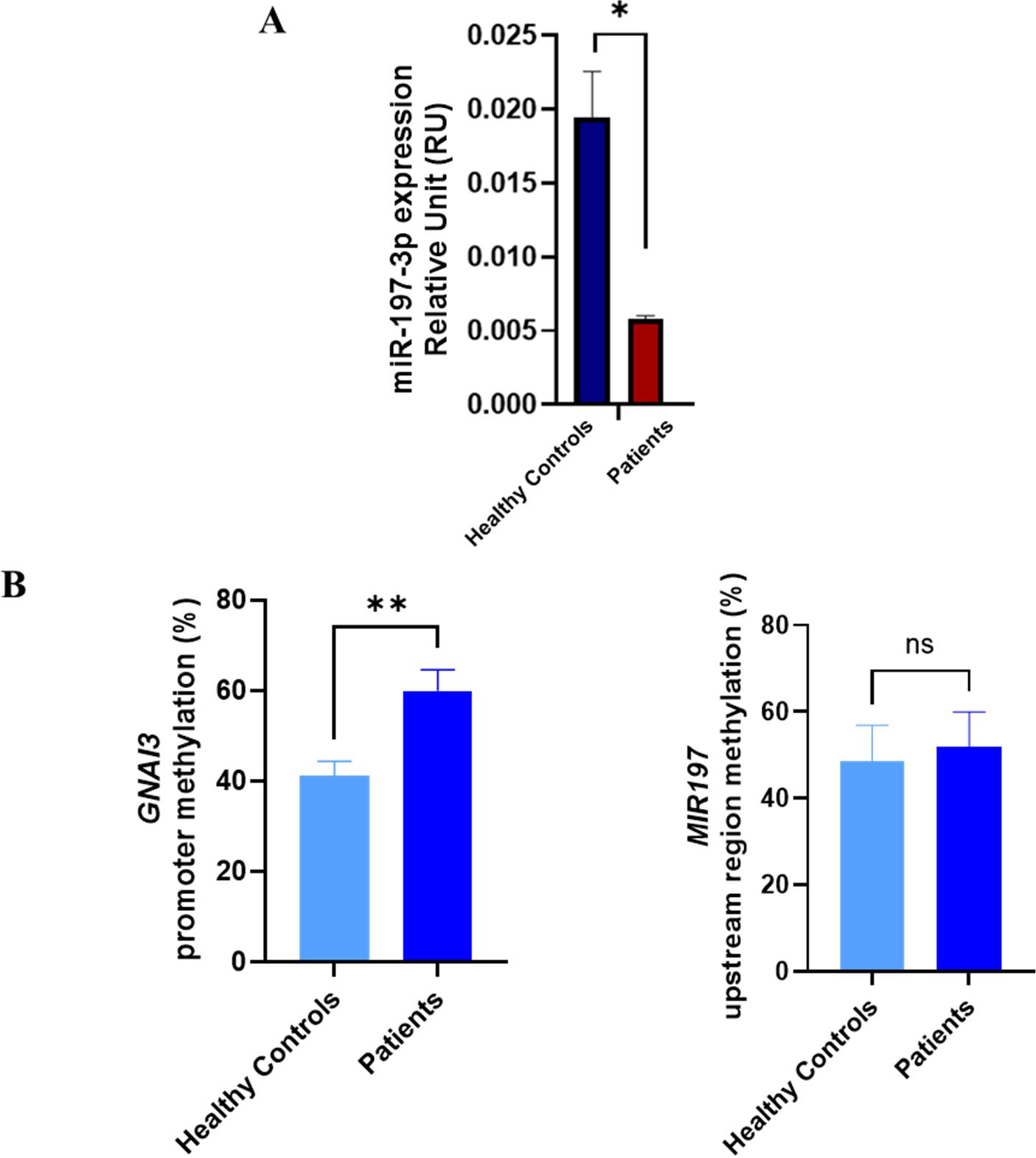
Expression levels of miR-197-3p were evaluated in PBMCs from FMF patients and healthy controls. miR-197-3p expression was significantly reduced in FMF patients compared with healthy controls under basal conditions (0.00500 ± 0.00393 vs. 0.01872 ± 0.00884, respectively; mean ± SD; *p = 0.0106) (Fig. 2A). In parallel, bisulfite sequencing analysis revealed increased DNA methylation within the GNAI3 promoter region in FMF patients compared with healthy controls (58.33 ± 4.37% vs. 44.00 ± 8.99%, respectively; mean ± SD; **p=0.0093). In contrast, methylation levels in the regions upstream of the MIR197 gene did not differ significantly between FMF patients and healthy controls (51.90 ± 8.07% vs. 48.55 ± 8.34%, respectively; mean ± SD; p=0.4957) (Fig. 2B). These findings suggest that reduced miR-197-3p expression in FMF may be linked to epigenetic regulation of the host gene GNAI3, while no significant methylation difference was detected in the examined upstream candidate regulatory region of MIR197.
Discussion
In this study, we investigated the molecular basis of miR-197-3p dysregulation in FMF, focusing on epigenetic mechanisms associated with its host gene, GNAI3. Our results demonstrate a significant reduction in miR-197-3p expression in FMF patients and suggest that this dysregulation is not driven by an independent MIR197 upstream candidate regulatory region but may be linked to host gene–dependent regulatory mechanisms.
Previous studies14-16 have demonstrated that miRNA dysregulation in FMF is heterogeneous and may be influenced by genotype, treatment status, and disease activity. Hortu et al.14 evaluated 15 inflammation-associated miRNAs and reported reduced expression of multiple miRNAs in FMF, with additional differences related to colchicine treatment and attack status. Kahraman et al.15 identified a distinct pattern involving both upregulated and downregulated miRNAs and further reported genotype-associated variation, including prominent changes in miR-107 among patients carrying homozygous M694V. In a mechanistically oriented study, Latsoudis et al.16 linked MEFV perturbation to altered miRNA expression and identified an association between miR-4520a, pyrin mutations, and RHEB/mTOR-related signaling. Together, these studies indicate that miRNA dysregulation is a recurring but molecularly heterogeneous feature of FMF. The present study differs from previous profiling approaches by focusing on a previously identified FMF-associated miRNA, miR-197-3p, and exploring a potential upstream epigenetic basis for its reduced expression. Our findings therefore extend the existing literature from the identification of altered miRNA profiles toward investigation of a possible host gene–associated regulatory mechanism.
Consistent with previous reports implicating miR-197-3p in the regulation of inflammatory pathways, we confirmed that miR-197-3p expression is significantly reduced in FMF patients compared with healthy controls under basal conditions.9 As a possible cause of this reduction, we identified differential DNA methylation within the promoter region of the GNAI3 gene in FMF patients compared with healthy controls. In contrast, no consistent methylation differences were detected in regions upstream of the MIR197 gene. Given that no independent MIR197 upstream candidate regulatory region has been described to date, these results are consistent with a host gene–dependent regulatory model in which epigenetic modifications of GNAI3 may indirectly influence miR-197-3p expression.
Intronic miRNAs are frequently co-regulated with their host genes, and alterations in host gene transcription can directly affect miRNA expression levels.17 Epigenetic mechanisms, particularly DNA methylation, have been shown to regulate both host gene transcription and intronic miRNA expression.18 miR-197-3p has previously been reported to exert anti-inflammatory effects by modulating inflammatory signaling pathways.8 Importantly, the present findings demonstrate an association rather than a causal relationship between increased methylation within the analyzed GNAI3 promoter region and reduced miR-197-3p expression. The current study did not directly assess GNAI3 transcription or experimentally manipulate DNA methylation. Therefore, it cannot be concluded from these data alone that altered GNAI3 promoter methylation causes the reduction in miR-197-3p expression. Functional studies combining GNAI3 expression analysis with targeted methylation editing, demethylation approaches, or promoter activity assays will be required to establish a direct regulatory relationship.
Dysregulated miRNA expression has been increasingly implicated in autoinflammatory and autoimmune diseases, including FMF.5 Reduced miR-197-3p expression may, in turn, impair its anti-inflammatory functions, thereby contributing to enhanced inflammatory responses characteristic of FMF.
This study should be considered a pilot molecular investigation. Although the cohort size is limited, statistically robust methylation differences (p = 0.0093) were identified. The analysis was intentionally restricted to M694V homozygous patients to reduce genetic variability. Therefore, the findings should not be generalized to FMF patients with other MEFV genotypes without further validation.
The principal limitation of this study is the small sample size. Accordingly, the findings should not be interpreted as establishing the general importance of miR-197-3p across the broader FMF population. Rather, they provide preliminary evidence of a possible association between altered methylation within the GNAI3 regulatory region and reduced miR-197-3p expression in a genetically homogeneous group of patients homozygous for the M694V variant. Taken together, our results provide evidence supporting a host gene–dependent regulatory mechanism underlying miR-197-3p dysregulation in FMF. This study adds to the growing body of literature highlighting the role of miRNA regulation in autoinflammatory diseases and underscores the importance of considering host gene–miRNA interactions when investigating epigenetic regulatory networks.
Given the demonstrated anti-inflammatory role of miR-197-3p, its downregulation may contribute to excessive inflammasome activation and cytokine production in FMF. Future studies should explore upstream regulators of MIR197 transcription and post-transcriptional processing pathways.
Ethical approval
The study was approved by Hacettepe University Non-Interventional Clinical Research Ethics Committee (date: 20.09.2022, number: 2022/14-35). Written informed consent was obtained from all participants or their legal guardians prior to enrollment, and the study was conducted in accordance with institutional ethical standards.
Source of funding
The authors declare that the study is funded by the Hacettepe University Scientific Research Projects Coordination Unit, grant number: BAP Project No. TKB-2023-20399.
Conflict of interest
The authors declare that there is no conflict of interest.
References
- Özen S, Batu ED, Demir S. Familial Mediterranean fever: recent developments in pathogenesis and new recommendations for management. Front Immunol 2017; 8: 253. https://doi.org/10.3389/fimmu.2017.00253
- Masters SL, Simon A, Aksentijevich I, Kastner DL. Horror autoinflammaticus: the molecular pathophysiology of autoinflammatory disease (*). Annu Rev Immunol 2009; 27: 621-668. https://doi.org/10.1146/annurev.immunol.25.022106.141627
- Ozen S, Demirkaya E, Amaryan G, et al. Results from a multicentre international registry of familial Mediterranean fever: impact of environment on the expression of a monogenic disease in children. Ann Rheum Dis 2014; 73: 662-667. https://doi.org/10.1136/annrheumdis-2012-202708
- Krol J, Loedige I, Filipowicz W. The widespread regulation of microRNA biogenesis, function and decay. Nat Rev Genet 2010; 11: 597-610. https://doi.org/10.1038/nrg2843
- O’Connell RM, Rao DS, Baltimore D. microRNA regulation of inflammatory responses. Annu Rev Immunol 2012; 30: 295-312. https://doi.org/10.1146/annurev-immunol-020711-075013
- Lucherini OM, Obici L, Ferracin M, et al. First report of circulating microRNAs in tumour necrosis factor receptor-associated periodic syndrome (TRAPS). PLoS One 2013; 8: e73443. https://doi.org/10.1371/journal.pone.0073443
- Kamiya Y, Kawada JI, Kawano Y, et al. Serum microRNAs as potential biomarkers of juvenile idiopathic arthritis. Clin Rheumatol 2015; 34: 1705-1712. https://doi.org/10.1007/s10067-015-2922-1
- Akkaya-Ulum YZ, Akbaba TH, Tavukcuoglu Z, et al. Familial Mediterranean fever-related miR-197-3p targets IL1R1 gene and modulates inflammation in monocytes and synovial fibroblasts. Sci Rep 2021; 11: 685. https://doi.org/10.1038/s41598-020-80097-4
- Akkaya-Ulum YZ, Balci-Peynircioglu B, Karadag O, et al. Alteration of the microRNA expression profile in familial Mediterranean fever patients. Clin Exp Rheumatol 2017; 35(Suppl 108): 90-94.
- Gulyaeva LF, Kushlinskiy NE. Regulatory mechanisms of microRNA expression. J Transl Med 2016; 14: 143. https://doi.org/10.1186/s12967-016-0893-x
- Jaenisch R, Bird A. Epigenetic regulation of gene expression: how the genome integrates intrinsic and environmental signals. Nat Genet 2003; 33(Suppl): 245-254. https://doi.org/10.1038/ng1089
- Gay S, Wilson AG. The emerging role of epigenetics in rheumatic diseases. Rheumatology (Oxford) 2014; 53: 406-414. https://doi.org/10.1093/rheumatology/ket292
- Ugurlu S, Kilinc OC, Karacan I, Parlar K. Familial Mediterranean fever: a comprehensive review of pathogenesis, genetics, and epigenetic regulation. Balkan Med J 2026; 43: 3-14. https://doi.org/10.4274/balkanmedj.galenos.2025.2025-11-133
- Hortu HO, Karaca E, Sozeri B, et al. Evaluation of the effects of miRNAs in familial Mediterranean fever. Clin Rheumatol 2019; 38: 635-643. https://doi.org/10.1007/s10067-017-3914-0
- Kahraman CY, Egin ME, Tatar A, Turkez H, Mardinoglu A. The assessment of selected miRNA profile in familial Mediterranean fever. Biomed Res Int 2021; 2021: 6495700. https://doi.org/10.1155/2021/6495700
- Latsoudis H, Mashreghi MF, Gruen J, et al. Differential expression of miR-4520a is associated with gain of function mutations in familial Mediterranean fever (FMF). Pediatr Rheumatol Online J 2015; 13(Suppl 1): O10. https://doi.org/10.1186/1546-0096-13-S1-O10
- Kim YK, Kim VN. Processing of intronic microRNAs. EMBO J 2007; 26: 775-783. https://doi.org/10.1038/sj.emboj.7601512
- Chuang JC, Jones PA. Epigenetics and microRNAs. Pediatr Res 2007; 61: 24R-29R. https://doi.org/10.1203/pdr.0b013e3180457684
Copyright and license
Copyright © 2026 The Author(s). This is an open access article distributed under the Creative Commons Attribution License (CC BY), which permits unrestricted use, distribution, and reproduction in any medium or format, provided the original work is properly cited.