Abstract
Background. Gonadotropin-releasing hormone-independent precocious puberty (GnRH-independent PP) due to a human chorionic gonadotropin (hCG)-secreting mediastinal germ cell tumor is an exceptionally rare presentation, particularly in the context of Klinefelter syndrome. This case highlights the diagnostic challenges posed by GnRH-independent PP and underscores the importance of considering extragonadal germ cell tumors in the differential diagnosis of virilization with suppressed gonadotropins.
Case Presentation. A 7-year-9-month-old boy presented with rapidly progressive virilization, including pubic hair development, penile enlargement, and voice deepening. Laboratory evaluation revealed markedly elevated testosterone, estradiol, β-hCG, and alpha-fetoprotein (AFP) levels, with suppressed gonadotropins and a prepubertal GnRH stimulation response. Imaging studies demonstrated a large anterior mediastinal mass containing fat and calcifications, while scrotal ultrasonography showed no testicular lesion. The mediastinal mass was completely resected and histopathology confirmed a mixed germ cell tumor composed of seminoma, yolk sac tumor, and Grade II immature teratoma with β-hCG–positive trophoblastic foci. Karyotype analysis revealed 47,XXY, consistent with Klinefelter syndrome. Following surgery and four cycles of bleomycin, etoposide, and cisplatin (BEP) chemotherapy, tumor markers normalized rapidly, and no recurrence was detected during long-term follow-up.
Conclusions. This case illustrates a rare presentation of GnRH-independent PP due to a β-hCG-secreting mediastinal mixed germ cell tumor in a child with previously undiagnosed Klinefelter syndrome. Routine karyotype analysis should be considered in male patients with mediastinal germ cell tumors. In children presenting with unexplained virilization and suppressed gonadotropins, extragonadal germ cell tumors and their biochemical markers (β-hCG, AFP) must be carefully evaluated. Early recognition, timely surgical intervention, metabolic monitoring, and multidisciplinary care are critical for optimizing outcomes.
Keywords: gonadotropin-releasing hormone-independent precocious puberty, germ cell tumor, Klinefelter syndrome, β-hCG, AFP
Introduction
Precocious puberty (PP) is defined as the onset of breast development before the age of eight in girls and testicular enlargement before the age of nine in boys, corresponding to pubertal development occurring 2–2.5 standard deviations earlier than population norms.1 PP is classified into two major categories based on the underlying mechanism: gonadotropin-dependent central precocious puberty, which results from early activation of the hypothalamic–pituitary–gonadal axis, and gonadotropin-releasing hormone-independent precocious puberty (GnRH-independent PP), which arises from mechanisms unrelated to central axis stimulation. GnRH-independent PP is considerably less common than central precocious puberty and is usually caused by exposure to sex steroids, or by excessive production of sex hormones from the adrenal glands or gonads.1
One of the important etiological groups of GnRH-independent PP is tumors that secrete human chorionic gonadotropin (hCG). Owing to its structural homology with luteinizing hormone (LH), hCG stimulates LH receptors in the testes, leading to increased testosterone production by Leydig cells. This results in virilization without testicular enlargement. In male children, the coexistence of markedly elevated hCG levels and rapidly progressive virilization should raise strong suspicion for hCG-secreting germ cell tumors (GCTs) in the differential diagnosis. Among the leading causes of elevated hCG in the pediatric population are mediastinal mixed GCTs and choriocarcinomas.2
GCTs are rare malignancies in childhood, accounting for approximately 3–4% of all pediatric cancers. The majority of GCTs arise from gonadal tissue; however, nearly 5% occur at extragonadal sites.3 The mediastinum is the most common location for extragonadal GCTs, followed by the retroperitoneum and the central nervous system. Mediastinal GCTs are more frequently observed in males, are often characterized by elevated hCG or alpha-fetoprotein (AFP) levels, and have been reported to present with precocious puberty at a higher rate compared with other subtypes.4
Genetic and chromosomal abnormalities play a notable role in the development of extragonadal GCTs.4 Klinefelter syndrome (KS) is the most common sex chromosome abnormality in males, and micropenis, gynecomastia, and delayed puberty constitute the most frequent clinical presentations in childhood. Individuals with KS have an increased risk of malignancy compared with the general population, with a particularly strong association reported between KS and mediastinal extragonadal GCTs.3 The risk of developing mediastinal GCT is estimated to be 20–30 times higher in males with KS, and approximately 8% of male patients with primary mediastinal GCT have been found to have KS.5 Precocious puberty is not a typical feature of KS; however, it may rarely occur in the presence of an hCG-secreting GCT.6
In this case report, we present a 7-year-9-month-old boy who was admitted with signs of precocious puberty and was diagnosed with GnRH-independent PP secondary to an hCG-secreting mediastinal GCT. Karyotype analysis revealed KS. This case is presented to highlight this extremely rare clinical association reported in the literature.
Case presentation
A 7-year-9-month-old male patient presented with complaints of genital hair development, rapid penile enlargement, and voice deepening that had been noticed one month earlier. His medical history revealed that he was a Syrian refugee who had previously been evaluated by a pediatric neurologist for delayed language development. As part of that evaluation, cranial magnetic resonance imaging (MRI), electroencephalography, thyroid function tests, liver function tests, renal function tests, and urinary organic acid analyses were all reported as normal. There was no family history of relevant diseases, and the parents were not consanguineous. On physical examination at presentation, the patient’s weight was 32 kg (+1.69 standard deviation score [SDS]), height was 135 cm (+1.64 SDS), and body mass index (BMI) was 17.6 kg/m2 (+1.12 SDS). Blood pressure was 100/60 mmHg, and cardiac, pulmonary, and abdominal examinations were unremarkable; no hyperpigmentation was noted. Pubertal assessment revealed a testicular volume of 4 mL bilaterally, as measured using a Prader orchidometer, Tanner stage 2 pubic hair, no axillary hair, and a stretched penile length of 9 cm.
Laboratory evaluation at presentation revealed normal thyroid function tests, electrolytes, and liver function tests. Despite suppressed gonadotropin levels (follicle-stimulating hormone [FSH] 0.3 IU/L, LH <0.07 IU/L), the total testosterone concentration was markedly elevated (1442 ng/dL; reference range: 0–25.9). Given the bilateral testicular volume of 4 mL, a GnRH stimulation test was performed to exclude central precocious puberty; the peak levels of FSH and LH were measured at 2.02 IU/L and 0.47 IU/L, respectively. Further investigations for GnRH-independent PP showed cortisol 9.07 μg/dL, adrenocorticotropic hormone (ACTH) 55.7 pg/mL, 17-hydroxyprogesterone 1.1 ng/mL, dehydroepiandrosterone sulfate (DHEAS) 32.4 mcg/dL, estradiol 102 ng/mL (reference range: <11–39), β-hCG 314 IU/L (reference range: <5 IU/L), and AFP 586 ng/mL (reference range: 0–8.2). Bone age corresponded to a value of 9 years, indicating an advancement of 1 year and 3 months relative to chronological age.
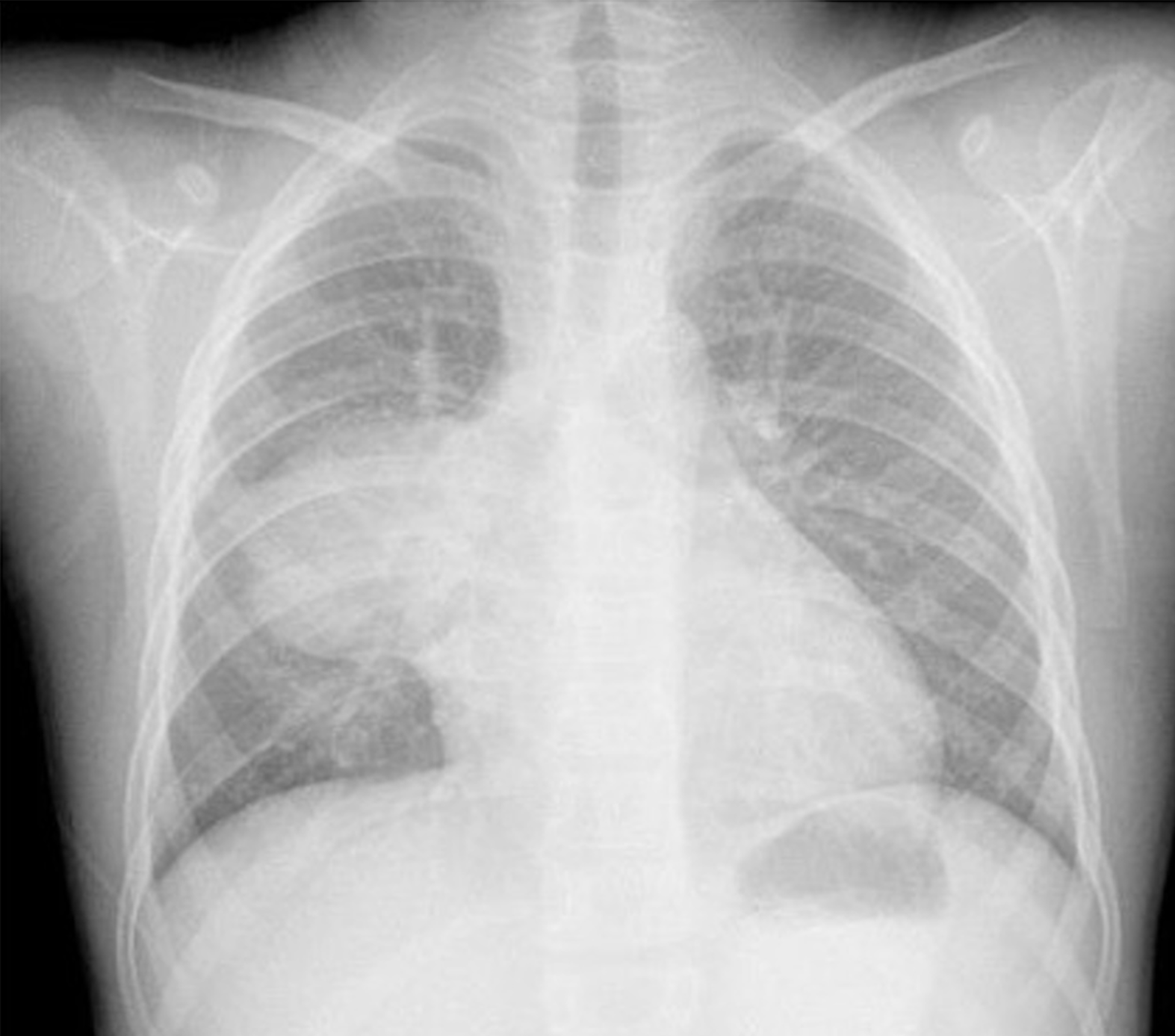
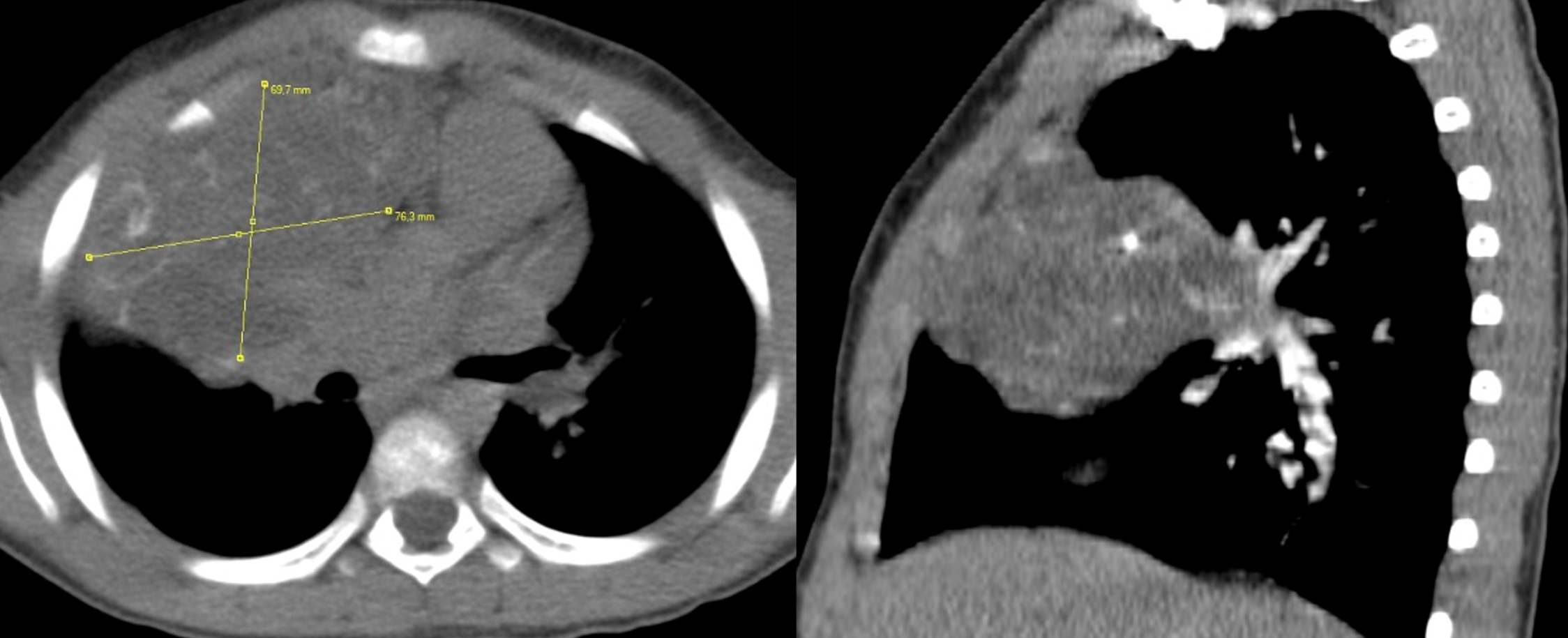
As part of the etiologic evaluation for GnRH-independent PP secondary to hCG secretion, scrotal ultrasonography revealed testicular volumes near the upper limit for age and compatible with early pubertal development; however, no intratesticular mass was detected. Cranial, pituitary, and abdominal MRI findings were normal. Posteroanterior chest radiography demonstrated asymmetric mediastinal widening with tracheal deviation, without evidence of pulmonary infiltration or pleural/pericardial effusion (Fig. 1). Subsequent thoracic computed tomography (CT) showed a 10.5 × 7 × 7 cm anterior mediastinal mass occupying the right hemithorax. The mass exhibited no significant enhancement following contrast administration and showed no signs of invasion into adjacent structures. It extended from the thymic region toward the right hemithorax, compressing the right middle lobe. The lesion contained areas of fat density and coarse calcifications, and thymic tissue could not be clearly distinguished. No pathological lymph nodes or vascular abnormalities were identified in the mediastinum (Fig. 2).
The patient’s clinical and laboratory findings were considered consistent with a GCT secreting β-hCG and AFP. The lesion was extragonadal in origin and located in the mediastinum. Given the known association between this tumor group and karyotypic abnormalities, a karyotype analysis was performed, which revealed a 47,XXY pattern consistent with KS.
The mediastinal mass was completely resected via thoracotomy performed by the thoracic surgery team. The patient, who had no preoperative respiratory distress, experienced no intraoperative or postoperative complications. Pathologic evaluation of the mediastinal mass revealed a thymus-derived mixed GCT. Histologically, components representing all three germ layers were identified. Approximately 20% of the tumor consisted of immature neuroepithelial, immature glial, chondroid, and muscular tissues, consistent with a Grade II immature teratoma, accompanied by focal necrotic areas (5%). Surgical margins were reported as tumor-negative. Immunohistochemical analysis confirmed the mixed nature of the tumor, demonstrating D2-40, CD117, and placental alkaline phosphatase positivity in the seminomatous component; AFP, low-molecular-weight cytokeratin, and pancytokeratin positivity in the endodermal sinus (yolk sac) component; and strong β-hCG staining in focal trophoblastic areas suggestive of choriocarcinoma. The positivity of synaptophysin within glial regions supported the neuroepithelial component of the immature teratoma. The lesion was classified as a “mixed germ cell tumor composed of seminoma, yolk sac tumor, and Grade II immature teratoma with β-hCG–positive trophoblastic foci” on the basis of the findings of the pathology report.
Postoperative posteroanterior chest radiography demonstrated complete resolution of the mediastinal mass with no complications related to the surgery. Serial tumor marker measurements obtained on postoperative days 3, 7, and 30 showed a rapid and marked decline: β-hCG levels decreased to 82.6 IU/L, 6.4 IU/L, and <2 IU/L; AFP levels to 365 ng/mL, 108.2 ng/mL, and 3 ng/mL; and total testosterone levels to 800 ng/dL, 25.3 ng/dL, and <7 ng/dL, respectively. These findings indicated total resection of the tumor and the elimination of its β-hCG/AFP-producing components.
Post-surgically, the patient was placed under the care of the pediatric oncology unit and classified as having an intermediate-risk mediastinal mixed GCT. Accordingly, the patient received four cycles of bleomycin, etoposide, and cisplatin (BEP) chemotherapy following the standard treatment protocol. Neutropenia and sepsis that developed during therapy were managed appropriately according to established treatment guidelines.
During follow-up, the patient’s pubertal hormone profile remained appropriate for age, with FSH, LH, and testosterone levels within the expected ranges up to 10 years of age. Clinical examinations, laboratory studies, and imaging assessments revealed no evidence of tumor recurrence. The levels of tumor markers obtained at the initial presentation and throughout the postoperative follow-up period are summarized in Table I. A neuropsychological evaluation performed in the context of KS identified mild perceptual difficulties, while echocardiographic findings were normal.
| β-hCG, human chorionic gonadotropin; AFP, alpha-fetoprotein; LDH, lactate dehydrogenase. | |||||
| Table I. Tumor markers, total testosterone, and estradiol levels at initial presentation and during postoperative follow-up. | |||||
|
|
|
|
|
|
|
| β-hCG (IU/L) |
|
|
|
|
|
| AFP (ng/mL) |
|
|
|
|
|
| Total testosterone (ng/dL) |
|
|
|
|
|
| Estradiol (ng/mL) |
|
|
|
|
|
| LDH (U/L) |
|
|
|
|
|
At the most recent follow-up in April 2025, at 14 years and 1 month of age, no evidence of tumor recurrence was detected based on clinical, laboratory, and imaging findings, and pubertal development was appropriate for age. Laboratory evaluation revealed elevated gonadotropin levels, with FSH at 17.5 U/L (reference range: 1.24–10.37 U/L) and LH at 15 U/L (reference range: 0.2–5 U/L), despite a total testosterone level within the normal male range (449 ng/dL). These findings were considered consistent with hypergonadotropic compensated testicular dysfunction, likely reflecting the combined gonadotoxic effects of KS and BEP chemotherapy. Tumor markers remained within normal limits, with AFP at 1.15 ng/mL (reference range: 0–7 ng/mL) and β-hCG <5 IU/L (reference range: <5 IU/L). Androgen replacement therapy is planned for the later stages of adolescence due to underlying KS.
Informed consent was obtained from the legal guardian for the use of the patient’s information in the publication.
Discussion
This case highlights a rare etiology underlying GnRH-independent PP in a 7-year-9-month-old boy: a mediastinal GCT secreting β-hCG and AFP. Despite markedly elevated testosterone and estradiol levels, the suppression of gonadotropins indicated a GnRH-independent mechanism of puberty. The presence of a solid mediastinal lesion containing fat density and calcifications, the absence of testicular masses, and normal abdominal, cranial, and pituitary MRI findings effectively excluded primary testicular or central causes, strongly pointing toward an hCG-secreting mediastinal tumor as the source of the clinical picture. Although the patient’s testicular volume was borderline pubertal (4 mL bilaterally), this finding may be explained by the LH-like activity of β-hCG stimulating Leydig cell testosterone production despite suppressed pituitary gonadotropins. In contrast, the absence of true gonadotropin activation likely limited further testicular enlargement, which remained inconsistent with fully activated central puberty.7
Precocious puberty is not a typical feature of KS, although GCTs have been reported in these patients. While most cases of precocious puberty in KS arise from hCG-secreting GCTs, idiopathic central precocious puberty remains extremely rare among these patients.6 Although KS had not been previously diagnosed in our patient, the presence of a mediastinal tumor made this association noteworthy. Indeed, KS has been identified in at least 8% of male patients with primary mediastinal tumors, and the syndrome is reported to be nearly 50 times more prevalent among males diagnosed with mediastinal GCTs than in the general population.8 These data underscore the importance of routine karyotype analysis in male patients presenting with mediastinal GCTs.
The increased risk of mediastinal GCT development in individuals with KS is supported not only by epidemiological observations but also by several underlying biological mechanisms. The association between KS and mediastinal GCTs may involve genetic factors related to an extra X chromosome.3 Furthermore, the increased incidence of mediastinal GCTs in KS has been hypothesized to result from the malignant transformation of primordial germ cells misplaced during embryogenesis. This pathogenic association may be further influenced by the susceptibility locus at Xq27, as well as by sustained hypergonadotropism, which may promote germ cell proliferation.3 Additionally, disturbances in steroidogenesis and impaired androgen production may create a permissive environment for the proliferation of extragonadal germ cells. Collectively, these findings support the notion that KS provides a genetic substrate that predisposes individuals to the development of mediastinal GCTs.4
In this case, the markedly elevated level of β-hCG (314 IU/L) is consistent with the characteristic biochemical profile described in hCG-secreting mediastinal GCTs.3 As hCG shares an identical α-subunit and a highly similar β-subunit with LH, it stimulates Leydig cells, resulting in increased testosterone secretion. As a natural consequence of this mechanism, gonadotropins are suppressed through negative feedback.9 The suppressed gonadotropin levels observed in our patient, together with the blunted FSH/LH response on the GnRH stimulation test, strongly indicate hCG-mediated suppression of the hypothalamic–pituitary–gonadal axis.
The markedly elevated AFP level (586 ng/mL) indicated the presence of a non-seminomatous component within the tumor, a finding commonly associated with yolk sac tumors or embryonal carcinoma, and frequently observed in mixed GCTs.10 The areas of fat density and calcification detected on thoracic CT were consistent with a teratomatous component. When the biochemical markers and imaging findings were evaluated together, a clinical diagnosis of mixed GCT was strongly favored, and histopathologic examination confirmed this diagnosis. The identification of seminomatous, yolk sac, and trophoblastic components in the pathology samples was fully concordant with the patient’s biomarker profile.
In the literature, cases of precocious puberty secondary to hCG-secreting mediastinal tumors have commonly been associated with elevated testosterone and markedly increased β-hCG levels.3 For instance, in an 8.5-year-old KS case reported by Völkl et al., suppressed gonadotropins, elevated testosterone levels, and markedly elevated β-hCG and AFP levels were documented, and the tumor was histopathologically diagnosed as a mixed GCT.3 Similarly, in our case, the absence of any testicular mass on scrotal ultrasonography definitively supports the mediastinum as the primary site of the tumor.
Primary extragonadal malignant GCTs account for only 5% of all malignant GCTs; however, they may exhibit clinically aggressive behavior. Mediastinal non-seminomatous GCTs, in particular, tend to have a more aggressive course and lower chemotherapy response rates compared with their gonadal counterparts.10 Therefore, early diagnosis, timely surgical intervention, and a multidisciplinary treatment approach are crucial for improving prognosis. Treatment typically consists of complete surgical resection followed by cisplatin-based chemotherapy regimens.10 In the pediatric population, coordinated multidisciplinary management involving pediatric oncology, endocrinology, and thoracic surgery teams has been shown to positively influence both survival and long-term recurrence outcomes. In our case, complete tumor resection and the rapid normalization of postoperative tumor markers were considered indicators of favorable prognosis.
Primordial germ cells undergo a well-defined migratory process during embryogenesis, traveling from the yolk sac to the gonadal ridge. Defects in this migration pathway may result in germ cells becoming arrested at ectopic midline locations, including the pineal region, mediastinum, or retroperitoneum. Extragonadal GCTs are thought to arise from these ectopic primordial germ cells.4 This theory represents the most widely accepted embryological mechanism explaining the primary mediastinal location of the tumor in our patient. Extragonadal GCTs occur most commonly in the mediastinum, accounting for approximately 50–70% of all extragonadal GCT cases. Among mediastinal GCTs, the most frequent histologic type is mature teratoma.11
Conclusion
In summary, this case represents an extremely rare and noteworthy clinical presentation of GnRH-independent PP secondary to a β-hCG- and AFP-secreting mediastinal GCT, occurring in association with KS. The literature clearly demonstrates an increased association between KS and mediastinal GCTs, and routine karyotype analysis is of clinical importance in all male children diagnosed with mediastinal GCTs.
This case provides valuable insight into the diagnostic approach to similar clinical presentations and strongly emphasizes that, particularly in cases of unexplained precocious puberty accompanied by suppressed gonadotropins, extragonadal GCTs and their associated biochemical markers (β-hCG, AFP) must be carefully evaluated.
Ethical approval
Informed consent was obtained from the legal guardian for the use of the patient’s information in the publication.
Source of funding
The authors declare the study received no funding.
Conflict of interest
The authors declare that there is no conflict of interest.
References
- Faizah M, Zuhanis A, Rahmah R, et al. Precocious puberty in children: a review of imaging findings. Biomed Imaging Interv J 2012; 8: e6.
- Lozano C C, Molina P M. Teratoma tímico productor de Β-HCG: una causa infrecuente de pubertad precoz periférica. Rev Chil Pediatr 2018; 89: 373-379.
- Völkl TMK, Langer T, Aigner T, et al. Klinefelter syndrome and mediastinal germ cell tumors. Am J Med Genet A 2006; 140: 471-481. https://doi.org/10.1002/ajmg.a.31103
- Schmoll HJ. Extragonadal germ cell tumors. Ann Oncol 2002; 13(Suppl 4): 265-272. https://doi.org/10.1093/annonc/mdf669
- Bonouvrie K, van der Werff Ten Bosch J, van den Akker M. Klinefelter syndrome and germ cell tumors: review of the literature. Int J Pediatr Endocrinol 2020; 2020: 18. https://doi.org/10.1186/s13633-020-00088-0
- Maqdasy S, Barres B, Salaun G, et al. Idiopathic central precocious puberty in a Klinefelter patient: highlights on gonadotropin levels and pathophysiology. Basic Clin Androl 2020; 30: 19. https://doi.org/10.1186/s12610-020-00117-1
- Shibata N, Nyuzuki H, Sasaki S, Ogawa Y, Okada M, Nagasaki K. Peripheral precocious puberty in a girl with an intracranial hCG-producing tumor: case report and literature review. Endocr J 2021; 68: 1463-1467. https://doi.org/10.1507/endocrj.EJ21-0117
- Aguirre D, Nieto K, Lazos M, et al. Extragonadal germ cell tumors are often associated with Klinefelter syndrome. Hum Pathol 2006; 37: 477-480. https://doi.org/10.1016/j.humpath.2006.01.029
- Lin CM, Lee CT, Tung YC, et al. Endocrine dysfunction in Taiwanese children with human chorionic gonadotropin-secreting germ cell tumors. J Formos Med Assoc 2014; 113: 102-105. https://doi.org/10.1016/j.jfma.2012.04.007
- McKenney JK, Heerema-McKenney A, Rouse RV. Extragonadal germ cell tumors: a review with emphasis on pathologic features, clinical prognostic variables, and differential diagnostic considerations. Adv Anat Pathol 2007; 14: 69-92. https://doi.org/10.1097/PAP.0b013e31803240e6
- Gao Y, Jiang J, Liu Q. Extragonadal malignant germ cell tumors: a clinicopathological and immunohistochemical analysis of 48 cases at a single Chinese institution. Int J Clin Exp Pathol 2015; 8: 5650-5657.
Copyright and license
Copyright © 2026 The Author(s). This is an open access article distributed under the Creative Commons Attribution License (CC BY), which permits unrestricted use, distribution, and reproduction in any medium or format, provided the original work is properly cited.