Graphical Abstract
Abstract
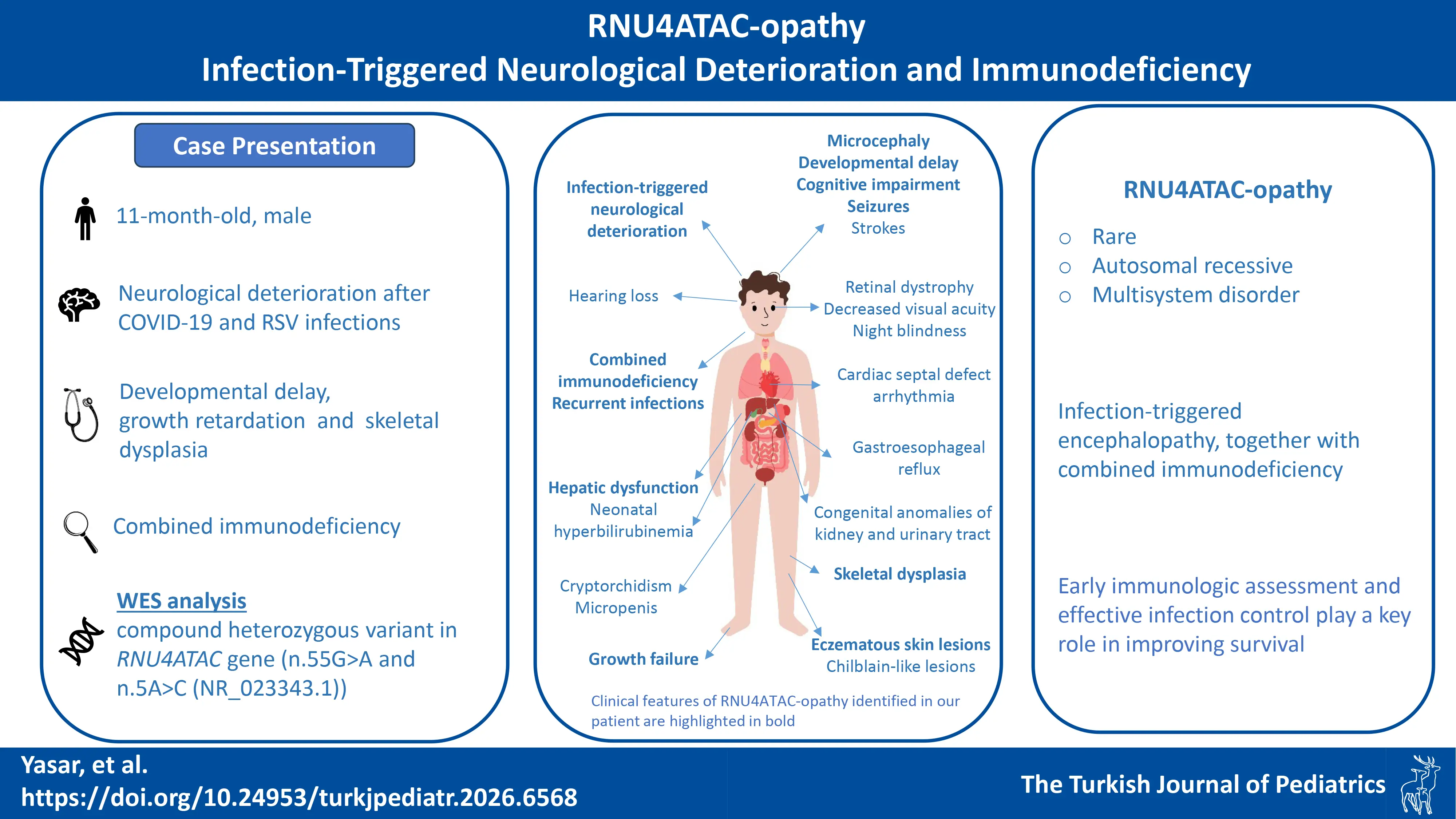
Introduction. RNU4ATAC-opathy is a rare autosomal recessive disorder characterized by a wide clinical spectrum, including brain anomalies, seizures, strokes, immunodeficiency, and cardiac defects. Other manifestations include ophthalmologic, dermatologic, renal, gastrointestinal, auditory, and endocrine involvement. Here, we report the identification of pathogenic RNU4ATAC variants through whole exome sequencing in an infant presenting with microcephaly, growth failure, and immunodeficiency.
Case Presentation. We describe an 11-month-old infant who had been followed since 4 months of age for growth failure and developmental delay. During follow-up, the patient experienced episodes of neurological deterioration triggered by febrile illnesses associated with SARS-CoV-2 and respiratory syncytial virus infections (RSV) at 9 and 11 months of age, respectively. Immunological evaluation showed normal immunoglobulin levels, a marked reduction in switched memory and memory B cells, impaired T-cell activation, and decreased T-cell subpopulations, suggesting combined immunodeficiency that may have contributed to increased susceptibility to viral infections. Whole exome sequencing subsequently identified compound heterozygous pathogenic variants in the RNU4ATAC gene, confirming the diagnosis of RNU4ATAC-opathy.
Discussion. RNU4ATAC-opathy should be considered in cases with microcephaly, growth failure, immunodeficiency, and skeletal abnormalities. Our case shows that RNU4ATAC-opathy may present as infection-related encephalopathy, together with combined T- and B-cell dysfunction. Early immunological assessment and timely initiation of prophylactic therapy could play a key role in improving survival. Genetic and immunological evaluations are essential for accurate diagnosis, early intervention, and effective management. Further research is needed to clarify the syndrome’s impact on immune function and guide targeted therapies.
Keywords: RNU4ATAC-opathy, immunodeficiency, encephalopathy, microcephaly
Introduction
RNU4ATAC-opathy encompasees the phenotypic spectrum associated with biallelic RNU4ATAC likely pathogenic or pathogenic variants, which include the clinical phenotypes of microcephalic osteodysplastic primordial dwarfism type I/III (MOPDI; OMIM#210710), Roifman syndrome (RFMN; OMIM #616651), and Lowry-Wood syndrome (LWS; OMIM #226960).1 RNU4ATAC-opathy is an autosomal recessive inherited disorder characterized by a broad clinical spectrum, including brain anomalies, seizures, strokes, immunodeficiency, and cardiac anomalies, as well as ophthalmologic, skin, renal, gastrointestinal, hearing, and endocrine involvement.1 MOPD1 is characterized by severe disease, prominent brain abnormalities, and early mortality within the first three years of life.2,3 RFMN is strongly associated with immunodeficiency, leading to frequent hospital admissions due to recurrent infections. In contrast, immunodeficiency has been reported less frequently in patients with LWS than in those with RFMN.3,4 In the 2024 update of the International Union of Immunological Societies Expert Committee classification, MOPD1 deficiency, caused by RNU4ATAC gene defects, has been included in the group of immuno-osseous dysplasias. The reported immunological features comprise decreased natural killer (NK) cell function, reduced total and memory B cells, hypogammaglobulinemia, and variably reduced specific antibodies.5 In this study, we report RNU4ATAC variants identified through whole exome sequencing (WES) in an infant presenting with microcephaly, growth failure, and immunodeficiency.
Case Presentation
A four-month-old male patient was admitted with acute gastroenteritis. He was born at 36 weeks with a birth weight of 1.5 kg (Z-score –3.32) to a non-consanguineous Turkish family, and a history of intrauterine growth restriction. He required a 33-day neonatal intensive care stay, including one day of intubation. No infections were reported until 4 months of age. Before this admission, he had been evaluated for microcephaly and growth failure, and was diagnosed with atopic dermatitis due to persistent eczematous lesions.
During the evaluation of growth failure, cytomegalovirus (CMV) IgM was positive and CMV PCR was 495 IU/mL; however, congenital CMV infection was not considered, given normal hearing, absence of retinitis, and unremarkable brain magnetic resonance imaging (MRI) findings. Neuroimaging revealed a thin corpus callosum and shallow gyral pattern (Fig. 1a and Fig. 1b). Metabolic tests are summarized in Supplementary Table S1.
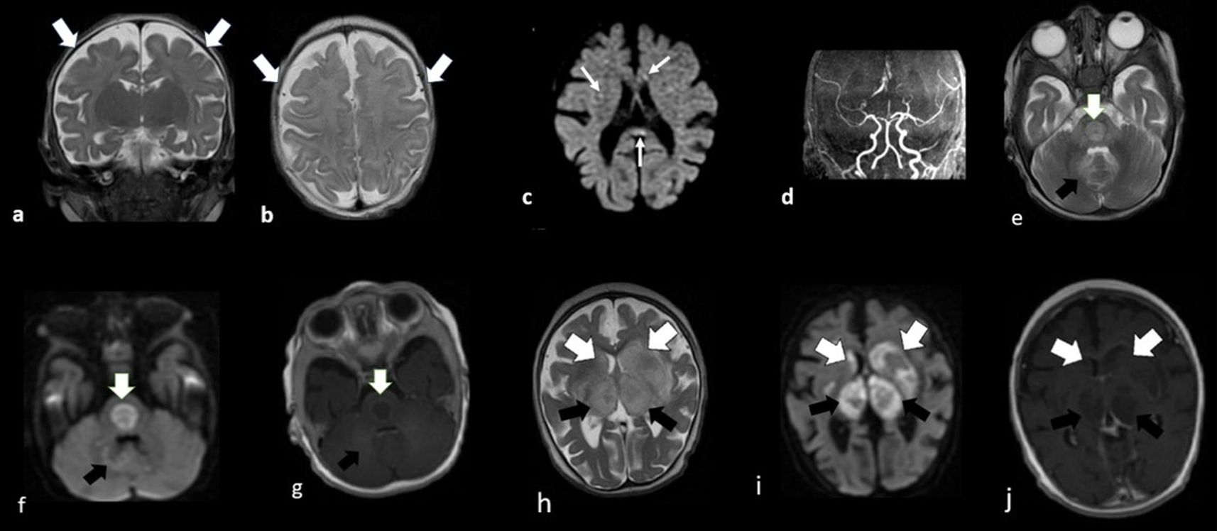
At 8 months, he was rehospitalized due to a generalized eczematous rash and fever. Examination showed diffuse xerosis with fine scaling, consistent with ichthyosis. Laboratory evaluation revealed leukocytosis, eosinophilia, anemia, elevated IgE, and elevated liver transaminases (Table I). Skin biopsy confirmed atopic/contact dermatitis, and the rash improved with antibiotics, supportive care, and moisturizers (Fig. 2c-e). During hospitalization, he developed diarrhea and a generalized tonic–clonic seizure. Diffusion-weighted imaging revealed punctate diffusion restriction in the splenium of the corpus callosum, periventricular subependymal regions, and right corona radiata, while MR angiography showed normal results (Fig. 1c and Fig. 1d). Electroencephalography revealed focal epileptiform anomalies originating from the frontal and frontocentral regions of the left hemisphere, and levetiracetam was started. A skeletal survey revealed mild spondyloepiphyseal dysplasia (Supplementary Fig. S1). The patient’s leukocytosis and eosinophilia resolved in the follow-up.
| ALP: Alkaline phosphatase; ALT: Alanine aminotransferase; AST: Aspartate aminotransferase; CRP: C-reactive protein; ESR: Erythrocyte sedimentation rate; GGT: Gamma-glutamyl transferase; MCV: Mean corpuscular volume; RBC: Red blood cell; RTE: Recent thymic emigrants. | ||
| Table I. The hematological, biochemical, and immunological results of the patient. | ||
| Tests |
|
|
| Hemoglobin (g/dL) |
|
|
| MCV (fL) |
|
|
| RBC (x 106/µL) |
|
|
| Leukocyte (x 103/µL) |
|
|
| Platelet (x 103/µL) |
|
|
| Neutrophil (x 103/µL) |
|
|
| Lymphocyte (x 103/µL) |
|
|
| Eosinophil (x 103/µL) |
|
|
| AST (U/L) |
|
|
| ALT (U/L) |
|
|
| ALP (U/L) |
|
|
| GGT (U/L) |
|
|
| Bilirubin (total) (mg/dL) |
|
|
| Bilirubin (direct) (mg/dL) |
|
|
| CRP (mg/L) |
|
|
| ESR (mm/h) |
|
|
| Immunoglobulins | ||
| IgA (g/L) |
|
|
| IgM (g/L) |
|
|
| IgG (g/L) |
|
|
| Total IgE (IU/mL) |
|
|
| Lymphocyte subsets | ||
| CD3, % |
|
|
| CD4, % |
|
|
| CD8, % |
|
|
| CD16-56, % |
|
|
| CD19, % |
|
|
| Lymphocyte activation | ||
| CD3 CD25 Cells, % |
|
|
| CD3 CD69 Cells, % |
|
|
| B Cell Panel | ||
| Memory B Cells, % |
|
|
| Switched Memory B Cells, % |
|
|
| Marginal Zone B Cells, % |
|
|
| Naive B Cells, % |
|
|
| Active B Cells, % |
|
|
| Plasmoblasts, % |
|
|
| Transitional B Cells, % |
|
|
| T Cell Panel | ||
| CD4 T Cells, % |
|
|
| CD4 Naive T Cells, % |
|
|
| CD4 Central Memory T Cells, % |
|
|
| CD4 Effector Memory T Cells, % |
|
|
| CD4 Temra T Cells, % |
|
|
| CD8 T Cells, % |
|
|
| CD8 Naive T Cells, % |
|
|
| CD8 Central Memory T Cells, % |
|
|
| CD8 Effector Memory T Cells, % |
|
|
| CD8 Temra T Cells, % |
|
|
| RTE (Recent Thymic Emigrants), % |
|
|
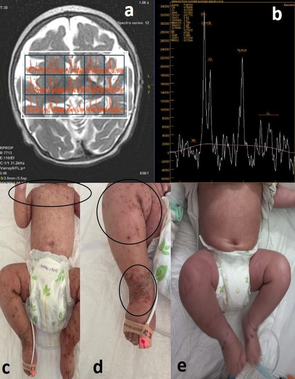
At the age of 9 months, he was hospitalized for dehydration due to gastroenteritis and coronavirus disease 2019 (COVID-19). He developed seizures and decorticate posturing. Emergency neuroimaging revealed hemorrhagic areas, raising suspicion of mitochondrial disease, acute hemorrhagic encephalomyelitis, and acute necrotizing encephalopathy (ANEC) as primary differential diagnoses (Fig. 1e-j). MR spectroscopy of the basal ganglia revealed an increased choline/creatine (Cho/Cr) ratio, decreased N-acetyl aspartate (NAA) values, and a lactate peak, leading to the suspicion of mitochondrial disease (Fig. 2a and Fig. 2b). A mitochondrial cocktail was started, consisting of thiamine (10 mg/kg/day), riboflavin (10 mg/kg/day), coenzyme Q10 (10 mg/kg/day), biotin (5 mg twice daily), carnitine (40 mg/kg/day), and vitamin E (20 IU/kg, three times per week).
The patient was monitored in the pediatric intensive care unit (PICU) after the clinical deterioration. Ceftriaxone and acyclovir treatments were initiated for encephalitis. Immunological investigations showed normal immunoglobulin levels, normal numbers of T, and B cells and decreased NK cell counts. However, lymphocyte activation was reduced, along with decreases in memory B cells, switched memory B cells, marginal zone B cells, CD8 naive T cells, CD8 central memory T cells, CD8 effector memory T cells, and CD4 central memory T cells (Table I). Intravenous immunoglobulin (IVIG) therapy was administered at 0.4 g/kg/day for 5 days, along with pulse steroid therapy (methylprednisolone 30 mg/kg/day) for 5 days. On the 10th day of intensive care, with clinical improvement under treatment, a follow-up MRI showed decreased edematous appearance and atrophic changes in the described areas. During this hospitalization, the patient lost the ability to sit unsupported and could no longer hold their head up. After discharge from the PICU, monthly IVIG therapy, acyclovir and trimethoprim-sulfamethoxazole (TMP-SMX) prophylaxis were started for a probable syndromic combined immunodeficiency diagnosis. Levetiracetam and the mitochondrial cocktail were continued.
At 11 months of age, the patient was hospitalized again due to respiratory syncytial virus (RSV) pneumonia requiring intubation. During hospitalization, transaminase levels were mildly elevated, without hepatosplenomegaly or hyperbilirubinemia, and resolved after ursodeoxycholic acid (UDCA) treatment. The patient’s echocardiography was normal, but sinus bradycardia was observed, and resolved without any treatment. During this hospitalization, WES revealed compound heterozygous pathogenic variants in the RNU4ATAC gene (see below), confirming the diagnosis of RNU4ATAC-opathy. Following the genetic diagnosis, the mitochondrial cocktail was discontinued.
At present, the patient is 36 months old, and continues to require recurrent hospitalizations due to lower respiratory tract infections. However, no further infection-triggered neurological deterioration has been observed for more than one year, possibly reflecting a beneficial effect of ongoing IVIG therapy. Neurological developmental delay persists, accompanied by physical inactivity and excessive weight gain. The patient remains on levetiracetam and valproic acid for epilepsy, along with antibacterial and antiviral prophylaxis and IVIG.
Genetic analysis
WES was performed at 10 months of age using DNA extracted from peripheral blood leukocytes with next-generation sequencing (NGS, Illumina) and the Illumina DNA Prep with Exome 2.0/2.5 Plus Enrichment Kit. The bioinformatic analysis of the raw data was performed and reported using the Varsome Clinical (Saphetor) software. The WES analysis revealed heterozygous n.55G>A and n.5A>C (RefSeq: NR_023343.1) variants in the 5’ stem-loop and Stem II structures of the RNU4ATAC gene, which is a non-coding spliceosomal gene. These variants were classified as “pathogenic” and as a “variant of uncertain significance” according to the American College of Medical Genetics and Genomics criteria, respectively. This biallelic configuration was validated by inspection of the NGS BAM file. Parental segregation analysis demonstrated that the n.5A>C variant (RefSeq: NR_023343.1) was inherited from the unaffected mother, while the father is wild type at both relevant nucleotide positions. The n.55G>A variant was absent in both parental samples and is therefore classified as de novo. Sanger sequencing confirmed both variants in the patient establishing the compound heterozygous state.
Written informed consent was obtained from the patient’s parents.
Discussion
This report describes a patient who was diagnosed with RNU4ATAC-opathy. MOPD1, LWS, and RFMN are overlapping syndromes associated with mutations in the RNU4ATAC gene.1 The clinical phenotype varies, but typically includes growth failure, microcephaly, skeletal dysplasia, and cognitive impairment.1,4 In our patient, the initial signs were microcephaly and growth failure, accompanied by combined immunodeficiency, eczematous lesions, and recurrent viral infections that triggered episodes of encephalopathy.
The n.55G>A variant, detected heterozygously in the patient, disrupts ncRNA function.4 It is rare (gnomAD: 0.0000885), is classified as pathogenic in ClinVar (ID: 30179), and has been reported in multiple affected cases in the homozygous state.6-8 These patients presented with the classical clinical spectrum of MOPD1, including severe microcephaly, growth restriction, skeletal dysplasia, neurodevelopmental impairment, and, in some cases, immunodeficiency and infection-triggered neurological deterioration.4,6,8 The patient’s other heterozygous variant, n.5A>C, is located within Stem II of RNU4ATAC. It is also rare (gnomAD: 0.00001), classified in ClinVar as a “variant of uncertain significance” (ID: 932367), and reported in one affected individual in the literature in a compound heterozygous state.9
Across RNU4ATAC-opathies, common immunological abnormalities include hypogammaglobulinemia, reduced memory B cells, recurrent infections, and variable T-cell defects, while NK cell counts (CD16+/CD56+) are usually within the normal range and functional NK assays were rarely performed.9-16 Reported cases of RNU4ATAC-opathy presenting with immunological involvement are summarized in Table II. MOPD1 is frequently associated with significant humoral defects, such as reduced B-cell subsets, impaired antibody production, poor vaccine responses, and in some cases, CD4+ T-cell lymphopenia, contributing to early mortality often secondary to infections.11,13-15 RFMN is characterized by a more consistent immunodeficiency phenotype, including profound hypogammaglobulinemia, reduced B-cell numbers, impaired specific antibody responses, and occasional NK cell reduction, frequently necessitating long-term immunoglobulin replacement therapy.10,12,16 Although described less often, LWS can also involve hypogammaglobulinemia and immune dysfunction.9 These overlapping findings support the concept of a shared immunological phenotype across the RNU4ATAC-opathies.
| AHEM: Acute Hemorrhagic Encephalomyelitis, ANE: Acute Necrotizing Encephalopathy, ARDS: Acute Respiratory Distress Syndrome, DIC: Disseminated Intravascular Coagulation, HHV-6: Human Herpesvirus 6, IVIG: Intravenous Immunoglobulin, LWS: Lowry Wood syndrome, MMR: Measles, Mumps, Rubella; MOPD1: Microcephalic Osteodysplastic Primordial Dwarfism Type I, MRSA: Methicillin-Resistant Staphylococcus aureus, P: number of patients, PCV13/PPV23: Pneumococcal Vaccines (13-valent conjugate / 23-valent polysaccharide), URTI: Upper Respiratory Tract Infection, VZV: Varicella Zoster Virus. | |||||
| Table II. Reported immunological findings in RNU4ATAC-opathy. | |||||
| Patient and syndrome | T-cells | B-cells | NK Cells | Immunoglobulin levels | Clinical immunological features |
| Merico et al.10, (2015) (P6) - Roifman | Normal T cells | ↓ Circulating B-cell, normal mature and memory B cells | Normal | Variable immunoglobulin level | Poor vaccine response |
| Kılıç et al.11, 2015 (P1) – MOPD1 | Not reported | B-cell related anomalies | Not reported | Hypogammaglobulinemia |
Recurrent infections (pneumonia, diarrhea) IVIG theraphy |
| Dinur Schejter et al.12, 2017 (P2) - Roifman |
↓ CD8+ T cells, Poor antigen response |
↓ CD19+ B cells | Normal | Hypogammaglobulinemia | Recurrent pneumonia, ear infections, atopy (asthma, eczema); improved with IVIG, Absent responses to tetanus, MMR, VZV, pneumococcus vaccines |
| Farach et al.9, 2018 (P3) - LWS | Not reported |
B-cell related anomalies (one patient) |
Not reported |
Hypogammaglobulinemia (one patient) |
Not detailed in 2 patients, Subclinical in one patient |
| Heremans et al.16, 2018 (P3) - Roifman |
↓ CD4+ T cells (in one patient), ↓ Total CD3+ T cells (one patient) |
↓ B cells | Normal | Hypogammaglobulinemia |
All patients experience recurrent viral infections, necessitating IVIG therapy. Herpes simplex infection (one patient), pneumococcal sepsis (one patient |
| Hagiwara et al.13, 2021 (P1)-MOPDI | Decreases in total lymphocytes, CD4+ T cells, and T cell regenerative activity. | ↓ B cells | Normal | Hypogammaglobulinemia |
Recurrent infections (respiratory and URTI), ARDS and DIC with MRSA infection. ANE with HHV-6 Poor vaccine response to varicella-zoster, MMR and influenza |
| Sirohi et al.14, 2023 (P2) – MOPDI-III | ↓ CD8+ T cells | ↓ CD19+ B cells | Normal | Hypogammaglobulinemia (one patient) |
Recurrent infection, paucity of plasma cells in gastrointestinal biopsies Impaired antibody response to PCV13/PPV23; protective diphtheria/tetanus titers |
| Gauthier et al.15, 2024 (P2, twin) – MOPDI | Normal T cells | Low naïve B cells, ↓ memory B cells and plasmablasts | Normal | Normal | Mild B-cell deficiency, recurrent infections |
| The patient |
↓ CD8 Naive T Cells, ↓ CD8 Central Memory T Cells, ↓ CD8 EffectorMemory T Cells, ↓ CD4 Central Memory T Cells |
↓ Memory B Cells, ↓ Switched Memory B Cells, ↓ Marginal Zone B Cells |
↓ CD16/56 | Normal |
ANE-AHEM with COVID-19, Recurrent infections (pneumonia), improved with IVIG |
Our patient demonstrated a broader and more complex immunological profile, with combined T- and B-cell abnormalities, normal immunoglobulin levels and reduced NK cells. The preserved serum Ig levels may reflect maternally transferred immunoglobulins. Decreased switched memory and memory B cells, along with impaired T-cell activation and reduced T-cell subpopulations, likely contributed to recurrent viral infections and infection-triggered neurological deterioration, as reported in similar cases.8,13 These observations highlight that even in the presence of normal immunoglobulin levels, detailed immunological evaluation is essential for accurate diagnosis and management. In patients with T-cell defects and antibody deficiencies, early initiation of IVIG therapy may help prevent recurrent infections and improve prognosis.
RNU4ATAC-opathy is associated with various radiographic findings, including multiple epiphyseal dysplasia (MED), spondyloepiphyseal dysplasia (SED), and spondyloepimetaphyseal dysplasia (SEMD).1,9 Historically, patients with MOPDI predominantly exhibit SEMD, while RFMN is characterized by SED, and LWS typically presents with MED.1,9 However, these syndromes overlap, leading to the manifestation of features across these conditions. Our patient exhibited mild SED, reflecting this overlap.
Nearly all patients with MOPD1 have brain abnormalities such as a thin corpus callosum, and an abnormal gyral pattern.17 Similarly, our case presented with these abnormalities during the initial admission. In the literature, a patient carrying the same n.55G>A variant as our patient presented with microcephaly, normal stature, and experienced status epilepticus and encephalitis triggered by infections. MRI findings, elevated serum and CSF lactate levels in this patient indicate the need for differential diagnosis of underlying mitochondrial disease.8 However, further evaluation, including muscle and liver biopsies and OxPHOS enzymology testing, excluded this diagnosis. In our case, when ANEC was diagnosed, MR spectroscopy raised suspicion of a metabolic disease. However, mitochondrial disease was definitively ruled out, as whole mitochondrial genome analysis yielded normal results. Encephalitis has also been reported in other MOPD1 cases carrying the n.55G>A variant.13,18,19
In the literature, RFMN and MOPD1 have been associated with dry skin and eczematous lesions.12,20 Our patient was diagnosed with atopic dermatitis and exhibited refractory eczematous lesions over a short period. Cardiac abnormalities can be seen in the RNU4ATAC-opathy spectrum, though rhythm problems have not been reported.1,17,18 Our patient’s echocardiography was normal, but sinus bradycardia was observed and resolved in follow-up. In addition, neonatal cholestasis and hepatosplenomegaly have been reported in the literature.21 Our case had mildly elevated transaminase levels that resolved with UDCA treatment.
A limitation of our report is the lack of functional assays to directly confirm the link between RNU4ATAC variants and immunodeficiency due to the patient’s unstable clinical condition and limited access to advanced analyses. Nevertheless, the immunophenotyping findings presented here expand the current understanding of the immunological spectrum of RNU4ATAC-opathies and highlight the importance of comprehensive immune assessment in all affected patients.
In conclusion, patients with RNU4ATAC-opathy may present with T-cell immunodeficiency and progressive neurological regression, often triggered by various viral infections. Immunological evaluation of these patients is critically important, as cases with varying degrees of immunodeficiency have been reported. Reducing the frequency of attacks and effectively controlling infections may significantly improve the patient’s prognosis. Further studies will be important to better explain the mechanisms of immune-mediated strokes and immunodeficiency in these patients.
Acknowledgements
We would like to thank Esra Kılıç and Elifcan Taşdelen for their valuable contributions to this case.
Ethical approval
Written informed consent was obtained from the patient and her legal guardians for publication of this case report and accompanying images. Ethical committee approval was not obtained, as it was not required for a single case report according to our institutional policy.
Source of funding
The authors declare the study received no funding.
Conflict of interest
The authors declare that there is no conflict of interest.
References
- Duker A, Velasco D, Robertson N, Jackson A, DeFelice M, Bober MB. RNU4atac-opathy. In: Adam MP, Bick S, Mirzaa GM, Pagon RA, Wallace SE, Amemiya A, editors. GeneReviews®. Seattle (WA): University of Washington; 1993.
- Edery P, Marcaillou C, Sahbatou M, et al. Association of TALS developmental disorder with defect in minor splicing component U4atac snRNA. Science 2011; 332: 240-243. https://doi.org/10.1126/science.1202205
- Shelihan I, Ehresmann S, Magnani C, et al. Lowry-Wood syndrome: further evidence of association with RNU4ATAC, and correlation between genotype and phenotype. Hum Genet 2018; 137: 905-909. https://doi.org/10.1007/s00439-018-1950-8
- Benoit-Pilven C, Besson A, Putoux A, et al. Clinical interpretation of variants identified in RNU4ATAC, a non-coding spliceosomal gene. PLoS One 2020; 15: e0235655. https://doi.org/10.1371/journal.pone.0235655
- Poli MC, Aksentijevich I, Bousfiha AA, et al. Human inborn errors of immunity: 2024 update on the classification from the International Union of Immunological Societies Expert Committee. J Hum Immun 2025; 1: e20250003. https://doi.org/10.70962/jhi.20250003
- He H, Liyanarachchi S, Akagi K, et al. Mutations in U4atac snRNA, a component of the minor spliceosome, in the developmental disorder MOPD I. Science 2011; 332: 238-240. https://doi.org/10.1126/science.1200587
- Shaheen R, Maddirevula S, Ewida N, et al. Genomic and phenotypic delineation of congenital microcephaly. Genet Med 2019; 21: 545-552. https://doi.org/10.1038/s41436-018-0140-3
- McMillan HJ, Davila J, Osmond M, et al. Whole genome sequencing identifies pathogenic RNU4ATAC variants in a child with recurrent encephalitis, microcephaly, and normal stature. Am J Med Genet A 2021; 185: 3502-3506. https://doi.org/10.1002/ajmg.a.62457
- Farach LS, Little ME, Duker AL, et al. The expanding phenotype of RNU4ATAC pathogenic variants to Lowry Wood syndrome. Am J Med Genet A 2018; 176: 465-469. https://doi.org/10.1002/ajmg.a.38581
- Merico D, Roifman M, Braunschweig U, et al. Compound heterozygous mutations in the noncoding RNU4ATAC cause Roifman Syndrome by disrupting minor intron splicing. Nat Commun 2015; 6: 8718. https://doi.org/10.1038/ncomms9718
- Kilic E, Yigit G, Utine GE, et al. A novel mutation in RNU4ATAC in a patient with microcephalic osteodysplastic primordial dwarfism type I. Am J Med Genet A 2015; 167A: 919-921. https://doi.org/10.1002/ajmg.a.36955
- Dinur Schejter Y, Ovadia A, Alexandrova R, et al. A homozygous mutation in the stem II domain of RNU4ATAC causes typical Roifman syndrome. NPJ Genom Med 2017; 2: 23. https://doi.org/10.1038/s41525-017-0024-5
- Hagiwara H, Matsumoto H, Uematsu K, et al. Immunodeficiency in a patient with microcephalic osteodysplastic primordial dwarfism type I as compared to Roifman syndrome. Brain Dev 2021; 43: 337-342. https://doi.org/10.1016/j.braindev.2020.09.007
- Sirohi N, Duker AL, Bober MB, DeFelice ML. Immune deficiency in microcephalic osteodysplastic primordial dwarfism type I/III. J Clin Immunol 2023; 43: 895-897. https://doi.org/10.1007/s10875-023-01447-1
- Gauthier LW, Gossez M, Malcus C, et al. B-cell immune deficiency in twin sisters expands the phenotype of MOPDI. Clin Genet 2024; 106: 476-482. https://doi.org/10.1111/cge.14571
- Heremans J, Garcia-Perez JE, Turro E, et al. Abnormal differentiation of B cells and megakaryocytes in patients with Roifman syndrome. J Allergy Clin Immunol 2018; 142: 630-646. https://doi.org/10.1016/j.jaci.2017.11.061
- Tabib A, Richmond CM, McGaughran J. Delineating the phenotype of RNU4ATAC-related spliceosomopathy. Am J Med Genet A 2023; 191: 1094-1100. https://doi.org/10.1002/ajmg.a.63110
- Abdel-Salam GMH, Abdel-Hamid MS, Issa M, Magdy A, El-Kotoury A, Amr K. Expanding the phenotypic and mutational spectrum in microcephalic osteodysplastic primordial dwarfism type I. Am J Med Genet A 2012; 158A: 1455-1461. https://doi.org/10.1002/ajmg.a.35356
- Abdel-Salam GMH, Emam BA, Khalil YM, Abdel-Hamid MS. Long-term survival in microcephalic osteodysplastic primordial dwarfism type I: evaluation of an 18-year-old male with g.55G>A homozygous mutation in RNU4ATAC. Am J Med Genet A 2016; 170A: 277-282. https://doi.org/10.1002/ajmg.a.37409
- Putoux A, Alqahtani A, Pinson L, et al. Refining the phenotypical and mutational spectrum of Taybi-Linder syndrome. Clin Genet 2016; 90: 550-555. https://doi.org/10.1111/cge.12781
- Berger A, Haschke N, Kohlhauser C, Amman G, Unterberger U, Weninger M. Neonatal cholestasis and focal medullary dysplasia of the kidneys in a case of microcephalic osteodysplastic primordial dwarfism. J Med Genet 1998; 35: 61-64. https://doi.org/10.1136/jmg.35.1.61
Copyright and license
Copyright © 2026 The Author(s). This is an open access article distributed under the Creative Commons Attribution License (CC BY), which permits unrestricted use, distribution, and reproduction in any medium or format, provided the original work is properly cited.