Abstract
Background. Retinopathy of prematurity (ROP) remains a leading cause of childhood blindness. The microRNA-146a (miR-146a) rs2910164 polymorphism, implicated in angiogenesis and inflammation, may influence ROP pathogenesis, but its role is undefined. This study aimed to investigate the association of the miR-146a rs2910164 polymorphism with ROP susceptibility, disease severity, and complications.
Methods. This retrospective case-control study included 196 preterm infants with ROP and 185 healthy preterm (HC) infants who were treated from January 2020 to December 2021 at our hospital. rs2910164 genotyping and miR-146a expression analysis using quantitative reverse transcriptase polymerase chain reaction were performed. Diagnostic potential was evaluated by receiver operating characteristic analysis. Associations with clinical features, disease severity (need for treatment for ROP), and complications were assessed.
Results. Infants with ROP demonstrated significantly lower gestational age/birth weight and higher rates of intraventricular hemorrhage, bronchopulmonary dysplasia, and necrotizing enterocolitis than the HC infants (all P<0.001). miR-146a expression was an excellent diagnostic biomarker for ROP (AUC :0.918, 95% CI: 0.87-0.96). The CC genotype and C allele conferred a reduced risk of ROP. Among patients with ROP, CC/GC carriers had a lower likelihood of developing severe ROP that required treatment versus GG homozygotes. GG carriers also showed more severe manifestations and complications.
Conclusion. The rs2910164 C allele is associated with protection from ROP susceptibility, correlates with upregulated miR-146a expression, and is further associated with milder disease manifestations, fewer complications, and a reduced need for clinical intervention. This polymorphism is a valuable predictive biomarker for ROP risk, severity, and prognosis, potentially mediated via miR-146a regulation.
Keywords: retinopathy of prematurity, miR-146a, rs2910164, genetic polymorphism, biomarker
Introduction
Retinopathy of prematurity (ROP) is a sight-threatening condition marked by abnormal proliferation of retinal blood vessels and associated inflammation, constituting a major contributor to visual impairment in children.1-3 While advancements in neonatal intensive care have markedly increased survival among extremely preterm and extremely low birth weight infants, this success has paradoxically elevated the incidence of ROP, imposing a substantial public health challenge.4,5 The central pathophysiological mechanism of ROP involves an initial arrest of retinal vascular development, followed by pathological neovascularization.6,7 Crucially, inflammatory processes are recognized as fundamental drivers in both retinal vascular injury and defective repair mechanisms, playing a pivotal role across all stages of ROP development.8
Research indicates that the microRNA-146a (miR-146a) rs2910164 G→C polymorphism is strongly linked to an elevated risk of various inflammatory and vascular conditions, including inflammatory bowel disease (IBD)9 and acute coronary syndrome / coronary heart disease (ACS/CHD).10-12 This genetic variant is also associated with heightened inflammatory activity and poorer clinical outcomes.13,14 Located within the stem-loop region of the miR-146a precursor15, this polymorphism disrupts mature miR-146a expression and function. Consequently, its ability to regulate inflammation is diminished, leading to varying inflammatory responses among individuals exposed to the same environmental factors.16 However, the specific contribution, clinical significance, and potential biomarker value of this polymorphism in ROP remain undefined. Considering the critical involvement of inflammatory pathways in ROP pathogenesis and the key regulatory role of miR-146a in inflammation1,2,9,10, dedicated investigation into its impact within the context of ROP is warranted.
Given that the functional genetic variant rs2910164 in the miR-146a gene may influence inflammatory and angiogenic pathways by modulating rs2910164 expression, thereby contributing to the pathogenesis of ROP, this study hypothesizes that the rs2910164 polymorphism is associated with ROP susceptibility, severity, related complications, and the necessity of therapeutic intervention, which serves as a proxy for severe disease. To systematically test this hypothesis, we will utilize a well-characterized cohort of preterm infants to comprehensively analyze the impact of this polymorphism on ROP risk, miR-146a expression levels, key clinical phenotypes, major prematurity-associated complications, and the requirement for treatment, reflecting disease severity.
Materials and Methods
Research design and sample data
This study employed a single-center, retrospective design. All serum samples were residual aliquots obtained from routine clinical biochemistry tests performed on hospitalized patients with ROP between January 2020 and December 2021. Following the completion of clinical testing, the samples were anonymized using a coding system and temporarily stored in accordance with our hospital’s Standard Operating Procedures for Clinical Specimen Management. They were subsequently transferred to the institutional biobank for long-term preservation at -80 °C. The storage and subsequent research use of these samples strictly complied with the Interim Regulations for the Use of Clinical Residual Samples in Scientific Research, established jointly by the Research Administration Department and the Institutional Review Board (IRB). Sample collection from January 2020 to December 2021 preceded the IRB application. Notably, these samples were not originally collected for the purposes of this study. The study protocol was submitted in March 2022, and ethical approval was granted on April 14, 2022 (Approval No. 2022030).
Participants and data collection
The study was approved by the Ethics Committee of Jiujiang Maternal and Child Health Care Hospital in Jiangxi Province. Written informed consent was obtained from the guardians of all participants. A total of 196 premature infants with ROP were enrolled in this retrospective study. The condition resolved spontaneously in 98 infants, while the other 98 required treatment. All cases were compared with 185 preterm controls without ROP. These control subjects were matched to the case group by mode of delivery and had no retinal pathology. Based on the prespecified clinically significant effect size (Cohen’s d = 0.5), the actual sample sizes (196 vs. 185) and α = 0.05, we assessed the statistical power of the study, which was 0.998. This indicates that the sample size in this study was sufficient to detect an effect of this magnitude.
Experienced pediatric ophthalmologists diagnosed and classified all ROP cases according to the International Classification of ROP (ICROP) guidelines. Inclusion in the study required the following: (1) gestational age below 37 weeks at delivery; (2) birth weight under 2,500 g; and (3) hospital admission within 24 hours of birth. Exclusion criteria encompassed neonates who: (1) died within the first 24 hours after admission; (2) had severe congenital malformations; and (3) were diagnosed with inherited metabolic diseases. Retrospective data collection included detailed demographic and clinical parameters: gestational age (weeks), birth weight (grams), mode of delivery (vaginal/cesarean section), sex, Apgar scores (at 1 and 5 minutes), duration of mechanical ventilation (days), and the occurrence of major complications such as intraventricular hemorrhage (IVH), bronchopulmonary dysplasia (BPD), and necrotizing enterocolitis (NEC).
Ethical statement
The research protocol was submitted in March 2022. Ethical approval was granted on April 14 of the same year (Approval No. 2022030). Due to the retrospective nature of the study, the anonymization of all data, and the use of residual samples that posed only minimal risk, the Ethics Committee granted an exemption from the requirement to obtain renewed informed consent. This decision was made in accordance with the national “Measures for the Ethical Review of Biomedical Research Involving Human Subjects.”
Furthermore, during the initial sample collection phase (2020-2021), all patients had previously provided written informed consent using a standard clinical form. This form included a clause permitting the use of anonymized samples for medical research purposes. Patients retained the right to refuse such use at that time. This study involved no interventions beyond standard clinical practice and was conducted in strict adherence to the ethical principles of the Declaration of Helsinki.
Genotyping
Genomic DNA was isolated from peripheral blood samples using a standard salting-out method. The rs2910164 (G>C) polymorphism within miR-146a was genotyped using a TaqMan allelic discrimination assay (Applied Biosystems, Foster City, CA, USA) on a real-time quantitative PCR system.
Analysis of miR-146a expression
The study quantified the relative expression levels of mature miR-146a in representative serum samples from HC and ROP participants using quantitative reverse transcription PCR (qRT-PCR). TRIzol reagent was employed for total RNA isolation. Complementary DNA (cDNA) synthesis was performed using miRNA-specific stem-loop reverse transcription (RT) primers. Using the 2−ΔΔCt method, we normalized expression levels to U6 small nuclear RNA (snRNA). Results are presented as relative expression levels normalized to a calibrator sample.
Statistics
Statistical analyses were performed using SPSS (version 26.0) and GraphPad Prism (version 9.0). Continuous variables were first examined for normality using the Shapiro–Wilk test and for homogeneity of variance using Levene’s test. For variables that were normally distributed with homogeneous variance, comparisons between the two groups were performed using the independent samples t-test, and the results were presented as the mean ± standard deviation. For variables that did not meet the assumptions of normality or homogeneity of variance, the Mann–Whitney U test was applied, and the results were reported as the median (interquartile range). Categorical variables were expressed as frequencies (%) and compared using the chi-square (χ2) test. Hardy-Weinberg equilibrium (HWE) was evaluated within the HC cohort. To assess associations between genotypes and ROP risk, odds ratios (OR) with 95% confidence intervals (CI) were calculated under codominant, dominant (GC+CC vs GG), recessive (CC vs. GG+GC), and allelic genetic models. The diagnostic potential of miR-146a expression for ROP was determined using receiver operating characteristic (ROC) curve analysis, yielding the area under the curve (AUC), sensitivity, and specificity. Multifactorial logistic regression was employed to determine the independent factors associated with the development of ROP. The goodness of fit of the model was evaluated using the Hosmer-Lemeshow test. A P value greater than 0.05 indicated that the model had a good fit. Statistical significance was defined as a two-tailed P< 0.05.
Results
Clinical characteristics of retinopathy of prematurity cases versus matched controls
Compared with the HC group, infants with ROP exhibited significantly lower gestational ages, lower birth weights, and reduced Apgar scores at both 1 and 5 minutes. Infants with ROP also required mechanical ventilation for a significantly longer duration. Furthermore, they developed IVH, BPD, and NEC more frequently (Table I).
| BPD, bronchopulmonary dysplasia; HC, healthy controls; IVH, intraventricular hemorrhage; NEC, necrotizing enterocolitis; ROP, retinopathy of prematurity; SD, standard deviation. | |||
| Table I. Comparative analysis of clinical characteristics between HC and infants with ROP. | |||
| Items |
|
|
|
| Mode of delivery, n (%) |
|
||
| Vaginal |
|
|
|
| Caesarean section |
|
|
|
| Gestational age at birth (weeks), mean±SD |
|
|
|
| Birth weight (g), mean±SD |
|
|
|
| Male sex, n (%) |
|
|
|
| Apgar score, median (Q1-Q3) | |||
| 1st minute |
|
|
|
| 5th minute |
|
|
|
| Duration of mechanical ventilation (days), mean±SD |
|
|
|
| IVH, n (%) |
|
|
|
| BPD, n (%) |
|
|
|
| NEC, n (%) |
|
|
|
Diagnostic significance of miR-146a expression
miR-146a expression was significantly lower in the ROP group than in the HC group (Fig. 1A). ROC analysis indicated that miR-146a levels effectively distinguished infants with ROP from healthy preterm controls, with an AUC of 0.918 (95% CI: 0.87-0.96, P <0.001), a sensitivity of 88.3% and a specificity of 84.3% (Fig. 1B). Notably, miR-146a levels did not significantly differ between GG genotype carriers and GC/CC genotype carriers within the HC group (Fig. 1C). However, within the ROP group, infants carrying the GG genotype exhibited significantly lower miR-146a expression than those with GC or CC genotypes (Fig. 1D).
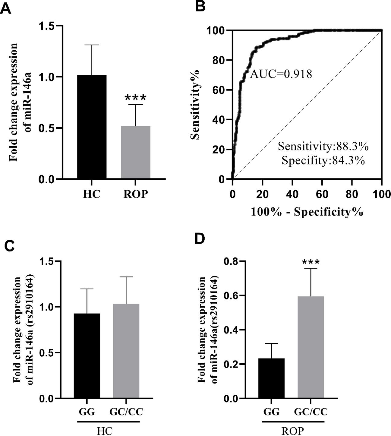
AUC, area under the curve; HC, healthy controls; ROP, retinopathy of prematurity.
Link between the rs2910164 polymorphism and susceptibility to retinopathy of ROP
The distribution of rs2910164 genotypes and alleles in the HC group and ROP groups is presented in Table II. In the HC group, the observed genotype frequencies were consistent with (HWE; P = 0.817). Significant differences in genotype frequencies were observed between the two groups. Under the allelic model, the minor C allele was significantly associated with a reduced ROP risk (OR = 0.745, 95% CI: 0.558–0.996, P = 0.047). Similarly, in the codominant model, the CC genotype conferred significant protection compared with the GG genotype (OR = 0.529, 95% CI: 0.291–0.963, P = 0.036). Moreover, under the dominant model (GC+CC vs. GG), individuals carrying the C allele (GC or CC genotype) exhibited a significantly lower risk of developing ROP than GG homozygotes (OR = 0.573, 95% CI: 0.333–0.985, P= 0.043). No significant association was detected in the recessive model.
Multifactorial analysis of the incidence of ROP
To control for confounding effects, all clinical variables listed in Table I were incorporated into a multivariate logistic regression model after confirming the absence of problematic multicollinearity (Supplementary Table S1). The Hosmer–Lemeshow goodness-of-fit test (χ2 = 5.481, df = 8, P = 0.705) indicated a satisfactory model fit. The adjusted results (Table III) demonstrated that certain clinical parameters and genetic polymorphisms were significantly associated with the development of ROP. Specifically, gestational age, birth weight, and Apgar scores at 1 and 5 minutes were protective factors against ROP, with higher values associated with lower risk. Conversely, prolonged mechanical ventilation and complications including IVH, BPD, and NEC significantly increased the risk of ROP. Notably, the miR-146a gene polymorphism at rs2910164 was significantly associated with ROP (OR = 3.295, 95% CI: 1.994–5.447, P < 0.001), suggesting that this genetic variant could be a strong hereditary risk factor. However, mode of delivery and the gender of the premature infants showed no statistically significant association with ROP. In summary, the occurrence of ROP was closely related to various perinatal clinical factors and the genetic background, with the rs2910164 polymorphism potentially playing an important role.
The association between rs2910164 and clinical characteristics as well as complications of ROP
Stratifying infants with ROP by genotype (GG vs. GC+CC) demonstrated significant differences in disease severity and complication markers (Table IV). Compared with infants carrying the C allele (GC/CC), those homozygous for the G allele (GG) were born at a significantly lower gestational age and had lower birth weights. Their Apgar scores at 1 and 5 minutes were also significantly lower. Additionally, GG homozygotes required mechanical ventilation for longer durations and experienced significantly higher rates of IVH, BPD, and NEC.
| BPD, bronchopulmonary dysplasia; IVH, intraventricular hemorrhage; NEC, necrotizing enterocolitis; ROP, retinopathy of prematurity; SD, standard deviation. | |||
| Table IV. Association of miR-146a rs2910164 polymorphism with clinical characteristics and complications in ROP. | |||
| Items |
|
|
|
|
|
|
||
| Mode of delivery, n (%) |
|
||
| Vaginal |
|
|
|
| Caesarean section |
|
|
|
| Gestational age at birth (weeks), mean±SD |
|
|
|
| Birth weight (g), mean±SD |
|
|
|
| Male sex, n (%) |
|
|
|
| Apgar score, median (Q1-Q3) | |||
| 1st minute |
|
|
|
| 5th minute |
|
|
|
| Duration of mechanical ventilation (days), mean±SD |
|
|
|
| IVH, n (%) |
|
|
|
| BPD, n (%) |
|
|
|
| NEC, n (%) |
|
|
|
Association between rs2910164 polymorphism and therapeutic response in ROP
We further analyzed the association between the rs2910164 polymorphism and the risk of progressing to severe ROP requiring treatment. As shown in Table V, patients possessing at least one C allele (GC or CC genotypes) exhibited a significantly reduced likelihood of needing treatment relative to GG homozygous patients. Under the codominant model, both the GC genotype (OR = 0.350, 95% CI: 0.159-0.769, P = 0.008) and the CC genotype (OR = 0.274, 95% CI: 0.119-0.631, P = 0.002) were associated with decreased treatment requirements compared with the GG genotype. The dominant model (GC/CC combined vs. GG) showed that the combined genotype was associated with lower odds of requiring treatment (OR = 0.316, 95% CI: 0.151-0.664, P = 0.002). Under the allelic model, the C allele was also associated with lower odds of requiring treatment (OR = 0.483, 95% CI: 0.322-0.724, P < 0.001). The recessive model showed no significant association.
Discussion
ROP is a sight-threatening disorder driven by pathological retinal neovascularization and concurrent inflammation, representing a leading cause of childhood visual impairment.1,2 Its pathogenesis encompasses hypoxia-induced angiogenic dysregulation, inflammatory pathway activation, and aberrant vascular remodeling. MiRNAs, as critical regulators of gene expression, contribute to ROP pathology by targeting key pathways including vascular endothelial growth factor (VEGF) signaling and inflammatory mediators.1,2,17 This study aimed to examine the potential association between the miR-146a rs2910164 polymorphism and susceptibility to ROP, as well as its clinical manifestations. Analysis of baseline characteristics revealed significant differences between patients with ROP and healthy preterm controls, providing crucial insights into the pathophysiological basis and risk factors for ROP. These differences underscore the complexity of ROP as a systemic disorder of prematurity, closely linked to perinatal stress, immature organ development, and secondary injury.
The study data suggest that infants developing ROP exhibit systemic vulnerability in critical growth parameters: reduced gestational age, lower birth weight, and diminished Apgar scores. This pattern aligns with global ROP epidemiology.18-22 As established markers of fetal maturity, diminished gestational age and birth weight directly correlate with impaired retinal vascularization. Premature birth can disrupt normal intrauterine retinal vascular development, particularly in the temporal peripheral region, leaving vascular endothelial cells exposed to various iatrogenic stressors and risk factors, which may trigger vascular occlusion.22,23 Low Apgar scores, indicative of perinatal asphyxia or stress, may potentiate hypoxia-reoxygenation injury and pathological angiogenic signaling. Collectively, these parameters define a “developmental threshold” for ROP onset, suggesting that retinal screening should be mandatory for infants with extremely low gestational ages and birth weights. The extended duration of mechanical ventilation observed in infants with ROP signals underlying severe pulmonary conditions, such as respiratory distress syndrome, and the need for prolonged oxygen supplementation.24 The repetitive hypoxia-hyperoxia exposure associated with ventilation chronically activates the retina, inducing oxidative stress. This inhibits normal vascular development while promoting the abnormal expression of pro-angiogenic factors such as VEGF. Additionally, research demonstrates a positive correlation between ventilation duration and circulating levels of pro-inflammatory cytokines, notably interleukin-6 (IL-6) and tumor necrosis factor-alpha (TNF-α).1 Such systemic inflammation can compromise the blood-retinal barrier, facilitate inflammatory cell infiltration into retinal tissue, and drive pathological neovascularization. Thus, mechanical ventilation represents a composite measure reflecting both oxygen toxicity and inflammatory load. The markedly higher prevalence of IVH, BPD, and NEC in infants with ROP underscores that ROP is not merely an isolated eye disease but rather a key component of multi-organ dysfunction in preterm infants. These complications interact with ROP in a detrimental cycle: primary conditions (e.g., BPD-related hypoxia) contribute to ROP pathogenesis, and concurrently, while the systemic inflammatory state linked to ROP can aggravate injury to the lungs, intestines, brain, and other organs. Consequently, effective ROP prevention and management demand a comprehensive approach to preterm infant care, extending well beyond isolated ophthalmological treatment.
A marked association was observed between the miR-146a gene polymorphism and ROP risk. The key results revealed that the C allele and GC/CC genotypes function as genetic protective factors against ROP, whereas the GG genotype significantly elevates ROP susceptibility. This genetic risk profile strongly correlates with the target gene’s expression level, which demonstrates outstanding predictive value for ROP. Collectively, these findings indicate a critical regulatory role for miR-146a in ROP pathogenesis. The heightened risk associated with the GG genotype may stem from insufficient miR-146a expression, thereby attenuating the suppression of inflammatory pathways and increasing vulnerability to aberrant retinal vascular proliferation under stressors such as oxygen therapy. Conversely, carriers of the C allele (GC/CC genotypes) likely sustain more stable miR-146a levels, bolstering retinal anti-inflammatory defenses. Notably, while the GG genotype was overrepresented in the ROP cohort, its distribution in healthy controls strictly adhered to Hardy-Weinberg equilibrium. This makes population stratification or genotyping artifacts less likely to be primary explanations for the observed association, thereby lending support to the potential validity of the genetic effect. These results, showing high predictive capability, offer additional validation of the promise of miR-146a as an ROP biomarker. Consistent with the known downregulation of miR-146a in this disease25, the findings lend support to the concept that microRNA activity modulates inflammatory processes in ROP.26,27
Furthermore, this investigation established a correlation between genotype and treatment requirement, offering deeper insight into the synergistic interplay between genetic susceptibility and environmental factors on ROP progression. Under a dominant genetic model, patients carrying the C allele (GC/CC genotypes) exhibited a substantially reduced risk of needing therapeutic intervention. This suggests that infants with the CC genotype may qualify for low-risk management protocols, potentially reducing the frequency of invasive screening. Compared with C allele carriers, the GG homozygote group was observed to have a lower gestational age and birth weight, which may suggest a tendency toward developmental immaturity. This group also required longer periods of mechanical ventilation, potentially indicating greater respiratory vulnerability. Additionally, they experienced higher rates of systemic complications, a pattern consistent with observations of reduced compensatory capacity in organ development. Importantly, these clinical phenotypes align closely with the “genetic susceptibility-environmental stress” synergy reported previously.21,28 Building upon this concept, this study pioneers risk quantification via genotyping. This advancement transforms prior hypotheses into practical predictive tools: infants with the GG genotype infants, particularly those with low birth weight or extended oxygen therapy, warrant more intensive monitoring.
Limitations
This study has several limitations. Most importantly, given the strong association between ROP and preterm birth, there are inherent and significant differences in key baseline characteristics such as gestational age and birth weight between the ROP case group and the control group. Although case-control methodologies are widely employed in exploratory research, they inherently carry a risk of confounding bias, with observed genetic associations potentially reflecting, in part, the impact of these robust clinical parameters. Adjustment for major clinical covariates via multivariate logistic regression maintained a significant and independent association between the miR-146a variant (rs2910164) and ROP, supporting an independent genetic contribution. Nonetheless, statistical correction cannot wholly substitute for a prospectively matched design based on clinical features, and residual or unmeasured confounding may persist. Thus, our results should be viewed as preliminary and hypothesis-generating, rather than as definitive proof of causation. Subsequent research is needed to verify these findings in prospectively matched cohorts (e.g., by gestational age and birth weight) or through larger, population-based studies, in order to better ascertain the predictive value of this genetic marker independent of clinical risk factors.
Conclusion
The findings suggest that the miR-146a rs2910164 G>C polymorphism may play a significant role in ROP. The G allele significantly heightens susceptibility compared with the protective C allele which is associated with a better prognosis and reduced treatment requirements. This heightened risk is linked to decreased miR-146a expression, increased disease severity, and higher frequencies of systemic complications such as IVH, BPD, and NEC. Genotyping of rs2910164 can thus be applied to ROP risk stratification in preterm infants. Additionally, miR-146a possesses potential as both a diagnostic biomarker and therapeutic target, especially for high-risk GG genotype infants. This supports early prediction, a better understanding of individual variation, and the development of precision strategies targeting miR-146a.
Ethical approval
The study was approved by Ethics Committee of Jiujiang Maternal and Child Health Care Hospital (date: April 14, 2022, number: 2022030).
Source of funding
The authors declare the study received no funding.
Conflict of interest
The authors declare that there is no conflict of interest.
References
- Albanese GM, Visioli G, Alisi L, et al. Retinopathy of prematurity and microRNAs. Biomedicines 2025; 13: 400. https://doi.org/10.3390/biomedicines13020400
- Gong R, Han R, Zhuang X, et al. MiR-375 mitigates retinal angiogenesis by depressing the JAK2/STAT3 pathway. Aging (Albany NY) 2022; 14: 6594-6604. https://doi.org/10.18632/aging.204232
- Sharafi SM, Ebrahimiadib N, Roohipourmoallai R, Farahani AD, Fooladi MI, Khalili Pour E. Automated diagnosis of plus disease in retinopathy of prematurity using quantification of vessels characteristics. Sci Rep 2024; 14: 6375. https://doi.org/10.1038/s41598-024-57072-4
- Eshetu B, Gashaw M, Solomon S, et al. Bacterial isolates and resistance patterns in preterm infants with sepsis in selected hospitals in Ethiopia: a longitudinal observational study. Glob Pediatr Health 2020; 7: 2333794X20953318. https://doi.org/10.1177/2333794X20953318
- Yu H, Fu J, Feng Y. Utility of umbilical cord blood 25-hydroxyvitamin D levels for predicting bronchopulmonary dysplasia in preterm infants with very low and extremely low birth weight. Front Pediatr 2022; 10: 956952. https://doi.org/10.3389/fped.2022.956952
- Chen X, Mangalesh S, He J, et al. Early single-examination optical coherence tomography biomarkers for treatment-requiring retinopathy of prematurity. Invest Ophthalmol Vis Sci 2024; 65: 21. https://doi.org/10.1167/iovs.65.4.21
- Ren H, Su G, Xu S, Zhou L, Cai S. The effects of Combuxil and Leizumab on retinal function and serum interleukin-17A in premature infants with retinopathy. Comput Math Methods Med 2022; 2022: 6371994. https://doi.org/10.1155/2022/6371994
- Guo R, Cheng F, Meng XJ, et al. Abnormal expression of peripheral blood leukocyte surface markers in retinopathy of prematurity patients. J Multidiscip Healthc 2025; 18: 651-661. https://doi.org/10.2147/JMDH.S495761
- Ghorab RA, Fouad SH, Sherief AF, El-Sehsah EM, Shamloul S, Taha SI. MiR-146a (rs2910164) gene polymorphism and its impact on circulating MiR-146a levels in patients with inflammatory bowel diseases. Inflammation 2025; 48: 1193-1205. https://doi.org/10.1007/s10753-024-02108-0
- Qiao XR, Zheng T, Xie Y, et al. MiR-146a rs2910164 (G/C) polymorphism is associated with the development and prognosis of acute coronary syndromes: an observational study including case control and validation cohort. J Transl Med 2023; 21: 325. https://doi.org/10.1186/s12967-023-04140-4
- Song Y, Zhang L, Huang Y. Differential expression of peripheral circulating microRNA-146a between patients with atherosclerotic vulnerable plaque and stable plaque. Int Heart J 2023; 64: 847-855. https://doi.org/10.1536/ihj.23-006
- Bao Q, Li R, Wang C, et al. Association between microRNA-146a rs2910164 polymorphism and coronary heart disease: an updated meta-analysis. Medicine (Baltimore) 2022; 101: e31860. https://doi.org/10.1097/MD.0000000000031860
- Cho S, Dong J, Lu LF. Cell-intrinsic and -extrinsic roles of miRNAs in regulating T cell immunity. Immunol Rev 2021; 304: 126-140. https://doi.org/10.1111/imr.13029
- Carneiro VCS, Moreira ODC, Coelho WLDCNP, et al. miRNAs in neurological manifestation in patients co-infected with SARS-CoV-2 and herpesvírus 6 (HHV-6). Int J Mol Sci 2023; 24: 11201. https://doi.org/10.3390/ijms241311201
- Ali S, Haq TU, Hussain M, et al. Genetic variants rs2910164, rs4636297 and rs895819 may contribute to the onset of acute myocardial infarction in Pakistani population. PLoS One 2024; 19: e0296025. https://doi.org/10.1371/journal.pone.0296025
- Lustig A, Liu HB, Metter EJ, et al. Telomere shortening, inflammatory cytokines, and anti-Cytomegalovirus antibody follow distinct age-associated trajectories in humans. Front Immunol 2017; 8: 1027. https://doi.org/10.3389/fimmu.2017.01027
- Vishwakarma S, Kaur I. Molecular mediators and regulators of retinal angiogenesis. Semin Ophthalmol 2023; 38: 124-133. https://doi.org/10.1080/08820538.2022.2152706
- Sun H, Xia Z, Li M, et al. Identification of genetic factors underlying severe retinopathy of prematurity in preterm infants. Mol Vis 2025; 31: 33-43. https://doi.org/10.63500/mv_v31_33
- Gaba A, Singh H, Kaur R. Association of various postnatal risk factors with progression of retinopathy of prematurity (ROP) in preterm neonates: a case-control study. J Family Med Prim Care 2025; 14: 1368-1374. https://doi.org/10.4103/jfmpc.jfmpc_1493_24
- Gimenez LG, Gili JA, Elias DE, et al. Genetic susceptibility for retinopathy of prematurity and its associated comorbidities. Pediatr Res 2024; 96: 1325-1331. https://doi.org/10.1038/s41390-024-03068-9
- de Las Rivas Ramírez N, Luque Aranda G, Rius Díaz F, Pérez Frías FJ, Sánchez Tamayo T. Risk factors associated with retinopathy of prematurity development and progression. Sci Rep 2022; 12: 21977. https://doi.org/10.1038/s41598-022-26229-4
- Nair A, El Ballushi R, Anklesaria BZ, Kamali M, Talat M, Watts T. A review on the incidence and related risk factors of retinopathy of prematurity across various countries. Cureus 2022; 14: e32007. https://doi.org/10.7759/cureus.32007
- Hartnett ME. Pathophysiology of retinopathy of prematurity. Annu Rev Vis Sci 2023; 9: 39-70. https://doi.org/10.1146/annurev-vision-093022-021420
- Athikarisamy SE, Lam GC, Cooper MN, Strunk T. Retinopathy of prematurity and placental histopathology findings: a retrospective cohort study. Front Pediatr 2023; 11: 1099614. https://doi.org/10.3389/fped.2023.1099614
- Ovali F, Hakbilen M, Akalin I, Çelik G, Yildirim S. The association of microRNAs in the development of retinopathy of prematurity. J Neonatal Perinatal Med 2024; 17: 49-55. https://doi.org/10.3233/NPM-230029
- Huang K, Lin Z, Ge Y, et al. Immunomodulation of MiRNA-223-based nanoplatform for targeted therapy in retinopathy of prematurity. J Control Release 2022; 350: 789-802. https://doi.org/10.1016/j.jconrel.2022.08.006
- Zhou Y, Wang Z, Zhou H, et al. Identification and clinical significance of tsRNAs and miRNAs in PBMCs of treatment-requiring retinopathy of prematurity. Exp Eye Res 2023; 232: 109518. https://doi.org/10.1016/j.exer.2023.109518
- Bortea CI, Enatescu I, Dima M, et al. A prospective analysis of the retinopathy of prematurity correlated with the inflammatory status of the extremely premature and very premature neonates. Diagnostics (Basel) 2023; 13: 2105. https://doi.org/10.3390/diagnostics13122105
Copyright and license
Copyright © 2026 The Author(s). This is an open access article distributed under the Creative Commons Attribution License (CC BY), which permits unrestricted use, distribution, and reproduction in any medium or format, provided the original work is properly cited.