


Abstract
Background. Acute cerebellitis is an uncommon pediatric neuroinflammatory disorder, most often associated with viral or post-infectious mechanisms. Rubella-associated acute cerebellitis is exceptionally rare, and posterior reversible encephalopathy syndrome (PRES)-like radiological evolution has not been clearly established as a typical manifestation.
Case Presentation. We report a 3-year-old boy who presented with fever, vomiting, imbalance, marked truncal ataxia, dysmetria, and dysdiadochokinesis. Initial brain magnetic resonance imaging (MRI) showed bilateral cerebellar and vermian hyperintensities compatible with acute cerebellitis. Cerebrospinal fluid analysis was normal, and alternative metabolic, infectious, autoimmune/rheumatological, and paraneoplastic causes were excluded. Serum rubella serology demonstrated positive rubella IgM and IgG, supporting a recent infection. The patient received acyclovir, intravenous immunoglobulin, and pulse methylprednisolone. During treatment, he developed altered consciousness, and follow-up MRI revealed new PRES-like lesions in the posterior cerebral hemispheres without diffusion restriction or contrast enhancement. Because of progressive clinical deterioration, plasmapheresis was added, followed by marked clinical improvement and complete radiological resolution.
Conclusion. This case contributes to the literature by describing probable rubella-associated acute cerebellitis with subsequent PRES-like MRI changes, a rare and underreported radiological evolution. Clinically, it highlights the need for repeated neurological and radiological reassessment in pediatric patients with deteriorating cerebellitis and suggests that additional immunomodulatory therapy, including plasmapheresis, may be considered in selected progressive cases, while causality should be interpreted cautiously.
Keywords: acute cerebellitis, rubella, posterior reversible encephalopathy syndrome, PRES-like changes, pediatric neurology, MRI
Introduction
Acute cerebellitis (AC) is defined as an acute neurological disorder characterized by cerebellar ataxia or dysfunction associated with recent or concurrent infection, vaccination, or drug exposure, in which magnetic resonance imaging (MRI) demonstrates predominant cerebellar inflammation.1 Clinically, it may present with fever, headache, imbalance, ataxia, dysarthria, tremor, nystagmus, and hypotonia. The disease usually follows a self-limiting course, and clinical and radiological recovery can be achieved within a few weeks with supportive therapy.1,2 However, in some cases, AC may follow a fulminant course in which cerebellar edema can lead to permanent damage or even sudden death.3 The pathogenesis is thought to involve immune-mediated cerebellar inflammation and vasogenic edema triggered by infectious agents. The most common causes of AC include varicella-zoster (VZV), Epstein–Barr (EBV), influenza, enterovirus, cytomegalovirus (CMV), measles, mumps, and rubella viruses.4 Although rubella virus has been reported among the infectious agents associated with AC; rubella-associated AC appears to be very rare in the literature.5
Posterior reversible encephalopathy syndrome (PRES) is a clinicoradiological condition that typically presents with neurological symptoms such as seizures, headache, visual disturbances, and altered consciousness, together with predominantly posterior vasogenic edema on brain imaging.6 PRES has been associated with hypertension, infection, immunotherapy, and metabolic stress. Herein, we report a rare case of a 3-year-old boy who developed rubella-associated AC following a viral infection and subsequently exhibited PRES-like MRI findings during treatment, which completely resolved on follow-up. This case suggests that rubella-related AC, although typically benign, may occasionally follow an atypical course and that PRES-like changes may represent part of a broader immune-mediated response spectrum.
Case Presentation
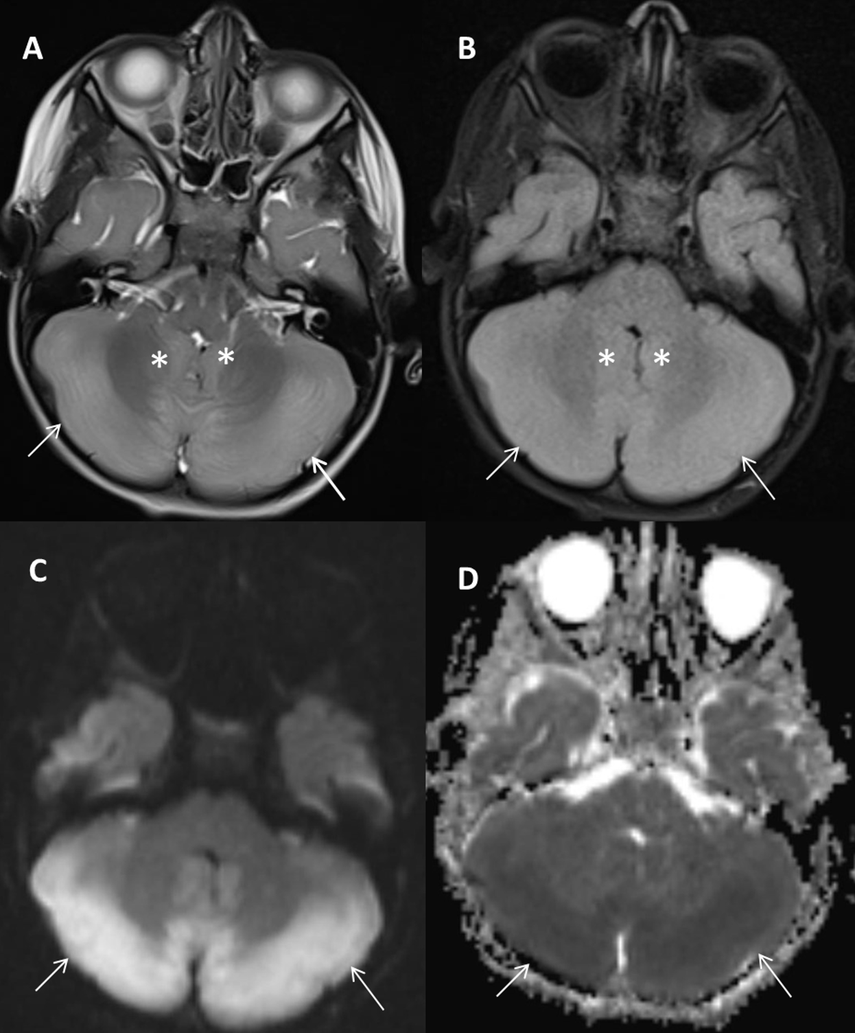
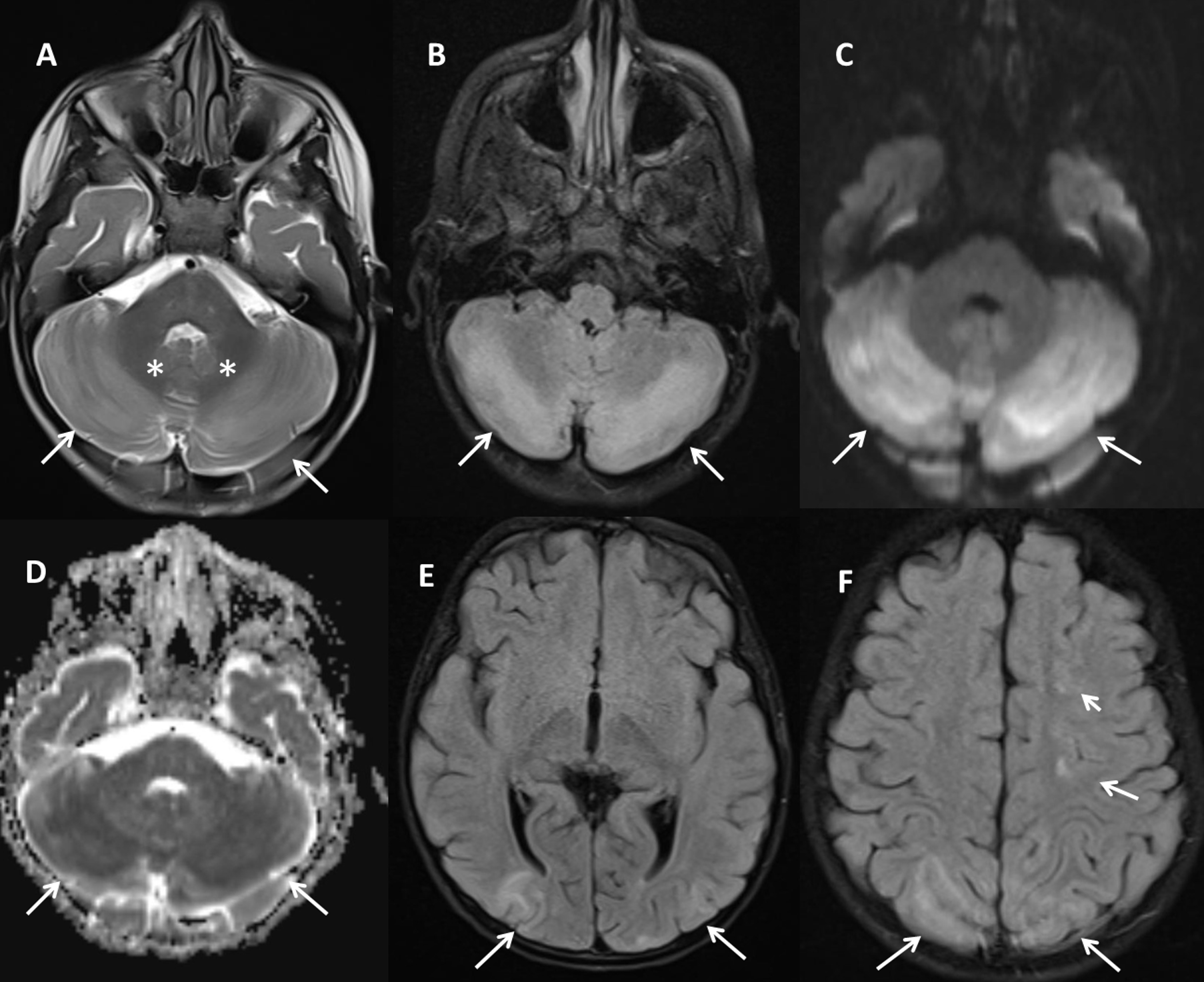
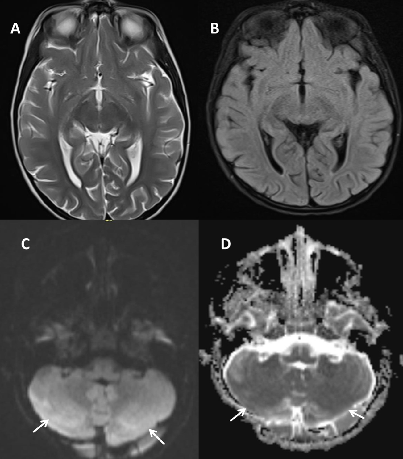
A 3-year-old boy with no significant past medical history was admitted with progressively worsening vomiting, imbalance, and fever for one week. On admission, he was alert, cooperative, and oriented. Body temperature was 37.8 °C, and blood pressure and other vital signs were within normal limits. Neurological examination revealed marked truncal ataxia and unsteadiness, accompanied by dysmetria and dysdiadochokinesis; however, tremor was not observed. Muscle tone was mildly decreased, deep tendon reflexes were normal, and no pathological reflexes were noted. Laboratory evaluation revealed normal complete blood count and biochemistry results. C-reactive protein (CRP) was within normal limits. Creatine kinase (CK) was elevated at 721 U/L (normal range, 30-200 U/L), attributed to muscle injury secondary to vomiting and falls. Metabolic screening, including serum glucose, electrolytes, liver and renal function tests, thyroid function tests, vitamin B12, and folate levels, was unremarkable. Autoimmune and rheumatological screening, including antinuclear antibody (ANA), anti-dsDNA, extractable nuclear antigen (ENA) profile, anti-neutrophil cytoplasmic antibody (ANCA), rheumatoid factor, and complement C3 and C4 levels, was unremarkable and did not suggest an underlying autoimmune or rheumatological disorder. Clinical assessment, routine laboratory investigations, and available imaging studies (brain MRI, abdomen-neck us, and chest X-ray) revealed no findings suggestive of an underlying malignancy. Cerebrospinal fluid (CSF) analysis revealed normal protein and glucose levels with no pleocytosis. Oligoclonal bands were negative, and the IgG index was within the normal range. CSF bacterial culture showed no growth. Viral PCR testing for HSV-1/2, VZV, CMV, and EBV was negative. Rubella virus PCR was not available. Serum viral serology showed positive rubella IgM (3.2 S/CO) and positive rubella IgG (119 IU/mL), supporting recent rubella infection. Initial brain MRI demonstrated marked hyperintense signals in both cerebellar hemispheres and the vermis on T2- and FLAIR-weighted sequences, with restricted diffusion on diffusion-weighted imaging (DWI), consistent with acute cerebellitis (Fig. 1). The patient was started on supportive care along with intravenous acyclovir (10 mg/kg/dose, three times daily), intravenous immunoglobulin (IVIG, 2 g/kg administered over 5 days) and high-dose intravenous methylprednisolone (30 mg/kg/day for 3 days). As clinical improvement remained limited, a contrast-enhanced follow-up MRI performed one week later revealed new hyperintense T2 and FLAIR signals in both cerebral hemispheres, predominantly in the subcortical white matter, in addition to the cerebellar lesions. There was no diffusion restriction or contrast enhancement. The findings were consistent with PRES-like changes (Fig. 2). At the time of the PRES-like MRI changes, the patient developed altered consciousness, whereas seizures and hallucinations were not observed. EEG was not performed at the time of the PRES-like MRI findings because no clinical seizures or seizure-like episodes were observed, although the patient had altered consciousness. No MRI examination was available immediately before the development of PRES-related radiological findings; therefore, a pre-PRES MRI demonstrating the absence of occipital and/or parietal involvement could not be provided. At this stage, plasmapheresis (four sessions on alternate days) was added to the treatment regimen. Following plasmapheresis, the patient showed marked clinical improvement. A follow-up MRI at three weeks demonstrated near-complete regression of the hyperintense signals in the posterior cerebellum and complete resolution of the cerebral lesions (Fig. 3). Neurological findings completely resolved, and no recurrence or new neurological symptoms were observed during a one-month follow-up. The patient was discharged without sequelae.
A signed informed consent form was obtained from the patient’s parents for the publication of this report.
Discussion
AC, first described by Westphal and Batten in the 19th century, is an inflammatory syndrome leading to cerebellar dysfunction.4 In 2007, the International Multiple Sclerosis Study Group defined cerebellitis as a clinically isolated syndrome that may occur as a primary infectious, postinfectious, or postvaccinal disorder.7 The most common clinical features include headache, vomiting, lethargy, altered mental status, coma, ataxia, and fever. AC represents a heterogeneous neuroinflammatory condition that may result from viral, bacterial, or immune-mediated mechanisms. Most pediatric cases of AC have a self-limited course with complete recovery following supportive treatment. However, in some patients, progressive cerebellar edema can lead to acute obstructive hydrocephalus, requiring urgent neurosurgical intervention. Therefore, early neuroimaging is essential in cases of clinical deterioration to evaluate for hydrocephalus. Some cases may also develop encephalopathy or other complications. Previous studies have reported that AC represents a more severe subgroup among childhood ataxias and that MRI findings correlate closely with the clinical course.1 Moreover, several studies have shown that cerebellar atrophy may develop on follow-up MRI even in hemispheres that initially appear normal.3 In our case, no cerebellar atrophy was observed on follow-up imaging. Although AC and acute cerebellar ataxia (ACA) are relatively common in pediatric practice, the effectiveness of antiviral and corticosteroid therapy remains controversial and insufficiently studied.8,9 International guidelines do not clearly recommend whether intravenous acyclovir and/or corticosteroids should be used in immunocompetent children with cerebellitis.10 Consequently, treatment decisions are often individualized according to disease severity and institutional protocols. Some studies have reported the combined use of acyclovir and corticosteroids in varicella-zoster-related cases, with variable clinical responses.11 In our patient, intravenous acyclovir, corticosteroids, and IVIG were administered; however, significant clinical and radiological improvement was achieved only after plasmapheresis. This finding suggests that immunomodulatory therapies such as IVIG or plasmapheresis may be more effective in atypical or progressive cases with clinical deterioration despite initial treatment. In our patient, plasmapheresis was initiated because of progressive clinical deterioration despite initial treatment, rather than because of the absence of improvement alone. Therefore, the case should not be interpreted as definitively refractory to IVIG or pulse methylprednisolone. The possible continuing therapeutic effects of these treatments cannot be excluded. Although rubella-associated AC often follows a benign or self-limited course, severe or fulminant presentations have also been described.5 In our patient, rubella seropositivity was accompanied by AC that evolved into PRES-like MRI changes during treatment, suggesting that rubella infection may trigger broader immune-mediated and endothelial processes. The absence of hypertension or metabolic abnormalities supports the hypothesis that PRES-like changes may result from post-infectious immune dysregulation and endothelial dysfunction rather than hemodynamic instability.12 However, a treatment-related contribution should also be considered. The initial MRI showed no occipital or parietal cortical/subcortical involvement suggestive of PRES; however, PRES-like posterior cerebral hemisphere changes developed after the administration of acyclovir, IVIG, and pulse methylprednisolone. PRES has rarely been reported after IVIG therapy and during pulse methylprednisolone treatment in pediatric patients.13,14 Acyclovir is more commonly associated with neurotoxicity, particularly in patients with renal dysfunction, and its clinical manifestations may overlap with PRES, making the differential diagnosis challenging.15 In the present case, the temporal association between treatment and the emergence of PRES-like findings indicates that a treatment-related contribution cannot be excluded. Nevertheless, because the patient received multiple potentially relevant therapies and PRES is a multifactorial clinicoradiological syndrome, the findings could not be attributed to a single medication with certainty. The complete clinical and radiological recovery following plasmapheresis further supports the potential role of immune-mediated mechanisms and suggests that immunomodulatory treatment may be beneficial in selected cases.
To the best of our knowledge, PRES-like radiological findings directly associated with rubella infection have not been clearly reported in the literature. Although rubella-related encephalitis/encephalopathy has been described, PRES-like parieto-occipital involvement is not a well-established radiological manifestation of rubella infection. Therefore, the findings in our patient may represent a rare or underreported manifestation; however, causality cannot be definitively established because PRES is multifactorial and the patient also received multiple potentially relevant treatments.
The possibility of coincidental or nonspecific anti-rubella IgM positivity should be considered. However, other possible causes, including metabolic, infectious, autoimmune/rheumatological, and paraneoplastic etiologies, were excluded through detailed diagnostic evaluation. In this context, the combination of acute cerebellar syndrome, compatible MRI findings, positive serum rubella IgM and IgG serology, and the absence of an alternative etiology supported the diagnosis of probable rubella-associated acute cerebellitis. However, false-positive results for rubella IgM can occur after other viral infections, particularly parvovirus B19, and may also be due to positive rheumatoid factor.16 In our patient, however, other infection parameters were negative.
This report has several limitations. As a single case report, it cannot establish a definitive causal relationship between rubella infection, acute cerebellitis, and subsequent PRES-like MRI changes. Although positive serum rubella IgM and IgG supported recent infection, nonspecific or false-positive IgM seropositivity cannot be completely excluded, and rubella virus was not isolated from the CSF. Other metabolic, infectious, autoimmune/rheumatological, and paraneoplastic causes were excluded; however, an unrecognized contributing mechanism cannot be entirely ruled out. EEG was not performed when PRES-like findings developed, limiting the assessment of possible non-convulsive seizure activity. In addition, because PRES-like changes emerged after acyclovir, IVIG, and pulse methylprednisolone treatment, a treatment-related contribution cannot be excluded, although the findings could not be attributed to a single agent with certainty.
In conclusion, rubella-associated acute cerebellitis is rare and may exceptionally be accompanied by PRES-like cerebral involvement. Clinical deterioration should prompt careful neurological and radiological reassessment for complications such as hydrocephalus and posterior cerebral involvement. In selected cases with progressive worsening despite initial treatment, additional immunomodulatory therapy, including plasmapheresis, may be considered. However, the causal relationship between rubella infection, treatment exposure, and PRES-like changes should be interpreted cautiously.
Ethical approval
Written consent was obtained from the patients’ parents.
Source of funding
The authors declare the study received no funding.
Conflict of interest
The authors declare that there is no conflict of interest.
References
- Emelifeonwu JA, Shetty J, Kaliaperumal C, et al. Acute cerebellitis in children: a variable clinical entity. J Child Neurol 2018; 33: 675-684. https://doi.org/10.1177/0883073818777673
- Kornreich L, Shkalim-Zemer V, Levinsky Y, Abdallah W, Ganelin-Cohen E, Straussberg R. Acute cerebellitis in children: a many-faceted disease. J Child Neurol 2016; 31: 991-997. https://doi.org/10.1177/0883073816634860
- Yildirim M, Gocmen R, Konuskan B, Parlak S, Yalnizoglu D, Anlar B. Acute cerebellitis or postinfectious cerebellar ataxia? Clinical and imaging features in acute cerebellitis. J Child Neurol 2020; 35: 380-388. https://doi.org/10.1177/0883073820901407
- Gökçe Ş, Kurugol Z, Aslan A, Çiçek C. Corrigendum to “a rare cause of childhood cerebellitis-influenza infection: a case report and systematic review of literature”. Case Rep Pediatr 2018; 2018: 5781843. https://doi.org/10.1155/2018/5781843
- Arya V, Gehlawat VK, Singh A, Mittal K, Kaushik JS. acute cerebellitis as a rare treatable cause of obstructive hydrocephalus. J Pediatr Neurosci 2019; 14: 162-164. https://doi.org/10.4103/jpn.JPN_43_19
- Triplett JD, Kutlubaev MA, Kermode AG, Hardy T. Posterior reversible encephalopathy syndrome (PRES): diagnosis and management. Pract Neurol 2022; 22: 183-189. https://doi.org/10.1136/practneurol-2021-003194
- Kamate M, Chetal V, Hattiholi V. Fulminant cerebellitis: a fatal, clinically isolated syndrome. Pediatr Neurol 2009; 41: 220-222. https://doi.org/10.1016/j.pediatrneurol.2009.03.018
- Heininger U, Seward JF. Varicella. Lancet 2006; 368: 1365-1376. https://doi.org/10.1016/S0140-6736(06)69561-5
- Fritzler MJ, Zhang M, Stinton LM, Rattner JB. Spectrum of centrosome autoantibodies in childhood varicella and post-varicella acute cerebellar ataxia. BMC Pediatr 2003; 3: 11. https://doi.org/10.1186/1471-2431-3-11
- Lancella L, Esposito S, Galli ML, et al. Acute cerebellitis in children: an eleven year retrospective multicentric study in Italy. Ital J Pediatr 2017; 43: 54. https://doi.org/10.1186/s13052-017-0370-z
- Bozzola E, Bozzola M, Tozzi AE, et al. Acute cerebellitis in varicella: a ten year case series and systematic review of the literature. Ital J Pediatr 2014; 40: 57. https://doi.org/10.1186/1824-7288-40-57
- Fugate JE, Rabinstein AA. Posterior reversible encephalopathy syndrome: clinical and radiological manifestations, pathophysiology, and outstanding questions. Lancet Neurol 2015; 14: 914-925. https://doi.org/10.1016/S1474-4422(15)00111-8
- Incecik F, Hergüner MO, Altunbasak S, Yıldızdas D. Reversible posterior encephalopathy syndrome due to intravenous immunoglobulin in a child with Guillain-Barré syndrome. J Pediatr Neurosci 2011; 6: 138-140. https://doi.org/10.4103/1817-1745.92841
- İncecik F, Hergüner MÖ, Yıldızdaş D, Yılmaz M, Mert G, Horoz ÖÖ, Altunbaşak Ş. Posterior reversible encephalopathy syndrome due to pulse methylprednisolone therapy in a child. Turk J Pediatr 2013; 55: 455-457. https://doi.org/10.24953/turkjpediatr.2013.1533
- Haraguchi S, Watanabe Y. Differential diagnosis of posterior reversible encephalopathy syndrome and acyclovir neurotoxicity in children: a literature review of acyclovir neurotoxicity. Brain Dev Case Rep 2024; 2: 100007. https://doi.org/10.1016/j.bdcasr.2024.100007
- Best JM, O’Shea S, Tipples G, et al. Interpretation of rubella serology in pregnancy-pitfalls and problems. BMJ 2002; 325: 147-148. https://doi.org/10.1136/bmj.325.7356.147
Copyright and license
Copyright © 2026 The Author(s). This is an open access article distributed under the Creative Commons Attribution License (CC BY), which permits unrestricted use, distribution, and reproduction in any medium or format, provided the original work is properly cited.


