Abstract
Background. Background and objectives: Patent ductus arteriosus (PDA) is a common congenital cardiac condition, especially in premature infants, and is associated with significant morbidity and mortality. Artificial intelligence (AI) has emerged as a promising tool to aid clinicians in diagnosing PDA and guiding treatment decisions amidst this uncertainty.
Methods. A literature search (up to December 2025) was conducted in PubMed, Scopus, and Web of Science for peer-reviewed studies on AI (including machine learning, deep learning, computer vision, and predictive analytics) applied to PDA.
Results. Seventeen studies were included, predominantly involving preterm neonates. AI applications mainly targeted PDA diagnosis and outcome prediction using clinical and echocardiographic data. Imaging-based deep learning models generally outperformed tabular data approaches (with area under the curve often >0.80). Overall study quality was moderate, with limited external validation and data transparency.
Conclusions. AI has the potential to transform patent ductus arteriosus management by enabling earlier detection, more consistent decision-making, and individualized treatment planning. Integration of AI into PDA care may reduce practice variability, improve clinical outcomes, and serve as a model for the broader application of artificial intelligence in medicine.
Keywords: artificial intelligence, patent ductus arteriosus, machine learning, clinical decision support, echocardiography
Introduction
Patent ductus arteriosus (PDA) is a congenital cardiovascular condition defined by the failure of the fetal ductus arteriosus to close after birth, resulting in a persistent left-to-right shunt between the aorta and pulmonary artery.1 PDA is particularly common in preterm infants, with incidence inversely related to gestational age. In very low birth weight neonates, PDA is associated with substantial morbidity, including chronic lung disease, necrotizing enterocolitis, renal dysfunction, intraventricular hemorrhage, and increased mortality. In term infants and older children, persistent PDA may lead to chronic cardiac volume overload, while in adults, although uncommon, untreated large PDA can result in heart failure or infective endarteritis.2-4 Conversely, in certain congenital heart diseases, the PDA may be life-sustaining; for ductal-dependent lesions, maintaining ductal patency is critical, and procedures such as PDA stenting may be performed to ensure adequate pulmonary or systemic blood flow.5,6 Thus, PDA management spans a spectrum from intentional closure to intentional maintenance of patency, depending on the clinical context.
Despite its clinical importance, optimal PDA management remains highly controversial, particularly in preterm infants.7,8 No universal consensus exists regarding the treatment of hemodynamically significant PDA in this population. Therapeutic strategies range from conservative management (watchful waiting and supportive care), to pharmacological closure with nonsteroidal anti-inflammatory drugs (NSAIDs, such as indomethacin or ibuprofen, or acetaminophen), and surgical ligation or catheter-based closure.9,10 This lack of agreement is due in part to conflicting evidence on long-term benefits of PDA closure and the difficulty of determining which PDA is truly “pathological” (hemodynamically significant) versus benign.11-13 In older infants and children with PDA, treatment decisions (typically transcatheter device closure or surgery) are more straightforward once a PDA is identified, but selecting the optimal timing and device can still benefit from advanced planning tools.14,15 In ductal-dependent congenital heart disease, PDA stenting has emerged as a less invasive alternative to surgical shunts, but it is a technically complex procedure requiring careful planning due to highly variable ductal anatomy.6,16
Within this context of diagnostic and therapeutic uncertainty, artificial intelligence (AI) and machine learning (ML) have emerged as promising tools to support clinical decision-making in PDA. AI techniques can integrate large-scale clinical, imaging, and physiological data to identify subtle patterns, improve diagnostic accuracy, and generate individualized risk predictions.17,18 Potential applications include predicting spontaneous ductal closure, identifying PDAs likely to cause adverse outcomes, automated PDA detection using imaging or signal-based data, and procedural planning for interventions such as PDA stenting through anatomy-aware visualization and simulation.19-21 These applications align with broader trends toward data-driven decision support in high-acuity settings such as the neonatal intensive care unit.
Although an increasing number of studies have explored AI-based approaches for PDA diagnosis and management, a comprehensive synthesis of this evidence is currently lacking. This scoping review aims to systematically examine the application of AI in PDA across the full clinical spectrum, from diagnosis to therapeutic decision support and interventional planning, across all age groups and geographic regions. By summarizing current evidence, this review aims to clarify the evolving role of AI in improving PDA care and facilitating its integration into clinical practice.
Materials and Methods
This scoping review was conducted in accordance with the 2020 Preferred Reporting Items for Systematic Reviews and Meta-Analyses (PRISMA) Extension for Scoping Reviews.22
Search strategy
A comprehensive literature search was performed in PubMed, Scopus, and Web of Science, with supplementary searches in Google Scholar to identify additional relevant studies. The initial search was conducted in October 2025 and updated in December 2025 to capture the most recent publications. Search terms combined concepts related to patent ductus arteriosus (“patent ductus arteriosus,” “ductus arteriosus,” “PDA”) with artificial intelligence–related keywords, including “artificial intelligence,” “machine learning,” “deep learning,” “neural network,” “computer vision,” “predictive model,” “decision support,” and “automation.” Search strategies were adapted to each database. Only peer-reviewed journal articles were considered, and non-scholarly content was excluded. No restrictions on publication date, country, or language were applied at the search stage; however, all included studies were published in English.
Eligibility criteria
A scoping review approach was adopted to capture the breadth of AI applications in PDA care. Studies were eligible if they met the following criteria: (1) involved patients with PDA of any age, including isolated PDA or PDA associated with other congenital heart diseases; (2) applied AI techniques to PDA-related diagnosis, outcome prediction, decision support, or interventional planning; (3) reported relevant outcomes such as diagnostic performance metrics, predictive accuracy, or clinical utility; and (4) were original research articles, including retrospective or prospective studies, model development, validation, or technical feasibility studies. Review articles, conference abstracts without full texts, and non–peer-reviewed reports were excluded.
Study selection and data extraction
Titles and abstracts were independently screened by two reviewers, followed by full-text assessment of potentially eligible studies. Discrepancies were resolved by consensus. Studies that met the inclusion criteria and were included in the final analysis. From each study, data were extracted on publication year, country, patient population, AI methodology, application domain, and principal findings, including performance metrics where available.
Quality assessment
Included studies were grouped into thematic categories addressing AI applications in PDA diagnosis and AI-based treatment planning or clinical decision support. Given the scoping nature of the review, a formal risk-of-bias assessment was not performed. Instead, all included studies were evaluated for adherence to best practices in AI research in accordance with the DOME guidelines.23
Results
Search results
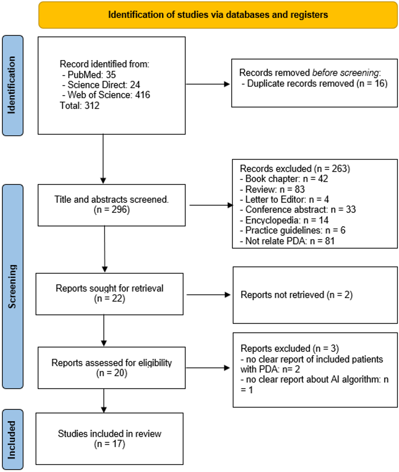
A total of 312 records were initially identified through searches of three electronic databases. After removal of 16 duplicate records, 296 articles remained for title and abstract screening according to the predefined inclusion criteria. Of these, 20 studies were retrieved for full-text assessment. Following detailed evaluation, 3 articles were excluded, and 17 studies were ultimately included in the review (Fig. 1).
Characteristics of included studies
The included studies showed wide geographic representation across nine countries, with China contributing the largest number (five studies), followed by the United States and South Korea (with three studies each).
Preterm neonates were the primary population in ten studies, reflecting the clinical significance of PDA in premature and very low birth-weight infants, while other studies involved term neonates or broader pediatric cohorts.
PDA diagnosis or screening and outcome prediction were each addressed in thirteen studies, whereas treatment planning or management was examined in four, with some overlap in objectives. Clinical and laboratory tabular data were the most frequently used inputs (11 studies), followed by echocardiographic images or video (five studies), while other modalities were rarely applied (Supplementary Table S1).
Applications of AI in PDA diagnosis
Seven studies evaluated AI for PDA diagnosis or screening. CNN-based models applied to echocardiographic images and video clips achieved expert level or superior diagnostic accuracy, including advanced architectures such as ResNet 50 and CHDNet for both grayscale and color Doppler imaging.20,24,25 Non-imaging approaches using heart sound signals, based on recurrent neural networks, residual convolution recurrent neural networks, and gradient boosting, consistently outperformed clinician auscultation, supporting their role as low cost and scalable screening tools.26-28 Models using routine clinical data, particularly random forest and extreme gradient boosting, enabled early prediction of PDA risk in preterm neonates.19
Six studies focused on PDA classification and severity assessment. Supervised machine learning models including random forest, light gradient boosting machine, multilayer perceptron, and logistic regression accurately predicted symptomatic and hemodynamically significant PDA earlier than standard clinical recognition.29-32 Unsupervised learning identified distinct hemodynamically significant PDA (hsPDA) subphenotypes with inflammatory or respiratory acidosis dominant profiles.33 Multimodal models combining chest X-ray imaging and clinical data improved severe PDA detection, while explainable AI approaches enhanced interpretability.34 Notably, simpler linear and multilayer perceptron models also demonstrated clinically meaningful performance for early hsPDA risk prediction in extremely preterm infants (Table I).
| AI: artificial intelligence, CNN: Convolutional Neural Network, hsPDA: hemodynamically significant PDA, PDA: patent ductus arteriosusi RF: Random Forest, VSD: ventricular septal defect, XGBoost:Extreme Gradient Boosting. | |||||
| Table I. AI applications in PDA diagnosis. | |||||
| First author (Year) | Target population | Diagnostic objective | Primary input modality | AI technique / model | Key diagnostic outcome |
| PDA diagnosis | |||||
| Howard Lei (2021)20 | Preterm neonates | PDA diagnosis | Echocardiogram video clip | Convolutional Neural Network (CNN) | Enhanced PDA detection from neonatal echocardiograms |
| Sergi Gómez-Quintana (2021)26 | Term and late-preterm neonates | PDA symptoms detection | Heart sounds signal | Extreme Gradient Boosting (XGBoost) | Strong potential for low-cost, scalable PDA screening |
| Jia Liu (2022) 27 | Infants | PDA diagnosis | Heart sound signals | Residual Convolution Recurrent Neural Network (RCRnet) | Outperformed expert auscultation in PDA screening |
| Hongbo Yang (2023)28 | Infants / children and adolescents | PDA diagnosis | Heart sounds signal | Recurrent Neural Network (RNN) | Improved detection accuracy for PDA and VSD |
| Jason Erno (2023) 24 | Preterm neonates | PDA diagnosis | Echocardiogram video clip | ResNet-50 CNN | Accurate detection of PDA using color Doppler echocardiography |
| Weimin Tan (2023)25 | Infants / children | PDA diagnosis | Echocardiography images | CHDNet | Expert-level PDA diagnosis on internal datasets |
| Ana Maria Cristina Jura (2025) 19 | Preterm neonates | PDA diagnosis | Clinical data | Random Forest (RF) and XGBoost | AI-based risk prediction may support early identification of high-risk neonates for PDA |
| PDA classification | |||||
| Jae Yoon Na (2021) 29 | Preterm neonates | predict symptomatic PDA | Clinical data | RF, Light Gradient Boosting Machine (L-GBM), Multilayer Perceptron (MLP), Support Vector Machine (SVM), k-Nearest Neighbors (k-NN) | Predicting symptomatic PDA occurrence and whether sPDA would be treated |
| Felipe Yu Matsushita (2023) 33 | Preterm neonates | hsPDA Subphenotyping | Clinical data | Unsupervised learning | Identify two distinct hsPDA subphenotypes: “Inflamed” vs “Respiratory acidosis” |
| Phillip Chang (2024) 34 | Preterm neonates | Severe PDA diagnosis | Chest X-ray images, clinical data |
XGBoost, ConvNeXt-based CNN, Grad-CAM++ SHAP |
AI models achieve high specificity to indentify explainable radiographic markers of severe PDA |
| Seoyeon Park (2024) 30 | Preterm neonates | Severe PDA diagnosis | Clinical data | RF |
Detect PDA symptoms earlier than routine clinical recognition Improve diagnostic accuracy of medical professionals |
| Erik Küng (2025) 31 | Extremely preterm neonates | hemodynamically significant (hs) PDA prediction | Clinical data | Linear perceptron, Multilayer perceptron, Logistic regression | Early identification of neonates at high risk for hsPDA |
| Oguzhan Ay (2025) 32 | Preterm neonates | hsPDA prediction | Clinical data | RF | Acurately predicts hPDA risk using non-imaging data |
Applications of AI in PDA outcome prediction and clinical decision support
Four studies investigated the application of AI to support PDA management, focusing on pharmacologic treatment optimization and outcome prediction. Overall, AI models demonstrated meaningful clinical utility in guiding therapy and anticipating treatment response or complications.
One early study showed that regression-based AI models using clinical and laboratory data could accurately predict continuous serum digoxin concentrations in neonates, supporting individualized dosing in PDA-related pharmacotherapy.35 Subsequent studies shifted toward predicting treatment response in hemodynamically significant PDA. Machine learning models, particularly random forest, integrating clinical, laboratory, and echocardiographic parameters achieved good predictive performance for closure after first line NSAID therapy, enabling early identification of likely responders and non-responders.36
Deep learning approaches further extended this capability. A convolutional neural network (CNN) model combining echocardiographic video sequences with clinical data successfully predicted pharmacologic PDA closure outcomes in preterm neonates, highlighting the added value of dynamic imaging features for treatment planning.21 In older infants and children, ensemble machine learning models applied to clinical data identified patients at risk of post-intervention thrombocytopenia, providing actionable information for peri-intervention monitoring and risk stratification (Table II).37
| AI: artificial intelligence, CNN: Convolutional Neural Network, hsPDA: hemodynamically significant PDA, NSAID: non-steroid anti-inflammatory drugs, PDA: patent ductus arteriosus, RF: Random Forest, XG: Extreme Gradient. | |||||
| Table II. AI applications in PDA outcome prediction clinical decision support. | |||||
| First author (Year) | Target population | Management objective | Primary input modality | AI technique/ model | Key significance |
| Shu-Hui Yao (2019)35 | Neonates | Prediction of continuous serum digoxin concentration | Clinical data/laboratory measurements | Regression | AI-assisted dosing support in PDA-related pharmacotherapy |
| Tai-Xiang Liu (2023)36 | Preterm neonates | Prediction of hsPDA closure after first-line NSAID therapy | Clinical data, laboratory data, echocardiographic measurements | RF | Good predictive ability for NSAID treatment response in hsPDA |
| Puneet Sharma (2025)21 | Preterm neonates | Prediction pharmacologic PDA closure outcomes | Echocardiogram video sequence and clinical data | CNN | Predicting response to PDA pharmacotherapy |
| Song-Yue Zhang (2025) 37 | Infants / children | Outcome prediction/Treatment planning | Clinical data | RF, XG, Adaptive Boosting (ADA), Logistic Regression | Identified children at risk of post-intervention thrombocytopenia |
Main AI algorithms employed in PDA research and model accuracy
A broad spectrum of AI algorithms was employed across studies, reflecting heterogeneity in data modalities and clinical objectives. Tree-based machine-learning methods, including random forest and gradient-boosting models, were most frequently applied to structured clinical and laboratory data. Convolutional neural networks were predominantly used for echocardiographic image or video analysis and consistently demonstrated strong diagnostic performance. Artificial neural networks or multilayer perceptrons were mainly utilized for outcome prediction using tabular datasets, whereas linear models and recurrent or hybrid architectures were less commonly applied, particularly for sequential data, and generally achieved moderate performance. One study adopted unsupervised clustering to identify PDA subphenotypes; although standard accuracy metrics were not applicable, clinically meaningful subgroup differentiation was reported.
Across studies, model performance showed consistent trends related to input data modality and task complexity. Imaging-based models trained on echocardiographic images or video sequences, largely using convolutional neural networks, tended to achieve higher discriminative performance than models relying solely on clinical or laboratory variables, frequently reporting area-under-the-curve values above 0.80 for PDA detection and classification of hemodynamically significant PDA. In contrast, models based on tabular clinical and laboratory data, most often implemented using tree-based algorithms, demonstrated moderate to high performance, with area under the curve (AUC) values typically ranging from approximately 0.75 to 0.85. Although performance was generally lower than that of imaging-based approaches, these models offered greater feasibility and scalability, particularly in resource-limited settings. Variability in reported accuracy across studies was partly attributable to differences in outcome definitions, feature selection, and validation strategies, underscoring that model performance was influenced more by data quality and modality than by algorithmic complexity alone.
Detailed characteristics of the included studies are provided in Supplementary Table S1.
Quality of included studies
Overall, the methodological quality of the included studies was moderate, with substantial variation across reporting and validation domains. A clear description of the AI methodology was provided in 94.4% of studies, and model interpretability or explanatory elements were reported in a similarly high proportion (94.4%). Most studies also reported appropriate data splitting strategies for model development and evaluation (88.9%) and included standard performance measures such as accuracy or area under the curve (88.9%).
In contrast, transparency and reproducibility were more limited. Only 11.1% of studies made data or code publicly available, restricting independent validation. Reporting of hyperparameter tuning procedures was inconsistent and documented in just 33.3% of studies, raising concerns regarding optimization transparency. Notably, none of the included studies adhered to a recognized best-practice reporting framework for AI research.
The quality assessment of the included studies is presented in Supplementary Table S2.
Discussion
Improved diagnosis and early detection
Artificial intelligence is based on data-driven computational models that learn complex patterns directly from data rather than relying on predefined rules.38 Unlike traditional statistical methods, AI algorithms can process high-dimensional, nonlinear, and multimodal inputs simultaneously, making them particularly well suited to neonatal physiology, where clinical signals are dynamic, interdependent, and often subtle.39,40 This capacity for representation learning allows AI-based systems to identify latent physiological signatures that are not readily apparent through standard threshold-based clinical assessment.41,42
In the context of PDA, AI contributes by integrating heterogeneous data sources including demographic variables, continuous vital signs, ventilatory parameters, imaging data, and biosignals into unified predictive frameworks. Machine learning models capture complex interactions among these variables, enabling earlier recognition of hemodynamically significant ductal patency before overt clinical instability occurs. Deep learning architectures further enhance this capability by automatically extracting hierarchical features from echocardiographic images, Doppler waveforms, and heart sounds, thereby reducing reliance on operator-defined measurements and subjective interpretation.24,26,27 Moreover, deep learning (DL)-based analysis of neonatal echocardiograms enables near real time detection and can be deployed on low-resource devices, supporting clinical application in settings with limited access to specialist expertise.20
Comparative evaluations indicate that AI-based systems are most effective when used as clinical decision-support tools rather than substitutes for clinician judgment. Studies by Ay et al. and Küng et al. showed that AI-based interpretation achieves diagnostic performance comparable to expert assessment, facilitating early identification of neonates at high risk for hemodynamically significant PDA.31,32 This early risk stratification can inform surveillance intensity, enable individualized treatment decisions, reduce overtreatment, and optimize resource utilization. Such consistency and accessibility are particularly valuable in neonatal intensive care units with limited access to experienced neonatologists or pediatric cardiologists.
Personalization of treatment decisions
Artificial intelligence facilitates personalized management of PDA by addressing the challenge of identifying ductus arteriosi that require treatment and selecting optimal therapeutic strategies. Sharma et al. demonstrated high discriminative performance in predicting response to pharmacologic closure, with the potential to reduce delays in definitive treatment, enable earlier referral for procedural intervention, and support individualized care in neonatal intensive care units.21 Accurate prediction of treatment response is clinically valuable, as early identification of likely non-responders may prevent ineffective drug exposure and related adverse effects, while recognition of likely responders can reduce unnecessary escalation to invasive intervention.36
In addition, Yao et al. reported that artificial neural network models outperformed traditional regression methods in predicting digoxin levels in neonates with patent ductus arteriosus, highlighting the feasibility of AI–assisted dosing support, particularly in settings where blood sampling is limited.35 These predictive tools may also reduce variability in clinical practice by providing individualized, evidence-based risk estimates that complement clinician judgment and inform the timing and intensity of intervention.19,30 However, these models function as clinical decision-support systems rather than autonomous decision-makers and must be interpreted within the broader clinical context, including hemodynamic status, comorbidities, and family preferences43-45
Enhancement of technical interventions
In contrast to the growing body of work on diagnostic and prognostic applications, AI-related innovations targeting technical and interventional aspects of PDA management remain at an early developmental stage. In this context, Zhang et al. demonstrated that an RF model could successfully identify children at increased risk of post-intervention thrombocytopenia following PDA closure.37 This approach highlights the potential role of AI in supporting procedural risk stratification, enhancing clinical trust, and facilitating anticipatory management and early intervention planning in interventional settings.
Building on recent progress in AI algorithms and interventional support systems, advanced image processing and three-dimensional reconstruction approaches are being developed to improve procedural guidance. These methods allow detailed depiction of cardiac anatomy, supporting thorough pre-procedural evaluation and informed selection of closure devices and interventional strategies.46,47 Computational modeling and simulation further enable patient-specific assessment of flow dynamics and device-tissue interactions, encouraging multidisciplinary collaboration and shared decision-making while reducing dependence on individual operator experience.48-50 Over time, incorporation of these technologies may expand access to transcatheter PDA closure by shortening the learning curve in centers with limited expertise.
The clinical role of artificial intelligence in PDA management
Although artificial intelligence has demonstrated strong performance in the detection of PDA and in outcome prediction, its clinical utility must be interpreted within the evolving framework of PDA management. In current neonatal practice, there is a growing preference for conservative management, particularly in preterm infants, driven by the high likelihood of spontaneous ductal closure and the absence of consistent long-term benefits from pharmacological or interventional therapies.51-53 Furthermore, the presence of PDA alone, including cases classified as hemodynamically significant, does not inherently justify treatment. Clinical decision-making is multifactorial and requires careful consideration of hemodynamic impact, associated comorbidities, timing of intervention, and interindividual variability in treatment response.8
A fundamental implication of this paradigm shift is that diagnostic accuracy alone does not directly translate into improved clinical outcomes. The central challenge in PDA management is not simply identifying the presence of a ductus, but determining which patients require intervention, when treatment should be initiated, and which patients are most likely to respond to therapy. In this context, traditional definitions of hemodynamically significant PDA based primarily on echocardiographic parameters are increasingly recognized as insufficient when used in isolation. Hemodynamic significance is dynamic and cannot be fully captured by anatomical and flow-based measurements alone.54,55
Within this framework, the clinical value of artificial intelligence lies not primarily in improving PDA detection, but in its ability to integrate multidimensional data and support individualized decision-making. AI-based systems should therefore be viewed as adjunctive tools that enhance risk stratification and clinical judgment rather than autonomous decision-makers.
Alignment between current AI research and clinical needs
Against this background, the current landscape of artificial intelligence research in PDA reveals a notable mismatch between technological development and clinical priorities. As demonstrated in this review, most studies have focused on diagnostic classification or identification of hemodynamically significant PDA. While these models often achieve high performance, their direct clinical impact remains limited if they do not address treatment decision-making.
In line with this observation, only a minority of studies have explored clinically relevant applications such as prediction of pharmacologic response 21,36, risk stratification for adverse outcomes 35, or individualized treatment planning 37. These areas are more closely aligned with real-world clinical uncertainty, where the primary question is not whether PDA exists, but whether intervention will improve patient outcomes. This gap between current research focus and clinical needs highlights the importance of shifting future AI development toward clinically meaningful endpoints. Models that incorporate longitudinal physiological data, treatment exposure, and patient-specific characteristics may provide more actionable insights and better support bedside decision-making in neonatal intensive care settings.
Beyond methodological focus, the overwhelming majority of studies focused on preterm neonates, reflecting the population in which PDA carries the greatest clinical burden. However, this narrow focus limits insights into the potential role of AI in older pediatric and adult populations, where disease characteristics and management strategies differ. Similarly, although the included studies spanned multiple countries, representation from low- and middle-income regions was limited. This lack of geographic diversity is clinically relevant, as AI-based diagnostic tools may offer substantial benefits in resource-constrained settings where access to echocardiography and specialist expertise is limited. Non-imaging approaches, such as heart sound analysis 28 or clinical data-driven models 29,30, are particularly promising in such environments, and prospective evaluation across diverse healthcare systems is therefore warranted.
In addition to these gaps, the current evidence base is constrained by study design limitations. Most studies were retrospective and focused on model development with internal validation, with few addressing real-time clinical implementation. As a result, the impact of AI-guided strategies on clinically meaningful outcomes remains uncertain. Randomized or pragmatic studies comparing AI-assisted management with standard care are needed to determine whether these tools can improve outcomes such as survival, bronchopulmonary dysplasia, or duration of respiratory support.
Furthermore, PDA physiology is inherently dynamic 56, yet most existing models rely on static snapshots of clinical, laboratory, or imaging data. In practice, the hemodynamic significance of PDA evolves over time in response to changing physiological conditions and therapeutic interventions. Future AI systems should therefore incorporate longitudinal data and time series modeling to provide continuously updated risk assessments and better capture disease trajectories.
Taken together, these limitations and gaps emphasize the need to reorient artificial intelligence research toward clinically meaningful applications. Future studies should prioritize decision support tools that integrate multidimensional and dynamic data to guide treatment selection, predict therapeutic response, and align with the current shift toward individualized and conservative management strategies.
Ethical and practical considerations
The integration of artificial intelligence into PDA management raises important ethical and practical considerations. Algorithmic bias may arise if training datasets are not representative of diverse populations or healthcare settings, potentially limiting model performance in underrepresented groups. Clear guidance will be required to define clinician responsibility when AI recommendations are incorporated into care decisions.57 Although AI tools function as supportive aids rather than decision-makers, accountability ultimately remains with the clinician.
Regulatory oversight will play a crucial role in ensuring the safety and efficacy of AI-based clinical tools. For high-stakes applications such as PDA diagnosis and treatment planning, rigorous validation and adherence to emerging regulatory frameworks will be necessary before routine clinical implementation.
Future directions
Future applications of artificial intelligence in PDA care are expected to move beyond isolated diagnostic tasks toward integrated, clinically meaningful decision support systems. In particular, multimodal AI platforms that combine physiologic monitoring, echocardiographic analysis, and treatment response prediction may enable a more comprehensive and individualized assessment of PDA at the bedside. Such systems have the potential to support earlier, more consistent, and context-aware clinical decision-making in neonatal intensive care settings.58
Real-time data integration represents another important direction. AI algorithms capable of continuously analyzing longitudinal physiological signals, including vital signs and respiratory parameters, may detect early hemodynamic changes associated with evolving PDA significance. This dynamic approach aligns more closely with the fluctuating nature of neonatal physiology and may allow earlier identification of clinically relevant deterioration, thereby facilitating timely intervention.59,60
Beyond short-term management, AI may also contribute to predicting longer-term outcomes associated with PDA and its treatment, including chronic lung disease and neurodevelopmental impairment. Such predictive models could help refine treatment thresholds by balancing immediate therapeutic benefits against potential long term risks, thereby supporting more individualized and evidence-informed care strategies.61 In addition, AI-assisted procedural planning and intraprocedural guidance are emerging areas of interest, particularly in transcatheter PDA closure, where anatomical variability and procedural complexity remain significant challenges.62
Importantly, the future application of AI-assisted evaluation of patient data must also be considered in the context of data quality and clinical expertise.63 The performance, reliability, and generalizability of AI models are fundamentally dependent on the quality, completeness, and representativeness of the underlying data used for training and validation. Inaccurate, biased, or heterogeneous datasets may lead to unreliable predictions and limit clinical applicability, particularly across different healthcare settings.64
In this context, specialist expertise remains essential for the appropriate interpretation of AI-generated outputs. AI systems are designed to support, rather than replace, clinical decision-making, and their outputs must be integrated with expert clinical judgment, particularly in complex conditions such as PDA where management decisions are highly individualized.65,66 The effective implementation of AI in clinical practice therefore requires not only robust data infrastructure but also close collaboration between data scientists and experienced clinicians to ensure safe, accurate, and clinically meaningful application.
Perspective for low- and middle-income countries
The potential clinical value of artificial intelligence in the management of patent ductus arteriosus is likely to be particularly significant in low- and middle-income countries, where access to advanced imaging technologies, continuous physiological monitoring, and specialized pediatric cardiology expertise remains limited.67,68 In these settings, delayed diagnosis and substantial variability in clinical decision-making frequently contribute to poorer outcomes. Artificial intelligence–based approaches, especially those utilizing non-imaging data such as clinical variables, heart sound analysis 28, or simplified echocardiographic inputs 24,25, represent scalable and cost-effective solutions to address these challenges. Machine learning models capable of integrating heterogeneous data sources may facilitate earlier identification of hemodynamically significant PDA and enable more consistent risk stratification, even in resource-constrained environments.33,34 Furthermore, deep learning–driven automation of image interpretation has the potential to reduce dependence on highly trained specialists, thereby enhancing diagnostic consistency across healthcare facilities.21,24 These characteristics are particularly well aligned with healthcare delivery models in low-resource settings, where tertiary centers are frequently overcrowded and the distribution of expertise is uneven. In this regard, AI has the potential to decentralize care, support task shifting, and optimize the use of limited clinical resources.69 Therefore, beyond its diagnostic and predictive capabilities, AI represents a feasible and transformative strategy to strengthen healthcare systems, improve equity in access to care, and ultimately enhance clinical outcomes in these regions.
Study limitations
This scoping review has several limitations that should be considered when interpreting the findings. As discussed above, the current body of evidence is limited by heterogeneity in study design, patient populations, and methodological approaches, reflecting the early stage of artificial intelligence research in patent ductus arteriosus.
In addition, most included studies were retrospective and primarily focused on model development with internal validation, with relatively few addressing external validation or prospective clinical implementation. This limits the ability to assess the real-world applicability and generalizability of the reported models.
Furthermore, issues related to transparency and reproducibility remain, as only a minority of studies provided access to underlying data or code, and reporting of model development and optimization was often incomplete. These limitations may hinder independent validation and broader clinical adoption.
Finally, as a scoping review, no formal risk-of-bias assessment or quantitative meta-analysis was performed. The findings should therefore be interpreted as a qualitative synthesis of the current literature. Overall, while artificial intelligence shows promise, its impact on clinically meaningful outcomes has not yet been established and requires further validation through well-designed prospective and outcome-oriented studies.
Conclusion
In conclusion, AI has the capacity to transform how we approach PDA – turning what has historically been a contentious and variable practice into a more data-driven, consistent, and optimized process. By leveraging AI for earlier detection, smarter decision-making, and meticulous intervention planning, we can improve the care of infants and patients with PDA, reducing morbidity and tailoring treatments to individual needs. As technology and medicine continue to intersect, the management of PDA can serve as a model for integrating artificial intelligence into the care of complex medical conditions, ultimately enhancing outcomes and saving lives.
Supplementary materials
Supplementary materials for this article are available online at https://doi.org/10.24953/turkjpediatr.2026.8122
Source of funding
The authors declare the study received no funding.
Conflict of interest
The authors declare that there is no conflict of interest.
Generative AI statement
During the preparation of this manuscript, ChatGPT (OpenAI, 2026) was used solely for language editing and proofreading to improve the grammar, clarity, and readability of the manuscript. The tool was not used for literature searching, data analysis, content generation, interpretation of results, or scientific writing. All content was reviewed and edited by the authors, who take full responsibility for the accuracy, integrity, and final content of the publication.
References
- Backes CH, Hill KD, Shelton EL, et al. Patent ductus arteriosus: a contemporary perspective for the pediatric and adult cardiac care provider. J Am Heart Assoc 2022; 11: e025784. https://doi.org/10.1161/JAHA.122.025784
- Kouris NT, Sifaki MD, Kontogianni DD, et al. Patent ductus arteriosus endarteritis in a 40-year old woman, diagnosed with transesophageal echocardiography. A case report and a brief review of the literature. Cardiovasc Ultrasound 2003; 1: 2. https://doi.org/10.1186/1476-7120-1-2
- Haboub M, Khaldi M, Drighil A. Infective endocarditis on a patent ductus arteriosus revealed by a ruptured pulmonary artery trunk aneurysm in the pericardium: a case report. Eur Heart J Case Rep 2025; 9: ytaf267. https://doi.org/10.1093/ehjcr/ytaf267
- Ambalavanan N, Aucott SW, Salavitabar A, Levy VY; Committee on Fetus and Newborn, Section on Cardiology and Cardiac Surgery. Patent ductus arteriosus in preterm infants. Pediatrics 2025; 155: e2025071425. https://doi.org/10.1542/peds.2025-071425
- Alwi M. Stenting the ductus arteriosus: case selection, technique and possible complications. Ann Pediatr Cardiol 2008; 1: 38-45. https://doi.org/10.4103/0974-2069.41054
- Downing TE, Boucek DM, Glatz AC, et al. Stenting the ductus arteriosus in neonates with ductal-dependent pulmonary blood flow: technical and anatomic considerations. Pediatr Cardiol 2026; 47: 2322-2334. https://doi.org/10.1007/s00246-025-03834-4
- Chan B, Singh Y. Personalized evidence-based management of patent ductus arteriosus in preterm infants. J Cardiovasc Dev Dis 2023; 11: 7. https://doi.org/10.3390/jcdd11010007
- Seyam O, Frishman WH, Aronow WS. Contemporary Management Strategies for Patent Ductus Arteriosus. Cardiol Rev. Published online February 13, 2026. https://doi.org/10.1097/CRD.0000000000001204
- Buvaneswarran S, Wong YL, Liang S, Quek SC, Lee J. Active treatment vs expectant management of patent ductus arteriosus in preterm infants: a meta-analysis. JAMA Pediatr 2025; 179: 877-885. https://doi.org/10.1001/jamapediatrics.2025.1025
- Parkerson S, Philip R, Talati A, Sathanandam S. Management of patent ductus arteriosus in premature infants in 2020. Front Pediatr 2021; 8: 590578. https://doi.org/10.3389/fped.2020.590578
- Reese J, Laughon MM. The patent ductus arteriosus problem: infants who still need treatment. J Pediatr 2015; 167: 954-956. https://doi.org/10.1016/j.jpeds.2015.08.023
- Jansen EJS, Hundscheid T, Onland W, Kooi EMW, Andriessen P, de Boode WP. Factors associated with benefit of treatment of patent ductus arteriosus in preterm infants: a systematic review and meta-analysis. Front Pediatr 2021; 9: 626262. https://doi.org/10.3389/fped.2021.626262
- Ghouse F, Idrobo Zapata C, Kasam Shiva PK, et al. Closing the gap: investigation of various approaches in the management of patent ductus arteriosus. Cureus 2023; 15: e45009. https://doi.org/10.7759/cureus.45009
- Forbes TL, Evans MG. Optimal elective management of patent ductus arteriosus in the older child. J Pediatr Surg 1996; 31: 765-767. https://doi.org/10.1016/s0022-3468(96)90127-x
- Choi GJ, Song J, Kim YS, Lee H, Huh J, Kang IS. Outcomes of transcatheter closure of ductus arteriosus in infants less than 6 months of age: a single-center experience. Korean J Pediatr 2018; 61: 397-402. https://doi.org/10.3345/kjp.2018.06548
- Moore JW. PDA stenting for ductal-dependent cyanotic congenital heart disease: history and view from 10,000 feet. Pediatr Cardiol 2024. https://doi.org/10.1007/s00246-024-03737-w
- Holt DB, El-Bokl A, Stromberg D, Taylor MD. Role of artificial intelligence in congenital heart disease and interventions. J Soc Cardiovasc Angiogr Interv 2025; 4: 102567. https://doi.org/10.1016/j.jscai.2025.102567
- Jone PN, Gearhart A, Lei H, et al. Artificial intelligence in congenital heart disease: current state and prospects. JACC Adv 2022; 1: 100153. https://doi.org/10.1016/j.jacadv.2022.100153
- Jura AMC, Popescu DE, Cîtu C, et al. Predicting risk for patent ductus arteriosus in the neonate: a machine learning analysis. Medicina (Kaunas) 2025; 61: 603. https://doi.org/10.3390/medicina61040603
- Lei H, Ashrafi A, Chang P, Chang A, Lai W. Patent Ductus Arteriosus (PDA) detection in echocardiograms using deep learning. Intelligence-Based Medicine 2022; 6: 100054. https://doi.org/10.1016/j.ibmed.2022.100054
- Sharma P, Gearhart A, Luo G, et al. Development and validation of a novel deep learning model to predict pharmacologic closure of patent ductus arteriosus in premature infants. J Am Soc Echocardiogr 2025; 38: 624-632. https://doi.org/10.1016/j.echo.2025.03.018
- Tricco AC, Lillie E, Zarin W, et al. PRISMA extension for Scoping Reviews (PRISMA-ScR): checklist and explanation. Ann Intern Med 2018; 169: 467-473. https://doi.org/10.7326/M18-0850
- Walsh I, Fishman D, Garcia-Gasulla D, et al. DOME: recommendations for supervised machine learning validation in biology. Nat Methods 2021; 18: 1122-1127. https://doi.org/10.1038/s41592-021-01205-4
- Erno J, Gomes T, Baltimore C, Lineberger JP, Smith DH, Baker GH. Automated identification of patent ductus arteriosus using a computer vision model. J Ultrasound Med 2023; 42: 2707-2713. https://doi.org/10.1002/jum.16305
- Tan W, Cao Y, Ma X, et al. Bayesian inference and dynamic neural feedback promote the clinical application of intelligent congenital heart disease diagnosis. Engineering 2023; 23: 90-102. https://doi.org/10.1016/j.eng.2022.10.015
- Gómez-Quintana S, Schwarz CE, Shelevytsky I, et al. A Framework for AI-assisted detection of patent ductus arteriosus from neonatal phonocardiogram. Healthcare (Basel) 2021; 9: 169. https://doi.org/10.3390/healthcare9020169
- Liu J, Wang H, Yang Z, Quan J, Liu L, Tian J. Deep learning-based computer-aided heart sound analysis in children with left-to-right shunt congenital heart disease. Int J Cardiol 2022; 348: 58-64. https://doi.org/10.1016/j.ijcard.2021.12.012
- Yang H, Pan J, Wang W, Guo T, Ma T. Application of artificial intelligence-based auxiliary diagnosis in congenital heart disease screening. Anatol J Cardiol 2023; 27: 205-216. https://doi.org/10.14744/AnatolJCardiol.2022.1386
- Na JY, Kim D, Kwon AM, et al. Artificial intelligence model comparison for risk factor analysis of patent ductus arteriosus in nationwide very low birth weight infants cohort. Sci Rep 2021; 11: 22353. https://doi.org/10.1038/s41598-021-01640-5
- Park S, Moon J, Eun H, Hong JH, Lee K. Artificial intelligence-based diagnostic support system for patent ductus arteriosus in premature infants. J Clin Med 2024; 13: 2089. https://doi.org/10.3390/jcm13072089
- Küng E, Göral K, Unterasinger L, et al. Machine learning-based tool to assess risk of hemodynamically significant PDA in extremely premature infants. Pediatr Res 2026; 100: 270-276. https://doi.org/10.1038/s41390-025-04489-w
- Ay O, Gunes S. Machine learning-based prediction of hemodynamically significant patent ductus arteriosus in preterm neonates: a pioneering insight. Front Pediatr 2025; 13: 1727418. https://doi.org/10.3389/fped.2025.1727418
- Matsushita FY, Krebs VLJ, de Carvalho WB. Identifying two distinct subphenotypes of patent ductus arteriosus in preterm infants using machine learning. Eur J Pediatr 2023; 182: 2173-2179. https://doi.org/10.1007/s00431-023-04882-9
- Chang P, Choi HS, Lee J, Kim HH. Extraction and evaluation of features of preterm patent ductus arteriosus in chest X-ray images using deep learning. Sci Rep 2024; 14: 29382. https://doi.org/10.1038/s41598-024-79361-8
- Yao SH, Tsai HT, Lin WL, Chen YC, Chou C, Lin HW. Predicting the serum digoxin concentrations of infants in the neonatal intensive care unit through an artificial neural network. BMC Pediatr 2019; 19: 517. https://doi.org/10.1186/s12887-019-1895-7
- Liu TX, Zheng JX, Chen Z, Zhang ZC, Li D, Shi LP. An interpretable machine-learning model for predicting the efficacy of nonsteroidal anti-inflammatory drugs for closing hemodynamically significant patent ductus arteriosus in preterm infants. Front Pediatr 2023; 11: 1097950. https://doi.org/10.3389/fped.2023.1097950
- Zhang SY, Zhang YD, Li H, et al. Explainable machine learning model for predicting decline in platelet count after interventional closure in children with patent ductus arteriosus. Front Pediatr 2025; 13: 1519002. https://doi.org/10.3389/fped.2025.1519002
- Mendoza Bolivar LE, Satzer M. Essential concepts in artificial intelligence: a guide for pediatric providers. Children (Basel) 2025; 12: 1386. https://doi.org/10.3390/children12101386
- Sullivan BA, Beam K, Vesoulis ZA, et al. Transforming neonatal care with artificial intelligence: challenges, ethical consideration, and opportunities. J Perinatol 2024; 44: 1-11. https://doi.org/10.1038/s41372-023-01848-5
- Ardic N, Dinc R. Artificial intelligence in healthcare: current regulatory landscape and future directions. Br J Hosp Med (Lond) 2025; 86: 1-21. https://doi.org/10.12968/hmed.2024.0972
- Zhou Y, Khasentino J, Yun T, et al. Applying multimodal AI to physiological waveforms improves genetic prediction of cardiovascular traits. Am J Hum Genet 2025; 112: 1562-1579. https://doi.org/10.1016/j.ajhg.2025.05.015
- Wan J, Xu S, Lin J, et al. AI-enhanced wearable technology for human physiological signal detection: challenges and future directions. Small 2025; 21: e04078. https://doi.org/10.1002/smll.202504078
- Labkoff S, Oladimeji B, Kannry J, et al. Toward a responsible future: recommendations for AI-enabled clinical decision support. J Am Med Inform Assoc 2024; 31: 2730-2739. https://doi.org/10.1093/jamia/ocae209
- Pardoux É, Kerasidou A. Compliance with clinical guidelines and AI-based clinical decision support systems: implications for ethics and trust. Sci Eng Ethics 2025; 31: 34. https://doi.org/10.1007/s11948-025-00562-z
- Weissman GE. Evaluation and regulation of artificial intelligence medical devices for clinical decision support. Annu Rev Biomed Data Sci 2025; 8: 81-99. https://doi.org/10.1146/annurev-biodatasci-103123-095824
- Miller RJH, Shanbhag A, Killekar A, et al. AI-defined cardiac anatomy improves risk stratification of hybrid perfusion imaging. JACC Cardiovasc Imaging 2024; 17: 780-791. https://doi.org/10.1016/j.jcmg.2024.01.006
- Negussie M, Sanchez N, Sidiq SA, et al. Applying artificial intelligence to cardiac MRI to diagnose congenital heart disease in low-resource settings such as Sub-Saharan Africa. Commun Med (Lond) 2025; 5: 473. https://doi.org/10.1038/s43856-025-01179-w
- Aminorroaya A, Biswas D, Pedroso AF, Khera R. Harnessing artificial intelligence for innovation in interventional cardiovascular care. J Soc Cardiovasc Angiogr Interv 2025; 4: 102562. https://doi.org/10.1016/j.jscai.2025.102562
- Dash D, Dash U, Abu-Dakka B. Artificial intelligence-driven techniques for interventional cardiology: a current appraisal. Cardiovasc Revasc Med 2025; 81: 154-163. https://doi.org/10.1016/j.carrev.2025.06.016
- Sadeghi AH, Mathari SE, Abjigitova D, et al. Current and future applications of virtual, augmented, and mixed reality in cardiothoracic surgery. Ann Thorac Surg 2022; 113: 681-691. https://doi.org/10.1016/j.athoracsur.2020.11.030
- Yang M, Chang YS, Ahn SY, Sung SI, Jo HS, Park WS. Conservative management of patent ductus arteriosus is feasible in the peri-viable infants at 22-25 gestational weeks. Biomedicines 2022; 11: 78. https://doi.org/10.3390/biomedicines11010078
- Seo YM, Kim SY, Youn YA. Improved outcomes following a conservative approach to hemodynamically significant patent ductus arteriosus: a comparison across two periods. Cardiology 2025; 150: 294-302. https://doi.org/10.1159/000541477
- Borràs-Novell C, Riverola A, Aldecoa-Bilbao V, Izquierdo M, Domingo M. Clinical outcomes after more conservative management of patent ductus arteriosus in preterm infants. J Pediatr (Rio J) 2020; 96: 177-183. https://doi.org/10.1016/j.jped.2018.10.004
- Sehgal A, McNamara PJ. The ductus arteriosus: a refined approach! Semin Perinatol 2012; 36: 105-113. https://doi.org/10.1053/j.semperi.2011.09.020
- Gillam-Krakauer M, Reese J. Diagnosis and management of patent ductus arteriosus. Neoreviews 2018; 19: e394-e402. https://doi.org/10.1542/neo.19-7-e394
- Pugnaloni F, Doni D, Lucente M, Fiocchi S, Capolupo I. Ductus arteriosus in fetal and perinatal life. J Cardiovasc Dev Dis 2024; 11: 113. https://doi.org/10.3390/jcdd11040113
- Smith H, Downer J, Ives J. Clinicians and AI use: where is the professional guidance? J Med Ethics 2024; 50: 437-441. https://doi.org/10.1136/jme-2022-108831
- Ardic N, Dinc R. Emerging trends in multi-modal artificial intelligence for clinical decision support: a narrative review. Health Informatics J 2025; 31: 14604582251366141. https://doi.org/10.1177/14604582251366141
- Fernandes Prabhu D, Gurupur V, Stone A, Trader E. Integrating artificial intelligence, electronic health records, and wearables for predictive, patient-centered decision support in healthcare. Healthcare (Basel) 2025; 13: 2753. https://doi.org/10.3390/healthcare13212753
- Bai Y, Gu B, Tang C. Enhancing real-time patient monitoring in intensive care units with deep learning and the internet of things. Big Data. Published online January 17, 2025. https://doi.org/10.1089/big.2024.0113
- Mohammadi I, Rajai Firouzabadi S, Hosseinpour M, et al. Using artificial intelligence to predict post-operative outcomes in congenital heart surgeries: a systematic review. BMC Cardiovasc Disord 2024; 24: 718. https://doi.org/10.1186/s12872-024-04336-6
- Mohsin SN, Gapizov A, Ekhator C, et al. The role of artificial intelligence in prediction, risk stratification, and personalized treatment planning for congenital heart diseases. Cureus 2023; 15: e44374. https://doi.org/10.7759/cureus.44374
- Yu KH, Healey E, Leong TY, Kohane IS, Manrai AK. Medical artificial intelligence and human values. N Engl J Med 2024; 390: 1895-1904. https://doi.org/10.1056/NEJMra2214183
- Norori N, Hu Q, Aellen FM, Faraci FD, Tzovara A. Addressing bias in big data and AI for health care: a call for open science. Patterns (N Y) 2021; 2: 100347. https://doi.org/10.1016/j.patter.2021.100347
- Sokol K, Fackler J, Vogt JE. Artificial intelligence should genuinely support clinical reasoning and decision making to bridge the translational gap. NPJ Digit Med 2025; 8: 345. https://doi.org/10.1038/s41746-025-01725-9
- Niyogi SG, Nag DS, Shah MM, et al. Role of artificial intelligence in congenital heart disease. World J Clin Pediatr 2025; 14: 105926. https://doi.org/10.5409/wjcp.v14.i3.105926
- Wurie HR, Cappuccio FP. Cardiovascular disease in low- and middle-income countries: an urgent priority. Ethn Health 2012; 17: 543-550. https://doi.org/10.1080/13557858.2012.778642
- Mulure N, Hewadmal H, Khan Z. Assessing barriers to primary prevention of cardiovascular diseases in low and middle-income countries: a systematic review. Cureus 2024; 16: e65516. https://doi.org/10.7759/cureus.65516
- Ciecierski-Holmes T, Singh R, Axt M, Brenner S, Barteit S. Artificial intelligence for strengthening healthcare systems in low- and middle-income countries: a systematic scoping review. NPJ Digit Med 2022; 5: 162. https://doi.org/10.1038/s41746-022-00700-y
Copyright and license
Copyright © 2026 The Author(s). This is an open access article distributed under the Creative Commons Attribution License (CC BY), which permits unrestricted use, distribution, and reproduction in any medium or format, provided the original work is properly cited.