Abstract
Background. Flexible bronchoscopy (FB) is widely used in children with atelectasis; however, the optimal timing of FB and the influence of underlying disease on radiographic resolution remain uncertain. We aimed to describe bronchoscopic and microbiological findings, subsequent management changes, radiographic outcomes, and factors associated with atelectasis resolution in children who underwent FB.
Methods. This retrospective cohort included 108 children (0–18 years) who underwent FB for atelectasis between 2017 and 2022 with chest radiograph-based follow-up. Outcomes were immediate radiographic response and final radiographic status. An exploratory Cox model assessed time to complete resolution and included detection-to-FB interval, macroscopic pathological findings, congenital heart disease, primary ciliary dyskinesia, immunodeficiency, asthma and cystic fibrosis.
Results. Median age at FB was 4.9 years (IQR: 2.5–8.8; range: 14 days to 18 years). 67/108 patients (62.0%) had underlying disorders. Macroscopic pathological findings were observed in 95.4%. Bronchoalveolar lavage cultures were positive in 75/105 patients (71.4%) and additional treatment was administered in 67/108 patients (62.0%). At final radiographic status, complete, partial, and no resolution were observed in 44/108 (40.7%), 30/108 (27.8%), and 34/108 patients (31.5%), respectively. In the exploratory Cox model, each 30-day increase in detection-to-FB interval was associated with a lower hazard of complete resolution (HR: 0.88, 95% CI: 0.80–0.98; p=0.018), but this association was attenuated after excluding intervals >365 days (p=0.169). Procedure-related complications occurred in 4/108 patients (3.7%).
Conclusions. In selected children undergoing FB for atelectasis, bronchoscopy frequently revealed clinically relevant bronchoscopic and microbiological findings, informed subsequent management, and was followed by partial or complete radiographic resolution in more than two-thirds of patients. Although the timing-related findings should be interpreted cautiously, particularly because the association was attenuated in sensitivity analysis, this study supports the role of FB as a complementary diagnostic and management-guiding tool in pediatric atelectasis.
Keywords: atelectasis, bronchoscopy, pediatrics, treatment outcome, time-to-treatment
Introduction
Atelectasis is a common respiratory complication in children, characterized by lung volume loss due to airway obstruction. While the etiology often involves endogenous mucus plugging or exogenous foreign bodies secondary to inflammatory processes1-4, the condition is generally reversible with appropriate management.2 Flexible bronchoscopy (FB) has become a crucial modality for both investigating the underlying etiology and providing therapeutic interventions.5
Despite the widespread use of FB, the optimal timing for the intervention remains controversial. Existing literature and expert opinions frequently suggest considering FB if atelectasis persists for more than two to three weeks despite conservative therapy.6 However, this empirical “wait-and-see” approach lacks robust evidence and may not be universally applicable, especially in urgent cases such as suspected foreign body aspiration. Furthermore, pediatric atelectasis encompasses a highly heterogeneous patient population. It remains unclear how distinct underlying pathophysiological mechanisms, such as those seen in cystic fibrosis (CF), primary ciliary dyskinesia (PCD), neuromuscular disorders, or congenital heart disease (CHD), affect the clinical response to FB. To date, most pediatric studies have relied on univariate analyses3,7,8, leaving a critical knowledge gap regarding how the timing of FB and underlying comorbidities are associated with atelectasis resolution.
Therefore, the primary objective of this study was to describe the bronchoscopic and microbiological findings and subsequent management changes in children who underwent FB for atelectasis. The secondary objectives were to evaluate radiographic outcomes after FB and to explore clinical, radiological, and bronchoscopic factors associated with atelectasis resolution during follow-up.
Material and methods
Study design and ethics
This retrospective observational cohort study was conducted at a tertiary pediatric pulmonology department. The study protocol was approved by the institutional non-interventional clinical research ethics committee (Project No: GO 21/1231, Decision No: 2021/19-20). Given the retrospective design of the study and the use of anonymized data extracted from medical records, the requirement for informed consent was waived in accordance with institutional ethical guidelines.
Study population and definitions
The study included pediatric patients (aged 0–18 years) diagnosed with atelectasis who underwent FB for diagnostic and therapeutic purposes between January 1, 2017, and December 31, 2022. Atelectasis was confirmed on posteroanterior and lateral chest radiographs (CXR). To differentiate atelectasis from consolidation, all baseline and follow-up CXRs were re-reviewed by a single investigator (a pediatric pulmonologist), with specific attention to definitive signs of volume loss (e.g., fissure displacement, mediastinal shift, diaphragmatic elevation). Patients were required to have complete follow-up data, including post-bronchoscopy imaging and clinical outcomes.
In pediatric respiratory practice, routine follow-up of atelectasis relies predominantly on CXR to adhere to the ALARA (As Low As Reasonably Achievable) principle for radiation safety. Consequently, to ensure a standardized and comparable evaluation of radiological resolution, patients whose diagnosis and follow-up were based solely on computed tomography (CT) findings without corresponding CXR evidence were excluded from the analysis.
Data collection and bronchoscopy procedures
Patient data were obtained retrospectively from the hospital’s electronic database and the national health system. The national health system is a comprehensive, mandated electronic registry that integrates all medical records and DICOM-format radiological images from every healthcare facility nationwide (including rural, private, and state hospitals). This centralized database allowed for the complete chronological tracking and independent re-evaluation of all X-rays, regardless of the patient’s location or admitting center.
At our institution, FB is generally indicated for pediatric atelectasis when the condition persists despite appropriate conservative management (e.g., chest physiotherapy, antibiotics) or when an underlying airway anomaly or foreign body aspiration is highly suspected. Conservative management before FB was individualized according to the patient’s clinical condition and suspected etiology and generally included chest physiotherapy/airway clearance therapy and antibiotics when bacterial infection was suspected. Because of the retrospective design and heterogeneous clinical background of the cohort, the exact frequency, intensity, and duration of these treatments were not standardized across all patients.
During the procedure, airway inspection and suctioning were performed in all patients, and bronchoalveolar lavage (BAL) was performed when clinically and technically feasible. BAL culture results were available for 105 of 108 patients. In the remaining three patients, BAL culture data were not available in the retrospective records; therefore, these patients were excluded from BAL culture percentage calculations. Data extracted included the timing, location, extent, and suspected cause of atelectasis; underlying comorbidities; and presenting clinical symptoms. Macroscopic findings observed during FB (e.g., purulent secretions, mucous plugs, anatomical anomalies) and BAL culture results were also recorded. In this study, bronchoscopic evidence suggestive of lower airway infection was defined as the macroscopic presence of purulent secretions during FB. BAL culture results were reported separately as microbiological yield, acknowledging that culture positivity may reflect infection or colonization, particularly in chronic suppurative lung diseases.
Radiological resolution of atelectasis was assessed on CXR using prespecified, objective signs of volume loss (e.g., fissure displacement, mediastinal shift, diaphragmatic elevation) to minimize misclassification with consolidation, consistent with our diagnostic approach.
Given the retrospective design, formal blinding to all clinical data was not feasible; however, the investigator reviewing the imaging was blinded to the precise timing of the FB and the patients’ subsequent clinical outcomes during the radiological evaluation. To further minimize observer bias, radiologic categorization was based strictly on the predefined, objective radiographic criteria rather than subjective global assessment.
Radiographic outcomes were evaluated at two predefined time points. The immediate radiographic response was defined as the status on the CXR obtained within 24 hours after FB. The final radiographic status was defined as the status on the last available follow-up CXR within the study period. At both time points, resolution was categorized as complete, partial, or no resolution. Complete resolution was defined as full re-expansion of the affected lobe or segment with disappearance of radiographic signs of volume loss; partial resolution was defined as definite radiographic improvement with residual volume-loss signs; and no resolution was defined as no meaningful radiographic improvement compared with baseline. For descriptive purposes, the time from FB to the first CXR demonstrating complete or partial resolution was also recorded when applicable.
Time to complete resolution was defined as the number of days from FB to the first CXR demonstrating complete radiographic resolution.
Statistical analysis
Analyses were performed using IBM SPSS Statistics for Windows, Version 26.0 (IBM Corp., Armonk, NY, USA). Normality of continuous variables was assessed using visual inspection (histograms and Q–Q plots) and the Shapiro–Wilk test. Continuous variables were summarized as mean (SD) when approximately normally distributed and as median (IQR) when skewed; categorical variables were presented as n (%). Between-group comparisons were performed using the independent-samples t-test or Mann–Whitney U test for two groups and one-way ANOVA or Kruskal–Wallis test for ≥3 groups, as appropriate. Associations between categorical variables were evaluated using the chi-square test (or Fisher’s exact test when needed). For within-subject comparisons of oxygen saturation before and after radiologic resolution, the paired-samples t-test or Wilcoxon signed-rank test was used depending on distribution. A two-sided p value <0.05 was considered statistically significant.
An exploratory Cox proportional hazards regression model was constructed to evaluate factors associated with time to complete radiographic resolution while accounting for variable follow-up durations and potential clinical confounders. The time-to-event variable was defined as the number of days from FB to the first CXR demonstrating complete radiographic resolution. Patients who did not achieve complete resolution were censored at their last available follow-up CXR. The multivariable model included seven prespecified variables: the interval from radiologic detection of atelectasis to FB, the presence of macroscopic pathological findings during FB, CHD primary ciliary dyskinesia, immunodeficiency, asthma, and cystic fibrosis. The interval from radiologic detection to FB was entered into the model as a continuous variable and rescaled per 30-day increase to improve clinical interpretability. Results are presented as hazard ratios (HRs) with 95% confidence intervals (CIs). The model included seven variables and 44 complete-resolution events, corresponding to an events-per-variable ratio of approximately 6.3. Because the interval from radiologic detection to FB included very prolonged values, a sensitivity analysis was performed by excluding patients with an interval longer than 365 days. The same Cox proportional hazards model was then repeated in this restricted cohort. Because of the retrospective design, limited event count, and absence of bootstrapping or penalized regression, the multivariable analysis was interpreted as exploratory.
Results
Study population and baseline characteristics
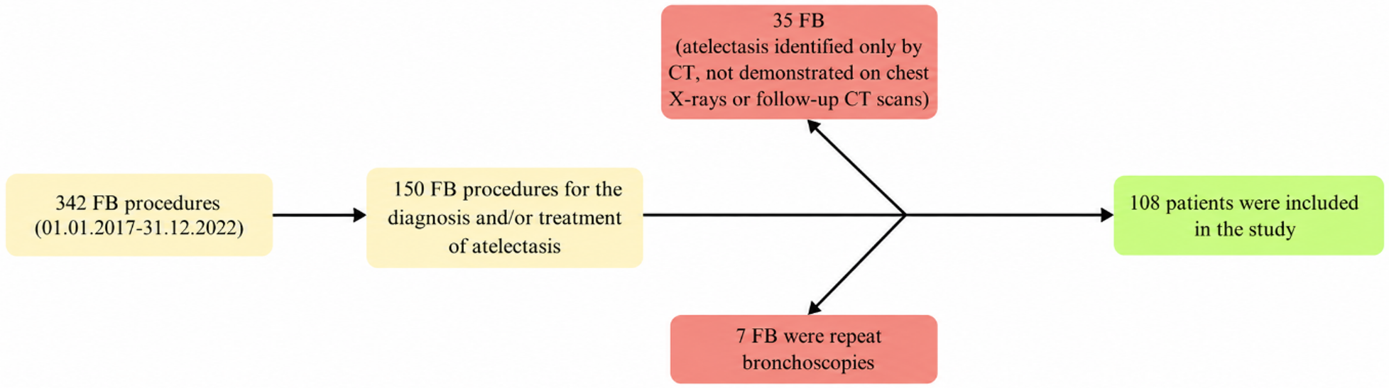
Between January 1, 2017, and December 31, 2022, a total of 342 FB procedures were performed at our pediatric pulmonology department. Of these, 150 (43.8%) were indicated for the diagnosis and/or management of atelectasis. After excluding 7 repeat procedures and 35 patients diagnosed solely via CT scans (due to the lack of appropriate CXR follow-up), 108 patients were included in the final analysis (Fig. 1).
The median age at the time of FB was 4.9 years (IQR: 2.5–8.8; range: 14 days to 18 years), with a slight female predominance (54.6%). Clinical symptoms were present in 95.4% of patients at admission, with dry cough being the most frequent (42.6%). Atelectasis was most frequently located in the right middle lobe (40.7%) and left lower lobe (40.7%). Radiologically, 51.9% of the cohort had additional findings accompanying atelectasis, most commonly inflammatory changes (consolidation/infiltration/ground-glass opacities) (33.3%). At the time of detection, the study population demonstrated marked clinical heterogeneity; 62% of patients had at least one underlying comorbidity. The most common underlying conditions were asthma (14.8%) and CHD (14.8%), followed by immunodeficiencies (8.3%), neuromuscular diseases (7.4%), and primary ciliary dyskinesia (PCD) (7.4%) (Table I). Based on the medical records at the time of initial presentation, patients with CHD exhibited a significantly higher rate of respiratory failure (62.5%, p<0.001), while those with neuromuscular diseases frequently presented with hypoxia (62.5%, p=0.004). These clinical manifestations were present concurrently with the initial radiological detection of atelectasis upon hospital admission.
|
PCD: Primary ciliary dyskinesia, CF: Cystic fibrosis, BPD: Bronchopulmonary dysplasia Some patients had more than one underlying disorder; therefore, subgroup counts are not mutually exclusive and may exceed the total number of patients with underlying disease. |
||
| Table I. Demographic and clinical characteristics of patients. | ||
| Variables |
|
|
| Sex | Female |
|
| Male |
|
|
| Radiological location of atelectasis | Right upper lobe of the lung |
|
| Right middle lobe of the lung |
|
|
| Right lower lobe of the lung |
|
|
| Left upper lobe of the lung |
|
|
| Left lower lobe of the lung |
|
|
| Right lung total |
|
|
| Left lung total |
|
|
| Pathological findings Accompanying atelectasis Radiologically | Number of patients with pathological findings |
|
| Radiographic inflammatory changes (consolidation/infiltration/ground-glass opacities) |
|
|
| Bronchiectasis |
|
|
| Chest deformity |
|
|
| Scoliosis |
|
|
| Dextrocardia |
|
|
| Symptoms at presentation with atelectasis | Number of patients with symptoms |
|
| Dry cough |
|
|
| High fever |
|
|
| Wet cough |
|
|
| Hypoxia |
|
|
| Respiratory failure |
|
|
| Wheezing |
|
|
| Respiratory distress |
|
|
| Other (fatigue, shortness of breath, runny nose, nasal congestion, difficulty swallowing) |
|
|
| Underlying disease at the time of atelectasis detection | Patients with underlying diseases |
|
| Asthma |
|
|
| Congenital heart disease |
|
|
| Immunodeficiency |
|
|
| Neuromuscular disease |
|
|
| PCD |
|
|
| CF |
|
|
| BPD |
|
|
| Wheezy infant |
|
|
Furthermore, the anatomical distribution of atelectasis demonstrated significant disease-specific radiological patterns. In patients diagnosed with asthma, the rate of right middle lobe atelectasis was statistically significantly higher (75.0%, p=0.006). Among patients diagnosed with CHD, the rate of total left lung atelectasis was statistically significantly higher (25.0%, p=0.026).
Bronchoscopic findings and therapeutic interventions
The median interval from radiological detection of atelectasis to FB was 55 days (IQR: 24.3–152.5; range: 1–2000 days). In our practice, FB is typically performed when atelectasis persists despite conservative management or when airway obstruction is suspected. FB revealed macroscopic pathological findings in 95.4% of patients. Purulent secretions, as a bronchoscopic finding suggestive of lower airway infection, were the most common macroscopic abnormality, observed in 72.2% of the cohort (Table II). Additional structural anomalies, such as bronchomalacia and tracheomalacia, were significantly more prevalent in patients with underlying CHD compared to those without (p=0.002).
|
FB: Flexible Bronchoscopy, BAL: Bronchoalveolar Lavage, MRSA: methicillin-resistant Staphylococcus aureus, MSSA: Methicillin-Sensitive Staphylococcus aureus, IV: Intravenous. Percentages for BAL culture results were calculated among patients with available BAL culture data (n=105). Percentages for bronchoscopic findings and post-bronchoscopy treatments were calculated using the total cohort as the denominator (n=108). |
||
| Table II. Bronchoscopic findings and post-bronchoscopy treatments in children undergoing flexible bronchoscopy for atelectasis. | ||
| Variables |
|
|
| Macroscopic findings during FB | Purulent secretions (bronchoscopic) |
|
| Bronchomalacia |
|
|
| Airway anomaly |
|
|
| Tracheomalacia |
|
|
| Mucus plug |
|
|
| Normal macroscopy |
|
|
| Foreign body |
|
|
| BAL culture | Growth in BAL culture |
|
| Haemophilus influenzae |
|
|
| Moraxella catarrhalis |
|
|
| Streptococcus pneumoniae |
|
|
| Pseudomonas aeruginosa |
|
|
| MSSA |
|
|
| Aspergillus |
|
|
| Candida |
|
|
| Klebsiella pneumoniae |
|
|
| Stenotrophomonas maltophilia |
|
|
| MRSA |
|
|
| Serratia marcescens |
|
|
| Additional treatments administered after FB | Number of patients receiving additional treatment |
|
| IV antibiotics |
|
|
| Oral antibiotics |
|
|
| Antireflux therapy |
|
|
| Inhaled ipratropium bromide |
|
|
BAL culture results were available in 105 patients, of whom 75/105 (71.4%) had positive cultures. Haemophilus influenzae was the most frequently isolated pathogen, detected in 23/105 patients (21.9%). Following FB, additional treatment was administered in 67/108 patients (62.0%), including intravenous antibiotics in 35/108 patients (32.4%) and oral antibiotics in 25/108 patients (23.1%) (Table II).
Resolution of atelectasis after flexible bronchoscopy and associated conditions
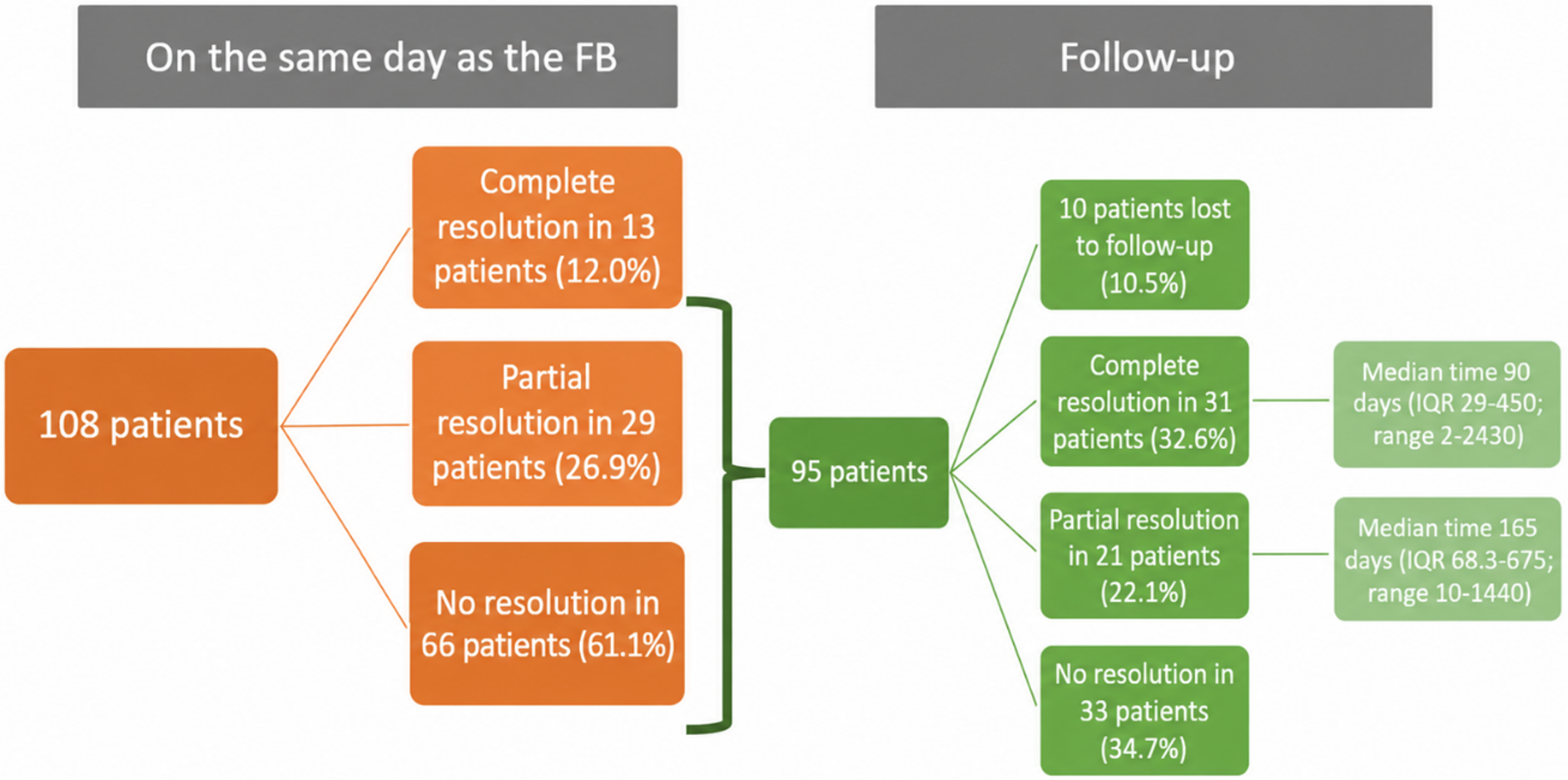
On the same day as the FB, complete resolution of atelectasis was observed in 12.0% of patients (n=13), partial resolution in 26.9% (n=29), and no resolution in 61.1% (n=66). The underlying diseases and FB findings of patients with partial or complete resolution are detailed in Table III.
| Values are presented as n (%). Percentages are calculated within each immediate radiographic response group. P values were calculated using the Pearson chi-square test. Some patients had more than one underlying disorder and/or more than one bronchoscopic finding; therefore, row totals may exceed the number of patients in a given subgroup. | ||||
| Table III. Immediate radiographic response of atelectasis after flexible bronchoscopy according to underlying disorders and bronchoscopic findings. | ||||
| Variables |
|
|
|
|
| Underlying disorders | ||||
| Asthma (n=16) |
|
|
|
|
| Congenital heart disease (n=16) |
|
|
|
|
| Immunodeficiency (n=9) |
|
|
|
|
| Neuromuscular disease (n=8) |
|
|
|
|
| Primary ciliary dyskinesia (n=8) |
|
|
|
|
| Cystic fibrosis (n=6) |
|
|
|
|
| Bronchopulmonary dysplasia (n=3) |
|
|
|
|
| Wheezy infant (n=3) |
|
|
|
|
| Bronchoscopic findings | ||||
| Purulent secretions (bronchoscopic) (n=78) |
|
|
|
|
| Bronchomalacia (n=25) |
|
|
|
|
| Airway anomaly (n=25) |
|
|
|
|
| Tracheomalacia (n=18) |
|
|
|
|
| Mucus plug (n=17) |
|
|
|
|
| Foreign body (n=4) |
|
|
|
|
| Normal macroscopy (n=5) |
|
|
|
|
Out of the 95 patients who did not achieve complete resolution on the same day as FB, 10 did not attend their initial outpatient follow-up appointments. Among the 85 patients who did not achieve complete resolution on the immediate post-FB CXR and had subsequent follow-up imaging, complete resolution was documented in 31/85 patients (36.5%), partial resolution in 21/85 patients (24.7%), and no resolution in 33/85 patients (38.8%).
The median time from FB to complete resolution was 90 days (IQR: 29–450; range: 2–2430 days), while the median time from FB to partial resolution was 165 days (IQR: 68.3–675; range: 10–1440 days). At final radiographic status, complete resolution was observed in 44/108 patients (40.7%), partial resolution in 30/108 patients (27.8%), and no resolution in 34/108 patients (31.5%). (Table III and Fig. 2). The final status of atelectasis in patients who missed their initial follow-up appointments was successfully determined based on other available medical records in the national database.
Twelve patients had an interval from radiologic detection to FB longer than 365 days. All had at least one underlying disorder, most commonly PCD (5/12). Macroscopic bronchoscopic abnormalities were present in 11/12 patients, most frequently purulent secretions (10/12). Regarding final radiographic outcomes, partial resolution was observed in 4/12 patients, whereas no radiographic resolution was observed in 8/12 patients. No patient in this subgroup achieved complete radiographic resolution. Initial univariate analyses revealed statistically significant differences in the time interval between the detection of atelectasis and the performance of FB. The interval from radiologic detection of atelectasis to FB was significantly shorter among patients who achieved partial or complete resolution at final radiographic status than among those with no resolution [median (IQR): 34 (17–121) vs 124 (49–330) days; p=0.001] (Table IV). Regarding other clinical factors, patients without additional pathological findings on CXR or CT had a significantly higher rate of complete resolution than those with additional pathological findings [28/51 (54.9%) vs 16/54 (29.6%); p=0.006]. Similarly, the complete resolution rate was higher among patients without scoliosis than among those with scoliosis [43/98 (43.9%) vs 1/7 (14.3%); p=0.034].
| Values are presented as median (IQR). Immediate radiographic response was defined as the status on the chest radiograph obtained within 24 hours after FB. Final radiographic status was defined as the status on the last available follow-up chest radiograph within the study period. P values were calculated using the Mann–Whitney U test. FB, flexible bronchoscopy; IQR, interquartile range. | |||||
| Table IV. Interval from radiologic detection of atelectasis to flexible bronchoscopy according to radiographic outcome. | |||||
| Outcome time point |
|
|
|
|
|
| Immediate radiographic response |
|
|
|
|
|
| Final radiographic status |
|
|
|
|
|
An exploratory Cox proportional hazards regression model was used to evaluate factors associated with time to complete radiographic resolution. The model included seven prespecified variables: the interval from radiologic detection of atelectasis to FB, the presence of macroscopic pathological findings during FB, CHD, PCD, immunodeficiency, asthma, and CF. The interval from radiologic detection to FB was entered into the model as a continuous variable and rescaled per 30-day increase to improve clinical interpretability. In the primary model, each 30-day increase in the interval from radiologic detection to FB was associated with a lower hazard of complete radiographic resolution (HR: 0.88, 95% CI: 0.80–0.98; p=0.018). CHD was also associated with a higher hazard of complete radiographic resolution (HR: 2.10, 95% CI: 1.05–4.20; p=0.036). Other variables in the model did not reach statistical significance.
In the sensitivity analysis excluding patients with an interval from radiologic detection to FB longer than 365 days, the association between the detection-to-FB interval and complete radiographic resolution was attenuated and was no longer statistically significant (HR per 30-day increase: 0.92, 95% CI: 0.81–1.04; p=0.169). In this restricted model, CHD remained associated with complete radiographic resolution (HR: 2.11, 95% CI: 1.05–4.23; p=0.035).
Finally, among patients who achieved partial or complete radiological resolution at their final follow-up, a statistically significant clinical improvement in oxygen saturation was observed. The mean oxygen saturation increased from 93.1% at the time of initial detection to 96.9% following resolution (p<0.001).
Recurrence and procedure safety
Atelectasis recurrence during the follow-up period was observed in 10 patients (9.3%), with a median time to recurrence of 49 days. The FB procedure demonstrated a favorable safety profile, with complications occurring in only 4 patients (3.7%). These included transient severe hypoxia in three patients with underlying CHD and mild bleeding in one patient, which necessitated the early termination of the procedure.
Discussion
This study describes the bronchoscopic findings, microbiological results, management changes, and radiographic outcomes of children who underwent FB for atelectasis in a tertiary pediatric pulmonology setting. The main finding was that FB provided clinically relevant information in most patients and was followed by management changes in a substantial proportion of the cohort. Partial or complete radiographic resolution was observed in more than two-thirds of patients at final radiographic status, although same-day resolution was less frequent. These findings suggest that, in selected children with persistent or clinically concerning atelectasis, FB may contribute to care not only through direct airway inspection and suctioning, but also by clarifying the underlying mechanism and guiding subsequent individualized treatment.
The timing-related findings should be interpreted cautiously. In the univariate analysis and in the primary exploratory Cox model, a longer interval from radiologic detection of atelectasis to FB was associated with a lower likelihood of complete radiographic resolution. However, this association was attenuated and was no longer statistically significant after excluding patients with intervals longer than 365 days. This suggests that the timing variable may partly reflect differences between acute atelectasis and chronic or recurrent atelectasis in medically complex patients rather than a direct causal effect of earlier FB alone. The subgroup with intervals longer than 365 days supports this interpretation: all had underlying disorders, most commonly primary ciliary dyskinesia, and none achieved complete radiographic resolution. Therefore, the Cox model findings should be considered exploratory and hypothesis-generating rather than definitive evidence that earlier FB independently determines resolution.
Our cohort’s clinical heterogeneity further underscores the necessity of understanding disease-specific mechanisms. We observed distinct anatomical patterns of atelectasis that were consistent with underlying morbidities. For instance, the higher frequency of total left lung atelectasis in patients with CHD may reflect mechanical compression. The close anatomical proximity of an enlarged left atrium or pulmonary artery in various CHD anomalies can externally compress the soft, cartilaginous left main bronchus of infants, leading to collapse and subsequent atelectasis.9-12 Correspondingly, we found significantly higher rates of structural airway anomalies (bronchomalacia, tracheomalacia) in this group. Furthermore, the predominant involvement of the right middle lobe in patients with asthma reflects the unique anatomical vulnerability of this lobe; its relatively narrow and long bronchus, combined with a sharp branching angle, makes it particularly susceptible to obstruction by the thick mucus plugs characteristic of asthmatic airway inflammation.13 Recognizing these distinct pathophysiological mechanisms emphasizes that pediatric atelectasis is not a single clinical entity; therefore, diverse etiologies should not be managed under a uniform therapeutic approach.
The bronchoscopic, microbiological, and radiographic findings also emphasize the practical value of FB in this population. Macroscopic pathological findings were present in most patients, and purulent secretions were the most frequent abnormality. BAL cultures were positive in a high proportion of patients with available culture data, with Haemophilus influenzae being the most commonly isolated pathogen. These findings are consistent with previous pediatric bronchoscopy series showing that FB can reveal infectious, obstructive, and structural contributors to persistent atelectasis.14-17 Same-day partial or complete radiographic resolution was observed in 42/108 patients (38.9%), which is lower than some previous reports describing immediate response rates as high as 87%.16,18 This difference may reflect the high clinical complexity and chronicity of our cohort. Nevertheless, partial or complete resolution increased to 74/108 patients (68.5%) at final radiographic status, and radiographic improvement was accompanied by a significant increase in oxygen saturation. Because conservative and post-procedural treatments were individualized, these improvements should not be attributed solely to the mechanical effect of FB. Rather, FB may be best viewed as a diagnostic and management-guiding procedure that can contribute to resolution through both direct airway clearance and subsequent targeted therapy. The overall complication rate was low; however, all episodes of transient severe hypoxia occurred in patients with CHD. Thus, children with CHD and other medically complex conditions warrant careful pre-procedural assessment, close monitoring, and subgroup-specific vigilance.19-21
This study has several limitations. Its retrospective, single-center design may introduce referral bias, and the cohort included only children who underwent FB; therefore, the findings cannot be generalized to all children with atelectasis or used to determine when FB should be performed compared with conservative management alone. Follow-up CXR were obtained at variable time points according to clinical judgment rather than a standardized imaging protocol, which may have introduced systematic bias into the time-to-resolution analysis. Radiographic assessment relied primarily on CXR and was performed by a single reviewer, although predefined objective criteria were used and the reviewer was blinded to procedural timing and subsequent outcomes. Conservative and post-procedural treatments were individualized, making it difficult to separate the direct mechanical effect of FB from the effect of bronchoscopy-guided medical therapy. Finally, the Cox model included a limited number of complete-resolution events, with an events-per-variable ratio of approximately 6.3; therefore, the multivariable findings should be interpreted as exploratory.
In conclusion, FB in selected children with atelectasis provides high bronchoscopic and microbiological yield, may guide clinically relevant management changes, and should be considered as a complementary diagnostic and management-guiding tool. Decisions regarding timing and expected benefit should remain individualized according to the patient’s underlying disease, clinical severity, and suspected mechanism of atelectasis.
Ethical approval
The study was approved by Hacettepe University Non-Interventional Clinical Research Ethics Committee (Decision No: 2021/19-20, Project No: GO 21/1231). The requirement for informed consent was waived due to the retrospective design.
Source of funding
The authors declare the study received no funding.
Conflict of interest
The authors declare that there is no conflict of interest.
References
- Peroni DG, Boner AL. Atelectasis: mechanisms, diagnosis and management. Paediatr Respir Rev 2000; 1: 274-278. https://doi.org/10.1053/prrv.2000.0059
- Carlsen KH, Smevik B. Atelectasis. In: Taussig LM, Landau LI, editors. Pediatric Respiratory Medicine. 2nd ed. Philadelphia: Mosby; 2008: 1005-1013. https://doi.org/10.1016/B978-032304048-8.50074-8
- Halter JM, Steinberg JM, Schiller HJ, et al. Positive end-expiratory pressure after a recruitment maneuver prevents both alveolar collapse and recruitment/derecruitment. Am J Respir Crit Care Med 2003; 167: 1620-1626. https://doi.org/10.1164/rccm.200205-435OC
- Eroğlu N, Kiper N. Atelektazi. In: Yurdakök M, editor. Yurdakök Pediatri. Ankara: Güneş Kitabevi; 2017: 2854.
- Wood RE, Gauderer MW. Flexible fiberoptic bronchoscopy in the management of tracheobronchial foreign bodies in children: the value of a combined approach with open tube bronchoscopy. J Pediatr Surg 1984; 19: 693-698. https://doi.org/10.1016/s0022-3468(84)80354-1
- Yüksel H, Bingöl A, Gülen F, editors. Pediatrik Fleksibl Bronkoskopi. Ankara: Ankara Nobel Tıp Kitabevleri; 2022.
- Duggan M, Kavanagh BP. Atelectasis. In: Wilmott RW, Boat TF, Bush A, et al., editors. Kendig & Chernick’s disorders of the respiratory tract in children. 8th ed. Philadelphia: Elsevier; 2012: 564-569. https://doi.org/10.1016/B978-1-4377-1984-0.00037-1
- Carlsen KH, Crowley S, Smevik B. Atelectasis. In: Wilmott RW, Deterding R, Li A, et al., editors. Kendig’s disorders of the respiratory tract in children. 9th ed. Philadelphia: Elsevier; 2019: 1027-1033. https://doi.org/10.1016/B978-0-323-44887-1.00070-5
- Lee SL, Cheung YF, Leung MP, Ng YK, Tsoi NS. Airway obstruction in children with congenital heart disease: assessment by flexible bronchoscopy. Pediatr Pulmonol 2002; 34: 304-311. https://doi.org/10.1002/ppul.10164
- Corno A, Giamberti A, Giannico S, et al. Airway obstructions associated with congenital heart disease in infancy. J Thorac Cardiovasc Surg 1990; 99: 1091-1098. https://doi.org/10.1016/s0022-5223(20)31466-5
- Sandhu G, Sharma D, Rajdev K, Habib S, El-Sayegh D. Bronchial obstruction caused by a dilated pulmonary artery. Cureus 2019; 11: e5354. https://doi.org/10.7759/cureus.5354
- Sachdev A, Chhawchharia R, Gupta D, Gupta N, Joshi R, Agarwal N. flexible fiber-optic bronchoscopy-directed interventions in children with congenital heart diseases. Indian J Crit Care Med 2020; 24: 340-343. https://doi.org/10.5005/jp-journals-10071-23419
- Soyer O, Ozen C, Cavkaytar O, Senyücel C, Dallar Y. Right middle lobe atelectasis in children with asthma and prognostic factors. Allergol Int 2016; 65: 253-258. https://doi.org/10.1016/j.alit.2015.12.002
- Zhang DJ, Zhao DY, Liang H, Tian M, Han Q. Application of flexible bronchoscopy in diagnosis and treatment of 104 children with pulmonary atelectasis. Zhonghua Er Ke Za Zhi 2010; 48: 767-770.
- Shin MY, Hwang JH, Chung EH, et al. Flexible bronchoscopic findings and therapeutic effects in atelectasis of children. J Korean Pediatr Soc 2002; 45: 1090-1096.
- IF-PUBMED-Talamoni HL, Pisapia ND, Buendía JA. Flexible fiberoptic bronchoscopy in children with persistent atelectasis: a case series report. Arch Argent Pediatr 2015; 113: e106-e108. https://doi.org/10.5546/aap.2015.e106
- Holmgren NL, Córdova M, Ortúzar P, Sánchez I. Role of flexible bronchoscopy in the re-expansion of persistent atelectasis in children. Arch Bronconeumol 2002; 38: 367-371. https://doi.org/10.1016/s0300-2896(02)75241-1
- Nussbaum E. Pediatric flexible bronchoscopy and its application in infantile atelectasis. Clin Pediatr (Phila) 1985; 24: 379-382. https://doi.org/10.1177/000992288502400703
- Field-Ridley A, Sethi V, Murthi S, Nandalike K, Li ST. Utility of flexible fiberoptic bronchoscopy for critically ill pediatric patients: a systematic review. World J Crit Care Med 2015; 4: 77-88. https://doi.org/10.5492/wjccm.v4.i1.77
- Sánchez I, Pesce C, Navarro H, Holmgren L, Bertrand P, Alvarez C. A ten years experience in flexible bronchoscopy in pediatric patients. Rev Med Chil 2003; 131: 1266-1272. https://doi.org/10.4067/S0034-98872003001100006
- Talamoni H, Buendia J, Serrud H, Morrone D, Langer L. Flexible fiberoptic bronchoscopy in children: a report on 8 years of experience. Am J Respir Crit Care Med 2014; 189: A4678.
Copyright and license
Copyright © 2026 The Author(s). This is an open access article distributed under the Creative Commons Attribution License (CC BY), which permits unrestricted use, distribution, and reproduction in any medium or format, provided the original work is properly cited.